Abstract
Background:
Sexual dimorphism refers to the differences in size, shape, etc., between males and females. The dentition's use in sex assessment has been explored and advocated owing to its strength and resistance to peri- and post-mortem insults.
Objectives:
The study evaluated permanent maxillary incisors and canines for sexual dimorphism and estimated the level of accuracy with which they could be used for sex determination.
Materials and Methods:
The study was conducted on 100 subjects (50 males, 50 females). The mesiodistal dimension of permanent maxillary incisors and canines was measured and the data were subjected to statistical analysis.
Result:
Univariate analysis revealed that all permanent maxillary incisors and canines exhibited larger mean values of mesiodistal dimension in males compared to females but only canines were found to be statistically significant for sexual dimorphism.
Conclusion:
The study showed maxillary canines exhibiting significant sexual dimorphism and can be used for sex determination along with other procedures.
Keywords: Anterior teeth, forensics odontology, mesiodistal dimension, sexual dimorphism
Introduction
Sex determination is one of the important parameters in forensic identification. Teeth being the central component of the masticatory apparatus of the skull are good sources of material for civil and medicolegal identification. Teeth provide resistance to damage in terms of bacterial decomposition and fire when rest of body is damaged beyond recognition which makes them valuable tool in forensic investigation.[1]
Sexual dimorphism refers to the systemic difference in the form (either in shape or size) between individuals of different sexes in the same species. Teeth of various species are known to exhibit sexual dimorphism.[2] The dentition in males is larger than in females in contemporary human populations.
Sex determination using dental features is primarily based upon the comparison of tooth dimensions in males and females, or upon the comparison of frequencies of nonmetric dental traits, like Carabelli's trait of upper molars. Mesiodistal and buccolingual diameters of the permanent tooth crown are the two most commonly used and researched features used in determining sex on the basis of dental measurements.[3]
Yuen et al. conducted a study on mesiodistal dimension of deciduous and permanent teeth of the Southern Chinese population and found that none of the primary teeth nor three of the permanent teeth were found to have significant sex differences in size. Percentage sexual dimorphism ranged from 0.06% to 1.97% for the primary teeth and from 0.36% to 5.27% for the permanent teeth.[4]
The aim of the present study was firstly to investigate whether there is any sexual dimorphism observed between mesiodistal (M-D) dimension of permanent maxillary incisors, canines, and secondly, the accuracy with which these could be employed for the determination of sex in population.
Materials and Methods
The study sample consisted of 100 dental students (50 males and 50 females) selected from D.J. College of Dental Sciences and Research, Modinagar belonging to North India, who were selected based on the following criteria:
Age-20-30 years.
Complete set of fully erupted teeth.
Peridontally healthy teeth.
Noncarious teeth.
Nonattrited and intact teeth.
Satisfactorily aligned maxillary teeth, no spacing or diastema, and no crowding.
No history or clinical evidence of crown restoration, orthodontic treatment, trauma.
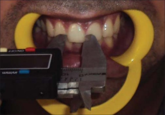
After obtaining informed consent, the maximum mesiodistal dimension of each tooth was measured between the anatomic contact points directly on the subject, with the help of a digital vernier caliper accurate to 0.01 mm (Mitutoyo Digital Caliper, Japan) held parallel to the occlusal plane. If it was difficult to place the vernier caliper, manual divider was used with very fine tips to measure the dimension; later we measured the divider distance with the same digital vernier caliper [Figure 1].
Figure 1.

Measuring the mesiodistal dimension of the maxillary right central incisor clinically on the patient with an electronic vernier caliper
All the measurements were done by a single examiner to eliminate interobserver error. Each reading was taken three times and the average of three values was obtained to minimize the intraobserver error. The data thus collected were subjected to statistical analysis. The SPSS software package version 17 was used for statistical analysis. The mean, range, and standard deviation were calculated for the size of the teeth. A two-sample t-test was used to test for statistical difference between means.
Results
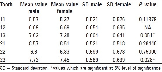
Table 1 shows detailed description of each tooth selected for study such as a mean value and standard deviation and P value both for males and females separately.
Table 1.
Mean and SD

Males showed greater mean mesiodistal dimensions for each tooth in comparison to females. Statistical analysis of permanent maxillary incisors and canines showed that the mesiodistal dimensions of only right and left maxillary canines were significantly different in males compared to those in females.
Several stepwise discriminant function statistics have been used to develop formulas to determine sex. The group centroids indicate the average discriminant scores for each sex.
Raw coefficients are the discriminant function coefficients used to calculate the discriminant score. To assess the sex, tooth dimensions are multiplied with the respective raw or unstandardized coefficients and added to the constant. If the values thus obtained were greater than the sectioning point the individual was considered a male and if less than the sectioning point the individual was considered female.
i.e., y = a + b (p1) +b(M2)
where a = constant of function between right and left maxillary canines, b = unstandardized coefficient of that particular tooth [Table 2].
Table 2.
Canonical discriminant function coefficient

Tables 3a–c shows the level of determining sex accurately in males and females when right and left maxillary canines are considered separately and in combination respectively.
Table 3a.
Accuracy of determination of sex using teeth 13

Table 3c.
Accuracy of determination of sex using teeth 13 and teeth 23

Table 3b.
Accuracy of determination of sex using teeth 23

When the level of accuracy for sex determination was measured using right maxillary canine separately, it was found that 44% females and 54% males were classified correctly whereas when the level of accuracy for sex determination was measured using left maxillary canine separately, 60% females and 60% males were classified correctly. When the level of accuracy for sex determination was measured using right and left maxillary canines together, it was found that 64% females and 58% males were classified correctly.
Table 4 shows the range of the measured mesiodistal dimension of right and left maxillary canines (which shows sexual dimorphism) for both males and females.
Table 4.
Percent of dimorphism

Percentage of dimorphism
The percentage of dimorphism is defined as the percentage by which the tooth size of males exceeds that of females. The percentage of dimorphism for each tooth was calculated using the following formula:
Percentage of dimorphism = {(Xm/Xf)–1} × 100
where Xm = mean male tooth dimension; Xf = mean female tooth dimension. Table 4 shows percent of dimorphism as observed for right and left maxillary canines.
Discussion
Gender determination in damaged and mutilated dead bodies or from skeletal remains constitutes the foremost step for identification in medico-legal examination and bioarcheology. Whenever it is possible to predict the sex, identification is simplified because then missing persons of only that sex need to be considered.[2]
Although the DNA profile gives accurate results yet measurement of linear dimensions such as arthopometric or odontometric parameters can be used for determination of sex in a large population because they are simple, reliable, inexpensive, and easy to measure.
Considering the fact that there are differences in odontometric features in specific populations, even within the same population in the historical and evolutional context, it is necessary to determine specific population values in order to make identification possible on the basis of dental measurements.[3] Thus the study evaluated mesiodistal dimension of permanent maxillary incisors and canines specific for males and females of the North Indian population.
Doris et al. have indicated that the early permanent dentitions provide the best sample for tooth size measurements because early adulthood dentition has less mutilation and less attrition in most individuals. Consequently, the effect of these factors on the actual mesiodistal tooth width would be minimum.[5] Thus only subjects in the 20-30 years’ age group were included in the study sample.
Various odontometric dimensions have been used for the purpose of sex estimation such as mandibular canine index,[6] buccolingual dimension of teeth,[7] and height of tooth.[8]
In this study, all the required dental measurements were taken directly on the subjects. As it was difficult to accurately measure the buccolingual width, of maxillary incisors, and canines, under indirect vision, only the mesio-distal width of these teeth was evaluated for sexual dimorphism.
Univariate analysis of the study showed that M-D dimensions of male dentition are greater than those of females which is in accordance with previous studies. Richardson et al. found that teeth of males tend to be larger than those of females for each type of tooth in both the arches.[9] Sanin and Savara reported differences in crown size patterns even among good occlusion cases.[10] Howe et al. in their study found combined mesiodistal width for males to be more compared to females.[11]
In this study, statistically significant dimorphism was exhibited by only two permanent maxillary anterior teeth, i.e., right and left maxillary canines. The Hashim and Murshid study in 1993 also showed that the canines were the only teeth to exhibit sexual dimorphism.[12]
Garn et al., studied the magnitude of sexual dimorphism by measuring the mesiodistal width of the canine teeth and showed that “the mandibular canine showed a greater degree of sexual dimorphism than the maxillary canine.”[13] However, Minzuno reported that maxillary canine showed a higher degree of sexual dimorphism compared to the mandibular canine in a Japanese population.[14]
Maxillary left canine in the study conducted by Pratibha et al. also exhibited a sexual dimorphism which is in accordance with the studies conducted on Turks by Iscan.[15] A study conducted by Otuyemi and Noar[16] shows dimorphism in maxillary canines bilaterally and another by Lund and Monstad[17] shows dimorphism of maxillary canine.
The multivariant analysis of the data showed that when combination of values for right and left maxillary canines was taken 64% females were classified correctly and 58% males were classified correctly. However the study conducted by Al-Rifaiy showed that an average of 65.5% individuals could be classified correctly.[18]
Various theories have been given to explain canine dimorphism.
According to Moss, it is because of the greater thickness of enamel in males due to the long period of amelogenesis compared to females.[19]
Because of Y chromosomes producing slower male maturation.[20]
The variation in the magnitude of dimorphism can be a result of various factors. Some authors have explained that such variation could be due to environmental influences on tooth size. Variation in food resources exploited by different populations has been explained as one such environmental cause. Others have suggested the interference of cultural factors with biological forces. There can be a complex interaction between a variety of genetic and environmental factors that are responsible for the variation in the magnitude of dimorphism. According to Garn et al., teeth have behaved in many ways through the course of evolution, ranging from reduction of the entire dentition to reduction of one group of teeth in relation to another.[21]
Conclusion
The study evaluated the use of a linear dimension (mesiodistal) of permanent maxillary incisors and canines because of simplicity and reliability. The study showed that right and left maxillary canines can be used for sex determination with 64% of accuracy in the case of females and 58% accuracy in the case of males. Thus this study indicates that maxillary canines show significant sexual dimorphism and can be used as an adjunct along with other accepted procedures for sex determination when fragmentary remains are encountered in mass disasters.
Footnotes
Source of Support: Nil
Conflict of Interest: None declared
References
- 1.Rao NG, Rao NN, Pai ML, Kotian MS. Mandibular canine index - a clue for establishing sex identity. Forensic Sci Int. 1989;42:249–54. doi: 10.1016/0379-0738(89)90092-3. [DOI] [PubMed] [Google Scholar]
- 2.Dahberg AA. Dental traits as identification tools. Dent Prog. 1963;3:155–60. [Google Scholar]
- 3.Iscan MY, Kedici PS. Sexual variation in bucco-lingual dimensions in Turkish dentition. Forensic Sci Int. 2003;137:160–4. doi: 10.1016/s0379-0738(03)00349-9. [DOI] [PubMed] [Google Scholar]
- 4.Yuen KK, So LL, Tang EL. Mesiodistal crown diameters of the primary and permanent teeth in Southern Chinese-a longitudinal study. Eur J Orthod. 1997;19:721–31. doi: 10.1093/ejo/19.6.721. [DOI] [PubMed] [Google Scholar]
- 5.Doris JM, Bernard BW, Kuftinec MM, Stom D. A biometric study of tooth size and dental crowding. Am J Orthod. 1981;79:326–36. doi: 10.1016/0002-9416(81)90080-4. [DOI] [PubMed] [Google Scholar]
- 6.Kaushal S, Patnaik VV, Sood V, Agnihotri G. Sex determination in north Indians using mandibular canine index. JIAFM. 2004;26:45–49. [Google Scholar]
- 7.Işcan MY, Kedici PS. Sexual variation in bucco-lingual dimensions in Turkish dentition. Forensic Sci Int. 2003;137:160–4. doi: 10.1016/s0379-0738(03)00349-9. [DOI] [PubMed] [Google Scholar]
- 8.Vodanović M, Demo Ž, Njemirovskij V, Keros J, Brkić H. Odontometrics: A useful method for sex determination in an archaeological skeletal population? J Archaeol Sci. 2007;34:905–13. [Google Scholar]
- 9.Richardson ER, Malhotra SK. Mesiodistal crown dimension of the permanent dentition of American Negroes. Am J Orthod. 1975;68:157–64. doi: 10.1016/0002-9416(75)90204-3. [DOI] [PubMed] [Google Scholar]
- 10.Sanin C, Savara BS. An analysis of permanent mesiodistal crown size. Am J Orthod. 1971;59:488–500. doi: 10.1016/0002-9416(71)90084-4. [DOI] [PubMed] [Google Scholar]
- 11.Howe RP, Mc Namara JA, Jr, O’Connor KA. An examination of dental crowding and its relationship to tooth size and arch dimension. Am J Orthod. 1983;83:363–73. doi: 10.1016/0002-9416(83)90320-2. [DOI] [PubMed] [Google Scholar]
- 12.Hashim MA, Murshid ZA. Mesiodistal tooth width: A comparision between Saudi males and females, part1. Egypt Dent. 1993;39:343–6. [PubMed] [Google Scholar]
- 13.Garn SN, Lewis AB, Swindler DR, Kerewsky RS. Genetic control of sexual dimorphism in tooth size. J Dent Res. 1967;46:963–72. doi: 10.1177/00220345670460055801. [DOI] [PubMed] [Google Scholar]
- 14.Minzuno O. Sex determination from maxillary canine by fourier analysis. Nihon Univ Dent J. 1990;2:139–42. [Google Scholar]
- 15.Iscan YM, Kedici SP. Sexual variation in buccolingual dimensions in Turkish dentition. Forensic Sci Int. 2003;137:160–4. doi: 10.1016/s0379-0738(03)00349-9. [DOI] [PubMed] [Google Scholar]
- 16.Otuyemi OD, Noar JH. A comparision of crown size dimensions of the permanent teeth in a Nigerian and British population. Eur J Ortho. 1996;18:623–8. doi: 10.1093/ejo/18.6.623. [DOI] [PubMed] [Google Scholar]
- 17.Lund H, Monstad H. Gender determination by odontometrics in Swedish population. J Forensic Odontostomatol. 1999;17:30–4. [PubMed] [Google Scholar]
- 18.Al-Rifaiy MQ, Abdullah MA, Ahraf I, Khan N. Dimorphism of mandibular and maxillary canine teeth in establishing sex identity. Saudi Dent J. 1997;9:17–20. [Google Scholar]
- 19.Vito CD, Sauders SR. A discriminant function analysis of deciduous teeth to determine sex. J Forensic Sci. 1990;35:845–58. [PubMed] [Google Scholar]
- 20.Acharya BA, Mainali S. Univariate sex dimorphism in the Nepalese dentition and use of discriminant functions in gender assessment. Forensic Sci Int. 2007;173:47–56. doi: 10.1016/j.forsciint.2007.01.024. [DOI] [PubMed] [Google Scholar]
- 21.Acharya BA. Sex determination potential of buccolingual and mesiodistal dimensions. J Forensic Sci. 2008;53:790–2. doi: 10.1111/j.1556-4029.2008.00778.x. [DOI] [PubMed] [Google Scholar]


