Abstract
Neutrophils play a key role in innate immunity, and the identification of new stimuli that stimulate neutrophil activity is a very important issue. In this study, we identified three novel peptides by screening a synthetic hexapeptide combinatorial library. The identified peptides GMMWAI, MMHWAM, and MMHWFM caused an increase in intracellular Ca2+ in a concentration-dependent manner via phospholipase C activity in human neutrophils. The three peptides acted specifically on neutrophils and monocytes and not on other non-leukocytic cells. As a physiological characteristic of the peptides, we observed that the three peptides induced chemotactic migration of neutrophils as well as stimulated superoxide anion production. Studying receptor specificity, we observed that two of the peptides (GMMWAI and MMHWFM) acted on formyl peptide receptor (FPR)1 while the other peptide (MMHWAM) acted on FPR2. Since the three novel peptides were specific agonists for FPR1 or FPR2, they might be useful tools to study FPR1- or FPR2-mediated immune response and signaling.
Keywords: calcium; chemotaxis; neutrophils; peptide library; peptides; receptors, formyl peptide
Introduction
Neutrophils play an important role in the regulation of immune responses, especially in the innate immune response (Kumar and Sharma, 2010). Neutrophils are recruited to infected areas or sites of injury according to a chemoattractant gradient (Borregaard, 2010; Kumar and Sharma, 2010). Recruited neutrophils can be activated by extracellular stimuli, resulting in the production of several reactive oxygen species (Dahlgren and Karlssson, 1999; Walther et al., 2000; Tiffany et al., 2001). It is a critical aspect of resting neutrophils that they must be activated for host defense. The activation of neutrophils can be induced by various different stimuli, including pathogen-derived molecules and several chemoattractants (Walther et al., 2000; Tiffany et al., 2001; Sabroe et al., 2003; Kobayashi, 2008). Among these, chemoattractants, including several chemokines that regulate the activities of neutrophils, have received much attention over the last decade. These chemoattractants bind to specific cell surface receptors, which are coupled to pertussis toxin (PTX)-sensitive G-protein(s), resulting in intracellular Ca2+ mobilization, cell migration, exocytosis, adhesion, and generation of bioactive lipids and/or reactive oxygen species (Walther et al., 2000; Tiffany et al., 2001; Kobayashi, 2008). Keeping in mind the important roles of chemoattractants in neutrophils, the identification of new chemoattractants and the characterization of their mechanisms of action are very much needed.
For the identification of new bioactive molecules, various different types of molecular diversities can be used. Positional scanning synthetic peptide combinatorial library (PS-SPCL), which is an easy and powerful tool for identifying peptide sequences in certain biological reactions, was developed by Houghten et al. (Houghten et al., 1991). Many groups have used this method for various purposes, including the identification of human immunodeficiency virus protease inhibitors, interleukin-8-specific antagonists, inhibitor for nuclear factor of activated T cells, and ligands for opioid receptors (Owens et al., 1991; Hayashi et al., 1995; Dooley et al., 1998; Aramburu et al., 1999). Further, we already identified several bioactive hexapeptide that stimulates superoxide anion production or arachidonic acid release by screening hexapeptide combinatorial libraries (Bae et al., 2001, 2003).
Here, we adopted the PS-SPCL method to identify novel peptides that can stimulate a Ca2+ increase in human neutrophils. We found that the peptides Gly-Met-Met-Trp-Ala-Ile-CONH2 (GMMWAI), Met-Met-His-Trp-Ala-Met-CONH2 (MMHWAM), and Met-Met-His-Trp-Phe-Met-CONH2 (MMHWFM) can stimulate human neutrophils, resulting in intracellular Ca2+ increase. We also investigated the functional roles of the peptides and the target receptors of these three peptides.
Results
Identification of peptides that stimulate Ca2+ increase in human neutrophils
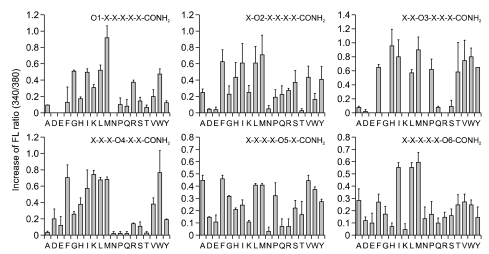
A total of 114 peptide pools (around 47 million peptides) from hexapeptide PS-SPCLs were screened to identify peptides that stimulate a Ca2+ increase in human neutrophils. As shown in Figure 1, we observed that each amino acid that was fixed at each position induced different levels of Ca2+ increase from the initial screening. The most active peptides at each position were as follows: Met (M) or Gly (G) in the 1st position, Met (M) in 2nd, His (H) or Met (M) in 3rd, Trp (W) in 4th, Ala (A) in 5th, and Met (M) or Ile (I) in 6th.
Figure 1.
Initial screening of PS-SPCLs for peptides stimulating intracellular Ca2+ increase in human neutrophils. Each panel shows the results obtained with the peptide pools with known amino acids at each of the six positions of the hexapeptide. The six positions were individually defined (O1, O2 etc.) by one of the 19 L-amino acids. The remaining five positions consist of mixtures (X) of the 19 L-amino acids (except for cysteine). Human neutrophils (1 × 10⁶ cells/assay) were used for each assay. Fluorescence ratio (340/38) was monitored as described under Methods. The results represent one of three independent experiments.
Peptides-induced Ca2+ increase is mediated via G-proteins and PLC
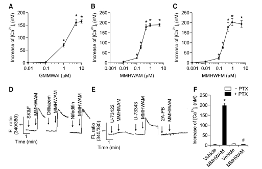
Based on the results of the initial screening of the peptide libraries, we synthesized three representative hexapeptides (GMMWAI, MMHWAM, and MMHWFM) and confirmed that stimulation of neutrophils with various concentrations of these three peptides induced a Ca2+ increase in a concentration-dependent manner with maximal activity of 0.5-5 µM (Figures 2A-2C).
Figure 2.
Effects of peptides on Ca2+ increase in human neutrophils. Fura-2-loaded human neutrophils were stimulated with various concentrations of GMMWAI, MMHWAM, and MMHWFM. The change in 340 nm/380 nm was monitored. The peak level of the increase in Ca2+ was monitored. Data are presented as means ± S.E. of four independent experiments (A-C). Fura-2-loaded human neutrophils were stimulated with 5 µM MMHWAM in the absence or presence of SK&F (10 µM), diltiazem (1 µM), nifidifin (1 µM), U-73122 (5 µM), U-73343 (5 µM), and 2A-PB (5 µM). The change in 340 nm/380 nm was monitored. The results are representative of three independent experiments (D, E). Human neutrophils were preincubated with or without 1 µg/ml of PTX for 4 h, after which fura-2 was loaded into the cells. Fura-2-loaded cells were stimulated with 5 µM MMHWAM. The peak level of the increase in Ca2+ was monitored. Data are presented as means ± S.E. of three independent experiments (F). *, P < 0.05, compared with the value obtained from the vehicle control; #, P < 0.05, significantly different from the -PTX control.
Intracellular Ca2+ increase can be induced by several different pathways. Firstly, the activation of some types of Ca2+ channels elicits intracellular Ca2+ increase in leukoyctic cells (Berridge, 1993; Burnashev, 1998; Zhu et al., 2010). Since we observed that the three novel peptides increased intracellular Ca2+ levels in human neutrophils, we examined the involvement of the cell surface Ca2+ channel. For this, we used several different Ca2+ channel-selective inhibitors. As shown in Figure 2D, MMHWAM-induced intracellular Ca2+ increases were not affected by preincubating human neutrophils with 1 µM nifedifine (voltage-sensitive L type Ca2+ channel inhibitor), 10 µM diltiazem (voltage-sensitive L type Ca2+ channel inhibitor), and 10 µM SK&F. These results indicate that MMHWAM increased Ca2+ concentration independent of the Ca2+ channel-dependent pathway in human neutrophils.
Another pathway for intracellular Ca2+ increase is mediated by the activation of PLC (Noh et al., 1995; Rhee, 2001). To determine the role of PLC in the MMHWAM-induced Ca2+ increase, we pretreated cells with a specific PLC inhibitor, U-73122, or with its inactive analogue, U-73343. As shown in Figure 2E, U-73122 but not U-73343 completely inhibited the MMHWAM-induced Ca2+ increase. 2-aminoethoxydiphenyl borate (2-APB), which is used to block IP3 receptor in cells (Maruyama et al., 1997), also completely inhibited the MMHWAM-induced Ca2+ increase in human neutrophils (Figure 2E). These results indicate that MMHWAM stimulated Ca2+ increase via PLC activation in human neutrophils. MMHWAM resulted in intracellular Ca2+ elevation not only in the presence of extracellular Ca2+ but also in the absence of extracellular Ca2+ (data not shown), supporting that the peptide induced Ca2+ increase via the activation of PLC in human neutrophils.
We also examined the effect of PTX, a specific inhibitor of Gi/o type G proteins, on the peptides-induced Ca2+ increase. When human neutrophils were preincubated with 1 µg/ml of PTX prior to stimulation with MMHWAM, the peptides-induced Ca2+ increase was almost completely inhibited (Figure 2F). These results indicate that MMHWAM stimulated Ca2+ increase via PTX-sensitive G proteins. We also observed that the other two peptides (GMMWAI and MMHWFM) stimulated Ca2+ increase via Gi protein and PLC but not the Ca2+ channel (data not shown).
Leukocyte-specific effects of the novel peptides
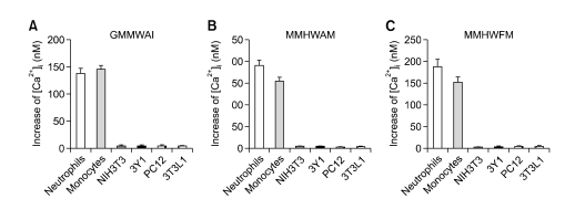
The fact that GMMWAI, MMHWAM, and MMHWFM stimulated human neutrophils led us to examine the effects of the peptides on other leukocytes such as monocytes. Stimulation of monocytes with the three peptides resulted in Ca2+ increase (Figure 3). The three peptides also enhanced Ca2+ levels in monocytes with a similar concentration dependency as observed for the Ca2+ increase (Figure 3 and data not shown). Next, we examined the effects of GMMWAI, MMHWAM, and MMHWFM on intracellular Ca2+ release in several non-leukocytic cell lines. NIH3T3 (NIH Swiss mouse embryo fibroblast), 3Y1 (Rat embryonic fibroblast), 3T3L1 (preadipocyte), and PC12 (rat adrenal pheochromocytoma) cells showed no response to the three peptides in terms of Ca2+ increase (Figure 3). From these results, we suggest that the effects of the three peptides were specific to human leukocytes.
Figure 3.
Cell specificity of the peptides. Prepared human primary monocytes, neutrophils, NIH3T3, 3Y1, 3T3L1, and PC12 cells were loaded with fura-2 and stimulated with three peptides at a concentration of 10 µM. The peak level of the Ca2+ increase was recorded. Data are presented are means ± S.E. from at least three independent experiments.
Chemotactic effects of the novel peptides on human neutrophils
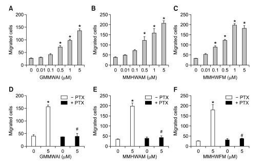
We demonstrated that the novel peptides stimulated Ca2+ increase in human neutrophils in a PTX-sensitive manner. Since peptides-induced and chemoattractant-induced neutrophil activation are similar to each other, we investigated whether or not the peptides exhibit chemotactic activity on human neutrophils. GMMWAI, MMHWAM, and MMHWFM induced migration of human neutrophils within a 0.1-5 µM concentration, showing concentration response curves (Figures 4A-4C). This implies that the three novel peptides induced chemotaxis in human neutrophils.
Figure 4.
Chemotactic effects of the peptides. Assays were performed using a modified Boyden chamber assay. Isolated human neutrophils (1 × 10⁶ cells/ml of serum-free RPMI 1640) were added to the upper wells of a 96-well chemotaxis chamber, and migration across the polycarbonate membrane with 3-µm pore size was assessed after 1.5 h of incubation at 37℃ (A-C). Human neutrophils were incubated with or without 1 µg/ml of PTX for 4 h. Then, the cells were used for chemotaxis assay using 5 µM GMMWAI, MMHWAM, or MMHWFM (D-F). The numbers of migrated cells were determined by counting them in high power field (400X). The data are presented as mean ± S.E. of three independent experiments, each performed in duplicate. *P < 0.05, compared with the value obtained from the vehicle control; #P < 0.05, significantly different from the -PTX control.
Various extracellular stimuli, including many chemoattractants, activate neutrophils via PTX-sensitive G-protein(s) (Badolato et al., 1995; Bae et al., 1999). Here, we also examined the involvement of PTX-sensitive G-protein(s) on peptide-induced neutrophil chemotaxis. Preincubation of human neutrophils with PTX (1 µg/ml) for 4 h caused complete inhibition of peptides-induced neutrophil chemotaxis (Figure 4D-F). The results indicate that GMMWAI, MMHWAM, and MMHWFM stimulated human neutrophil chemotaxis via PTX-sensitive G-protein(s).
MMHWAM and MMHWFM but not GMMWAI stimulate superoxide anion production from human neutrophils
In this study, we found that the three novel peptides stimulated not only intracellular Ca2+ increase but also chemotactic migration via PTX-sensitive G-protein(s). Neutrophils perform an important role in the innate immune response by producing a large amount of superoxide anion after stimulation with extracellular stimuli (Dahlgren and Karlsson, 1999). Superoxide anion is a well-known method of killing invading pathogens employed by human neutrophils (Rossi, 1986; Segal and Abo, 1993; Dahlgren and Karlsson, 1999). Here, we also tested the effects of the three novel peptides on the generation of superoxide anion from human neutrophils. Stimulation of human neutrophils with various concentrations of GMMWAI failed to induce superoxide anion production (Figure 5A). However, the other two novel peptides (MMHWAM and MMHWFM) strongly increased superoxide anion production from human neutrophils (Figures 5B and 5C).
Figure 5.
Effects of peptides on superoxide anion production in human neutrophils. Human neutrophils were stimulated with various concentrations of GMMWAI, MMHWAM, or MMHWFM, and the amount of generated superoxide was measured using cytochrome c reduction assay. The data are presented as mean ± S.E. of three independent experiments, each performed in duplicate. *P < 0.01 versus vehicle treatment.
Novel peptides stimulate formyl peptide receptor (FPR)1 or FPR2
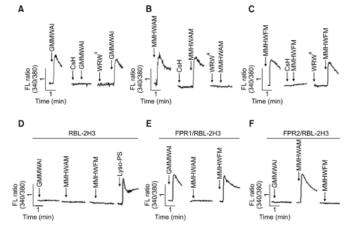
The three peptides showed similar effects on human neutrophils, in terms of Ca2+ increase and chemotactic migration via PTX-sensitive G-protein(s) (Figure 2F and data not shown). Formyl peptide receptors are representative chemoattractant receptors in human neutrophils (Ye et al., 2009). Here, we attempted to determine whether or not the three peptides acted through FPR1 and related receptors. For this purpose, we used FPR1 antagonist (CsH) (de Paulis et al., 1996) and FPR2 antagonist (WRW4) (Bae et al., 2004). As shown in Figures 6A and 6C, GMMWAI- and MMHWFM-induced Ca2+ increases were totally inhibited by CsH but not by WRW4. However, MMHWAM-induced Ca2+ increase was completely blocked by WRW4 but not by CsH (Figure 6B). These results suggest that GMMWAI and MMHWFM stimulated Ca2+ increases via FPR1 but not FPR2. On the other hand, MMHWAM stimulated a Ca2+ increase via FPR2 but not FPR1. We also used vector, FPR1-, or FPR2-expressing RBL-2H3 cells as previously reported (Lee et al., 2008). As shown in Figure 6E, stimulation of FPR1-expressing RBL-2H3 cells with the two novel peptides (GMMWAI and MMHWFM) elicited a dramatic increase in intracellular Ca2+. However, the two peptides did not induce an intracellular Ca2+ increase in vector- or FPR2-expressing RBL-2H3 cells (Figures 6D and 6F). These results strongly indicate that the two peptides (GMMWAI and MMHWFM) stimulated FPR1 but not FPR2, resulting in an increase in Ca2+. For MMHWAM, Ca2+ increase was observed in FPR2-expressing RBL-2H3 cells but not in FPR1-expressing RBL-2H3 cells (Figure 6E). The result indicates that MMHWAM acted through FPR2, increasing intracellular Ca2+.
Figure 6.
Role of FPR1 or FPR2 in novel peptide-induced Ca2+ increase. Isolated human neutrophils were incubated in the presence or absence of 10 µM CsH or WRW4 prior to Ca2+ measurement using 5 µM GMMWAI (A), 5 µM MMHWAM (B), or 5 µM MMHWFM (C). Vector- (D), FPR1- (E), or FPR2- (F) expressing RBL-2H3 cells (1 × 10⁶ cells/ml of serum-free RPMI 1640 medium) were stimulated with 5 µM GMMWAI, 5 µM MMHWAM, or 5 µM MMHWFM. The results represent one of two independent experiments.
Discussion
Since neutrophils perform important roles in early defense against invading pathogens and other harmful agents (Borregaard, 2010; Kumar and Sharma, 2010), the identification of agonists that enhance neutrophil function is of paramount importance. Here, we screened hexapeptide combinatorial libraries containing more than 47 million different peptide sequences, and we identified three novel hexapeptides (GMMWAI, MMHWAM, and MMHWFM) that stimulate intracellular Ca2+ increase in human neutrophils. GMMWAI and MMHWFM were shown to have selectivity on FPR1 but not on FPR2. In the case of MMHWAM, the peptide showed selectivity on FPR2 but not on FPR1.
Specific ligands are recognized by specific receptors, and this specificity results from a combination of subtle conformational and amino acid sequence differences. Previously some synthetic hexapeptides, WKYMVm, MFYLPM, HFYLPM, and HFYLPm, which act at two fMLF receptor subtypes have been described (Seo et al., 1997; Bae et al., 2001). In our previous reports, we demonstrated that the Y in the 3rd position of the hexapeptide and the M in the 6th position are important for stimulation of FPR family proteins (Bae et al., 2001). Here, we found that other peptides having different amino acids at the 3rd and 6th positions, such as H/M (for 3rd position) and I (for 6th position), showed stimulating activity on FPR family proteins. These results suggest that FPR1 has a broad spectrum of ligand recognition capacity. On the receptor specificity of MMHWAM and MMHWFM, we demonstrated that MMHWFM and MMHWAM acted on FPR1 and FPR2, respectively (Figures 6E and 6F). The result indicates that the 5th amino acid is important for the recognition of the peptides by FPR1 or FPR2.
Since GMMWAI, MMHWAM, and MMHWFM were identified by screening artificially synthesized peptides, we looked for sequence similarities between the novel peptides and known proteins by searching databases (SWISS-PROT and TrEMBL), and found that several bacterial proteins such as aminoacyltransferase femB contained the MMHWFM sequence. At this time, we cannot attach any significance to this sequence homology between our novel peptides and bacterial proteins. However, since FPRs recognize certain patterns derived from pathogens, the information that our peptides carry certain bacterial sequences may provide us useful insights into understanding the pattern involved in the recognition of pathogens.
Even though various chemokines and chemoattractants have been identified, only a few short peptides acting on human leukocytes have been known until now. The previously reported peptides WKYMVm (Seo et al., 1997) and fMLF (Spilberg et al., 1981) have been useful tools for studying neutrophil activation. Here, we demonstrated that the novel peptides GMMWAI, MMHWAM, and MMHWFM stimulate human neutrophils by acting on FPR1 or FPR2 (Figure 6). Therefore, these three peptides can also be used as agonists to study neutrophil activation and signaling.
Methods
Materials
Peripheral blood mononuclear cell (PBMC) separation medium (Histopaque-1077) and cytochrome c were purchased from Sigma (St. Louis, MO). Fura-2 pentaacetoxy-methylester (fura-2/AM) was purchased from Molecular Probes (Eugene, OR). PTX was purchased from Calbiochem (San Diego, CA).
Isolation of leukocytes
The Institutional Review Board (IRB) of Ajou University Hospital specifically approved this study (approval ID: AJIRB-MED-SMP-10-074). We obtained written informed consent from all participants involved in this study. Peripheral blood leukocytes were isolated from healthy donors. Human neutrophils were isolated according to the standard procedures of dextran sedimentation, hypotonic lysis of erythrocytes, and a lymphocyte separation medium gradient as described previously (Bae et al., 2001). PBMCs were separated on a Histopaque-1077 gradient. After two washings with HBSS without Ca2+ and Mg2+, the PBMCs were suspended in 10% FBS containing RPMI 1640 and incubated for 60 min at 37℃ to allow attachment of monocytes to the culture dish. The cells were washed five times with warm RPMI medium to washout lymphocytes, after which the attached monocytes were collected as described previously (Bae et al., 2001). The isolated human leukocytes were then used promptly.
Cell culture
NIH3T3 (NIH Swiss mouse embryo fibroblasts), 3Y1 (Rat embryonic fibroblasts), 3T3L1 (preadipocytes), and PC12 (rat adrenal pheochromocytoma) cells were obtained from the American Type Culture Collection (Rockville, MD) and maintained as recommended. The cells were maintained at about 1 × 10⁶ cells/ml under standard incubator conditions (humidified atmosphere, 95% air, 5% CO2, 37℃).
Initial screening of PS-SPCLs and measurement of intracellular Ca2+ increase
The hexapeptide libraries were obtained from the Peptide Library Support Facility of Pohang University of Science and Technology as described previously (Bae et al., 2001). Finally, 114 peptide pools (Cys was excluded in the construction of the libraries) were individually dissolved in water at a final concentration of 27 nM per peptide sequence in each pool. For initial screening of the PS-SPCLs, we measured the level of [Ca2+]i by using Grynkiewicz's method with fura-2/AM (Grynkiewicz et al., 1985). Briefly, prepared cells were incubated with 3 µM fura-2/AM at 37℃ for 50 min in fresh serum-free RPMI 1640 medium under continuous stirring. Then, 2 × 10⁶ cells were aliquoted for each assay in Ca2+-free Locke's solution (154 mM NaCl, 5.6 mM KCl, 1.2 mM MgCl2, 5 mM HEPES, pH 7.3, 10 mM glucose, and 0.2 mM EGTA) and incubated with the indicated concentrations of peptide pools (final 50 pM per peptide sequence for the initial screening). Fluorescence changes at the dual excitation wavelengths of 340 nm and 380 nm and the emission wavelength of 500 nm were measured, and the calibrated fluorescence ratio was translated into [Ca2+]i.
Chemotaxis assay
Chemotaxis assays were performed using multiwell chambers (Neuroprobe Inc., Gaithersburg, MD) (Bae et al., 2001). Briefly, prepared human neutrophils were suspended in RPMI 1640 at a concentration of 1 × 10⁶ cells/ml, and 25 µl of the suspension was placed into the upper well of a chamber separated by a 3 µm polyhydrocarbon filter from the lower well containing peptides. After incubation for 90 minutes at 37℃, non-migrated cells were removed by scarping, and cells that migrated across the filter were dehydrated, fixed, and stained with hematoxylin (Sigma, St. Louis, MO). Stained cells in five randomly chosen high power fields (400 X) in the well were then counted (Bae et al., 2001).
Measurement of superoxide anion production
Superoxide anion generation was determined by measuring cytochrome c reduction using a microtiter 96-well plate ELISA reader (Bio-Tek instruments, EL312e, Winooski, VT), as previously described (Bae et al., 2001). Human neutrophils (2 × 10⁶ cells in RPMI 1640 medium) were preincubated with 50 µM cytochrome c at 37℃ for 5 min and subsequently incubated with each peptide. Superoxide generation was determined by measuring light absorption changes at 550 nm over 5 min at 1 min intervals.
Statistical analysis
The results are expressed as mean ±S. E. of data obtained from the indicated number of experiments performed. Statistical significance was determined using the Student t test.
Acknowledgements
This research was supported by the Basic Science Research Program through the National Research Foundation of Korea (NRF) funded by the Ministry of Education, Science and Technology (2010-0020940), by National Research Foundation of Korea (NRF) grant funded by the Korean government (MEST) (No. 2009 0093198), and Samsung Research Fund, Sungkyunkwan University, 2011.
Abbreviations
- FPR
formyl peptide receptor
- GMMWAI
Gly-Met-Met-Trp-Ala-Ile-CONH2
- IP3
inositol-1,4,5-triphosphate
- MMHWAM
Met-Met-His-Trp-Ala-Met-CONH2
- MMHWFM
Met-Met-His-Trp-Phe-Met-CONH2
- PLC
phospholipase C
- PS-SPCL
positional scanning synthetic peptide combinatorial library
- PTX
pertussis toxin
References
- 1.Aramburu J, Yaffe MB, Lopez-Rodriguez C, Cantley LC, Hogan PG, Rao A. Affinity-driven peptide selection of an NFAT inhibitor more selective than cyclosporin A. Science. 1999;285:2129–2133. doi: 10.1126/science.285.5436.2129. [DOI] [PubMed] [Google Scholar]
- 2.Badolato R, Johnston JA, Wang JM, McVicar D, Xu LL, Oppenheim JJ, et al. Serum amyloid A induces calcium mobilization and chemotaxis of human monocytes by activating a pertussis toxin-sensitive signaling pathway. J Immunol. 1995;155:4004–4010. [PubMed] [Google Scholar]
- 3.Bae YS, Kim Y, Kim Y, Kim JH, Suh PG, Ryu SH. Trp-Lys-Tyr-Met-Val-D-Met is a chemoattractant for human phagocytic cells. J Leukoc Biol. 1999;66:915–922. doi: 10.1002/jlb.66.6.915. [DOI] [PubMed] [Google Scholar]
- 4.Bae YS, Bae H, Kim Y, Lee TG, Suh PG, Ryu SH. Identification of novel chemoattractant peptides for human leukocytes. Blood. 2001;97:2854–2862. doi: 10.1182/blood.v97.9.2854. [DOI] [PubMed] [Google Scholar]
- 5.Bae YS, Park EY, Kim Y, He R, Ye RD, Kwak JY, et al. Novel chemoattractant peptides for human leukocytes. Biochem Pharmacol. 2003;66:1841–1851. doi: 10.1016/s0006-2952(03)00552-5. [DOI] [PubMed] [Google Scholar]
- 6.Bae YS, Lee HY, Jo EJ, Kim JI, Kang HK, Ye RD, et al. Identification of peptides that antagonize formyl peptide receptor-like 1-mediated signaling. J Immunol. 2004;173:607–614. doi: 10.4049/jimmunol.173.1.607. [DOI] [PubMed] [Google Scholar]
- 7.Berridge MJ. Inositol trisphosphate and calcium signaling. Nature. 1993;361:315–325. doi: 10.1038/361315a0. [DOI] [PubMed] [Google Scholar]
- 8.Borregaard N. Neutrophils, from marrow to microbes. Immunity. 2010;33:657–670. doi: 10.1016/j.immuni.2010.11.011. [DOI] [PubMed] [Google Scholar]
- 9.Burnashev N. Calcium permeability of ligand-gated channels. Cell Calcium. 1998;24:325–332. doi: 10.1016/s0143-4160(98)90056-2. [DOI] [PubMed] [Google Scholar]
- 10.Dahlgren C, Karlsson A. Respiratory burst in human neutrophils. J Immunol Methods. 1999;232:3–14. doi: 10.1016/s0022-1759(99)00146-5. [DOI] [PubMed] [Google Scholar]
- 11.de Paulis A, Ciccarelli A, de Crescenzo G, Cirillo R, Patella V, Marone G. Cyclosporin H is a potent and selective competitive antagonist of human basophil activation by N-formyl-methionyl-leucyl-phenylalanine. J Allergy Clin Immunol. 1996;98:152–164. doi: 10.1016/s0091-6749(96)70237-3. [DOI] [PubMed] [Google Scholar]
- 12.Dooley CT, Ny P, Bidlack JM, Houghten RA. Selective ligands for the mu, delta, and kappa opioid receptors identified from a single mixture based tetrapeptide positional scanning combinatorial library. J Biol Chem. 1998;273:18848–18856. doi: 10.1074/jbc.273.30.18848. [DOI] [PubMed] [Google Scholar]
- 13.Hayashi S, Kurdowska A, Miller EJ, Albright ME, Girten BE, Cohen AB. Synthetic hexa- and heptapeptides that inhibit IL-8 from binding to and activating human blood neutrophils. J Immunol. 1995;154:814–824. [PubMed] [Google Scholar]
- 14.Houghten RA, Pinilla C, Blondelle SE, Appel JR, Dooley CT, Cuervo JH. Generation and use of synthetic peptide combinatorial libraries for basic research and drug discovery. Nature. 1991;354:84–86. doi: 10.1038/354084a0. [DOI] [PubMed] [Google Scholar]
- 15.Kobayashi Y. The role of chemokines in neutrophil biology. Front Biosci. 2008;13:2400–2407. doi: 10.2741/2853. [DOI] [PubMed] [Google Scholar]
- 16.Grynkiewicz G, Poenie M, Tsien RY. A new generation of Ca2+ indicators with greatly improved fluorescence properties. J Biol Chem. 1985;260:3440–3450. [PubMed] [Google Scholar]
- 17.Kumar V, Sharma A. Neutrophils: Cinderella of innate immune system. Int Immunopharmacol. 2010;10:1325–1334. doi: 10.1016/j.intimp.2010.08.012. [DOI] [PubMed] [Google Scholar]
- 18.Lee HY, Kim SD, Shim JW, Lee SY, Lee H, Cho KH, et al. Serum amyloid A induces CCL2 production via formyl peptide receptor-like 1-mediated signaling in human monocytes. J Immunol. 2008;181:4332–4339. doi: 10.4049/jimmunol.181.6.4332. [DOI] [PubMed] [Google Scholar]
- 19.Maruyama T, Kanaji T, Nakade S, Kanno T, Mikoshiba K. 2APB, 2-aminoethoxydiphenyl borate, a membrane-penetrable modulator of Ins(1,4,5)P3-induced Ca2+ release. J Biochem. 1997;122:498–505. doi: 10.1093/oxfordjournals.jbchem.a021780. [DOI] [PubMed] [Google Scholar]
- 20.Noh DY, Shin SH, Rhee SG. Phosphoinositide-specific phospholipase C and mitogenic signaling. Biochim Biophys Acta. 1995;1242:99–113. doi: 10.1016/0304-419x(95)00006-0. [DOI] [PubMed] [Google Scholar]
- 21.Owens RA, Gesellchen PD, Houchins BJ, DiMarchi RD. The rapid identification of HIV protease inhibitors through the synthesis and screening of defined peptide mixtures. Biochem Biophys Res Commun. 1991;181:402–408. doi: 10.1016/s0006-291x(05)81433-0. [DOI] [PubMed] [Google Scholar]
- 22.Rhee SG. Regulation of phosphoinositide-specific phospholipase C. Annu Rev Biochem. 2001;70:281–312. doi: 10.1146/annurev.biochem.70.1.281. [DOI] [PMC free article] [PubMed] [Google Scholar]
- 23.Rossi F. The O2- -forming NADPH oxidase of the phagocytes: nature, mechanisms of activation and function. Biochim Biophys Acta. 1986;853:65–89. doi: 10.1016/0304-4173(86)90005-4. [DOI] [PubMed] [Google Scholar]
- 24.Sabroe I, Prince LR, Jones EC, Horsburgh MJ, Foster SJ, Vogel SN, et al. Selective roles for Toll-like receptor (TLR)2 and TLR4 in the regulation of neutrophil activation and life span. J Immunol. 2003;170:5268–5275. doi: 10.4049/jimmunol.170.10.5268. [DOI] [PubMed] [Google Scholar]
- 25.Segal AW, Abo A. The biochemical basis of the NADPH oxidase of phagocytes. Trends Biochem Sci. 1993;18:43–47. doi: 10.1016/0968-0004(93)90051-n. [DOI] [PubMed] [Google Scholar]
- 26.Seo JK, Choi SY, Kim Y, Baek SH, Kim KT, Chae CB, et al. A peptide with unique receptor specificity: stimulation of phosphoinositide hydrolysis and induction of superoxide generation in human neutrophils. J Immunol. 1997;158:1895–1901. [PubMed] [Google Scholar]
- 27.Spilberg I, Mehta J, Daughaday C, Simchowitz L. Determination of a specific receptor for formyl-methionyl-leucyl-phenylalanine on th pulmonary alveolar macrophage and its relationship to chemotaxis and superoxide production. J Lab Clin Med. 1981;97:602–609. [PubMed] [Google Scholar]
- 28.Tiffany HL, Lavigne MC, Cui YH, Wang JM, Leto TL, Gao JL, et al. Amyloid-beta induces chemotaxis and oxidant stress by acting at formylpeptide receptor 2, a G protein-coupled receptor expressed in phagocytes and brain. J Biol Chem. 2001;276:23645–23652. doi: 10.1074/jbc.M101031200. [DOI] [PubMed] [Google Scholar]
- 29.Walther A, Riehemann K, Gerke V. A novel ligand of the formyl peptide receptor: annexin I regulates neutrophil extravasation by interacting with the FPR. Mol Cell. 2000;5:831–840. doi: 10.1016/s1097-2765(00)80323-8. [DOI] [PubMed] [Google Scholar]
- 30.Ye RD, Boulay F, Wang JM, Dahlgren C, Gerard C, Parmentier M, et al. International Union of Basic and Clinical Pharmacology. LXXIII. Nomenclature for the formyl peptide receptor (FPR) family. Pharmacol Rev. 2009;61:119–161. doi: 10.1124/pr.109.001578. [DOI] [PMC free article] [PubMed] [Google Scholar]
- 31.Zhu MX, Ma J, Parrington J, Calcraft PJ, Galione A, Evans AM. Calcium signaling via two-pore channels: local or global, that is the question. Am J Physiol Cell Physiol. 2010;298:C430–C441. doi: 10.1152/ajpcell.00475.2009. [DOI] [PMC free article] [PubMed] [Google Scholar]


