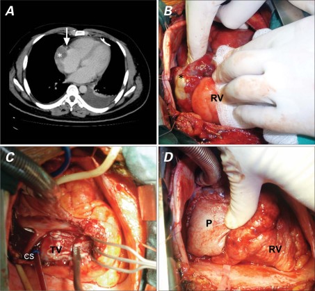
A 33-year-old man underwent echocardiographically guided decompression after presenting with cardiac tamponade in association with a bloody pericardial effusion. Chest computed tomography revealed an ill-defined heterogenous mass in the right atrium (Fig. 1A). Emergent surgery was performed after fresh blood gushed once again from the pericardial drain.
Fig. 1 A) Preoperative contrast-enhanced axial computed tomographic scan shows an irregular low-attenuation mass (asterisk) (55 × 27 mm) on the right atrium. Note the right coronary artery (arrow) encased in the atrioventricular groove. Intraoperative photographs show B) a discolored surface with active bleeding at the right atrium (asterisk), C) complete removal of the right atrial mass along with the atrioventricular groove, and D) reconstruction of the right atrium using bovine pericardium (P). CS = coronary sinus; RV = right ventricle; TV = tricuspid valve
The right atrial surface was occupied by a discolored mass, was ruptured, and was actively bleeding (Fig. 1B). The tumor was totally excised in such a manner that the margin included the partially normal right atrial wall (Fig. 1C). Right atrial reconstruction was completed with use of bovine pericardium (Fig. 1D). The gross surgical specimen (Fig. 2A) was diffusely infiltrated by a necrotic tumor with hemorrhage (Fig. 2B), compatible with malignancy. The bloody pericardial effusion was positive for malignancy. Photomicrographs of right atrial tissue samples showed high-grade angiosarcoma (Figs. 2C and 2D). Adjuvant therapy with docetaxel and gemcitabine was performed as soon as the patient recovered from surgery. Fourteen months after surgery, he was alive but had brain and lung metastases.
Fig. 2 Pathologic photographs. A) In gross examination, a 6 × 5.8 × 3.5-cm sample of resected right atrium is seen to be diffusely infiltrated by elastic and dark-brown tumor, marked by hemorrhage and necrosis. Hematoxylin and eosin staining shows B) tumor necrosis with hemorrhage (orig. ×5) and C) oval or plump spindle cells with slit-like or irregular vascular channels (orig. ×200). D) This tissue sample shows strong positive immunostaining for CD34, compatible with angiosarcoma (orig. ×200).
Comment
Malignant cardiac tumors, including angiosarcoma, are rare and are usually fatal. Primary cardiac angiosarcomas—such as our patient's tumor—are highly aggressive and locally invasive; fewer than half of the patients survive to 1 year.1–3 The tumor usually arises from the right atrium, with nonspecific symptoms and signs.4 Very rarely, the tumor presents with rupture, which leads to hemopericardium, cardiac tamponade, and a poor prognosis.5,6 Complete removal of the tumor and cardiac reconstruction is the mainstay of treatment and confers the best long-term outcome. In our patient, the surgery also controlled the bleeding and prevented death from cardiac tamponade; further, it provided a tissue specimen for diagnosis. The first cycle of adjuvant chemotherapy, with a combination of docetaxel and gemcitabine, provided more than a year of survival. Complete resection is of course the primary goal of the cardiac surgeon in such cases. However, even palliative cardiac resection can, with adjuvant therapy, increase the length of survival and improve the quality of life.
Footnotes
Address for reprints: Yao-Kuang Huang, MD, Division of Cardiac Surgery, Chang Gung Memorial Hospital, Chiayi Branch, No.6, W. Sec., Jiapu Rd., Puzi City, Chiayi County 61363, Taiwan, ROC
E-mail: huang137@mac.com
Section Editor: Raymond F. Stainback, MD, Department of Adult Cardiology, Texas Heart Institute at St. Luke's Episcopal Hospital, 6624 Fannin St., Suite 2480, Houston, TX 77030
References
- 1.Truong PT, Jones SO, Martens B, Alexander C, Paquette M, Joe H, et al. Treatment and outcomes in adult patients with primary cardiac sarcoma: the British Columbia Cancer Agency experience. Ann Surg Oncol 2009;16(12):3358–65. [DOI] [PubMed]
- 2.Gualis J, Castano M, Gomez-Plana J, Mencia P, Alonso D. Palliative surgical treatment of primary cardiac angiosarcoma complicated with recurrent tamponade. J Surg Oncol 2009; 100(8):744. [DOI] [PubMed]
- 3.Kim CH, Dancer JY, Coffey D, Zhai QJ, Reardon M, Ayala AG, Ro JY. Clinicopathologic study of 24 patients with primary cardiac sarcomas: a 10-year single institution experience. Hum Pathol 2008;39(6):933–8. [DOI] [PubMed]
- 4.Putnam JB Jr, Sweeney MS, Colon R, Lanza LA, Frazier OH, Cooley DA. Primary cardiac sarcomas. Ann Thorac Surg 1991;51(6):906–10. [DOI] [PubMed]
- 5.Koh TW, Kelly P, Timmis AD. Images in cardiology: Artefactual coronary artery lesions caused by effect of guidewire on tortuous coronary arteries: angiographic appearances during right coronary angioplasty. Heart 2001;86(6):655. [DOI] [PMC free article] [PubMed]
- 6.Sakaguchi M, Minato N, Katayama Y, Nakashima A. Cardiac angiosarcoma with right atrial perforation and cardiac tamponade. Ann Thorac Cardiovasc Surg 2006;12(2):145–8. [PubMed]


