Abstract
Purpose
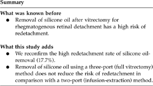
To assess the outcome of silicone oil removal after rhegmatogenous retinal detachment (RRD) surgery, and to compare results of a two-port (infusion-extraction) versus a three-port (full vitrectomy) approach.
Methods
Primary outcome measure was the rate of redetachment. Secondary outcome measures were visual acuity, rate of intraoperative and postoperative epiretinal membrane removal and complications.
Results
We included 147 consecutive cases. There were 15 cases of giant retinal tear, 26 cases of RRD without proliferative vitreoretinopathy (PVR) and 106 cases of RRD with PVR. The overall redetachment rate after silicone oil removal was 17.7%. In the group treated with the two-port technique (n=95), the retina redetached in 16 cases (16.8%), and in the group treated with the three-port technique (n=52), redetachment occurred in 10 cases (19.2%). This difference was not statistically significant (P=0.717; χ 2-test). There was a significantly higher redetachment rate in cases with a short oil tamponade duration of <2 months.
Conclusion
We reconfirm a relatively high redetachment rate after silicone oil removal. The risk of redetachment is not lower with the three-port compared with the two-port approach.
Keywords: retinal detachment, silicone oil, vitrectomy
Introduction
The role of silicone oil as a tamponade agent for complex rhegmatogenous retinal detachments (RRD) is well established.1, 2 The risk of redetachment in these complex cases is relatively high. Redetachment can occur under silicone oil tamponade, but more frequently, it occurs after removal of the silicone oil. Silicone oil can be extracted with different methods. We used to employ the two-port extraction method as our standard procedure. Recently, we have abandoned this method in favour of the three-port vitrectomy method. Potential advantages of the latter approach are better internal search for breaks, and ability to assess and treat epiretinal membranes (ERMs) causing tangential retinal traction. The present study was undertaken to compare the results of these two approaches.
Methods
The medical charts of patients that underwent silicone oil-extraction between June 2007 and June 2010 and had a minimum follow-up of 3 months were reviewed. We selected only those cases where the retina was fully attached and no clinically apparent macular pucker was visible at the last visit prior to the extraction. All patients were operated on at the Academic Medical Center, Amsterdam, a tertiary academic referral centre. The study was conducted in accordance with the recommendations of the Declaration of Helsinki and was approved by the institutional review board of the University of Amsterdam.
The main outcome measure was the rate of redetachment. Secondary outcome measures were rate of postoperative pucker formation and incidence of other complications. Parameters retrieved were patient characteristics, visual acuity (VA) before the preceding RRD operation, VA at final follow-up, duration of silicone oil tamponade, number of previous RRD surgeries, rate of intraoperative peeling, and postoperative complications.
In the two-port technique, two standard sclerotomies were made over the temporal pars plana after opening of the conjunctiva. One was fitted with a standard infusion line connected to the bottle containing BSS-plus (Alcon Laboratories, Fort Worth, TX, USA) and positioned at 40 cm above eye level, which corresponds to 30 mm Hg. Silicone oil was extracted through the second sclerotomy using a 20-gauge angiocatheter with its end cut-short on a shallow bevel. Extraction was performed manually using a 10-ml syringe attached to the angiocatheter. After removal of the oil, droplets of oil were typically removed from the anterior chamber through a corneal incision. After closure of the sclerotomies, the retinal periphery was checked using indirect ophthalmoscopy.
In the three-port technique, a standard three-port vitrectomy was performed with the Alcon Accurus or Constellation (Alcon Laboratories) and BIOM (binocular indirect ophthalmol microscope) wide-angle viewing system (Oculus, Inc., Wetzlar, Germany). Infusion pressure was set at 30 mm Hg and the silicone oil was extracted using the machine driven oil-extraction set (Alcon Laboratories) with the vacuum set at 600 mm Hg. After extraction of the oil, the anterior chamber was freed from oil droplets, and a minimum of three air-fluid exchanges were performed to ensure clearance of oil droplets. Then inspection of the retina was performed using a high-magnification contact lens, and if deemed necessary, by staining with membrane blue. If membranes were detected, these were peeled off. At the end, a thorough internal search of the retinal periphery was performed using the BIOM.
Statistical analysis was performed using SPSS software for Windows version 16.0 (SPSS Inc., Chicago, IL, USA) for Chi-square and Mann–Whitney tests. For analysis, VA was converted to logMAR values, whereby counting fingers was converted to 1.40 logMAR and hand movements to 2.70 logMAR.
Results
We included 147 consecutive cases in 142 patients. Baseline characteristics are shown in Table 1 and intraoperative characteristics are shown in Table 2. The two- and three-port technique was used in 95 and 52 cases, respectively. Using the two-port technique, the retina redetached in 16 cases (16.8%), and using the three-port, redetachment occurred in 10 cases (19.2%). This difference was not statistically significant (P=0.717; χ2-test). Unsupported breaks were found in 2 out of 95 cases (2%) in the two-port cases and treated with cryocoagulation. In the three-port cases, unsupported retinal breaks were found in 2 out of 52 cases (4%) and treated with endolaser. This difference in break incidence was not statistically significant (P=0.444; Fischer's exact test). No severe complications as choroidal haemorrhage or endophthalmitis occurred in this series.
Table 1. Removal of silicone oil baseline characteristics (n=147).
| Mean patient age (years) | 60.6±30.9 |
| Mean follow-up (months) | 10.8±9.9 |
| Patient gender | |
| Male | 91 (61.9%) |
| Female | 56 (38.1%) |
| Type of RRD | |
| Normal | 26 (17.7%) |
| Giant retinal tear | 15 (10.2%) |
| PVR | 106 (72.1%) |
| Retinectomy performed | |
| Yes | 101 (68.7%) |
| No | 46 (31.3%) |
| Type of silicone oil | |
| 1000 cts | 141 (97.2%) |
| Siluron 2000 | 4 (2.8%) |
| No data | 2 |
| Mean duration of oil tamponade (months) | 4.8±3.0 |
Table 2. Removal of silicone oil intraoperative characteristics (n=147).
| Type of anaesthesia | |
| Local | 109 (75.2%) |
| General | 36 (24.8%) |
| No data | 2 |
| Method of extraction | |
| Two-port | 95 (64.6%) |
| Three-port | 52 (35.4%) |
| Intraoperative peel during ppv (n=52) | |
| Yes | 41 (79%) |
| No | 11 (21%) |
| Retinal breaks | |
| Yes | 5 (3.4%) |
| No | 142 (96.6%) |
The duration of oil tamponade was significantly related to redetachment rate. Cases that had silicone oil-tamponade for <2 months (n=10) had a 43% redetachment rate. Cases with a tamponade duration of 2 months or more (n=133) had a 15% redetachment rate. This difference in risk was statistically significant (P=0.009; χ2-test). The different types of RRD had different redetachment rates. Of the 15 cases of giant retinal tear, 1 redetached (7%). Of the 26 cases of RRD without proliferative vitreoretinopathy (PVR), 3 redetached (11%). Of the 106 cases of PVR, 22 redetached (21%). This difference was not statistically significant (P=0.271; χ2-test). Of the 147 cases, 63 were primary RRD surgeries and 84 were repeat surgeries. Of the primary operations, 11 redetached (17%) and of the repeat surgeries 15 cases redetached (18% P=0.950; χ2-test; no significant difference). Of the 52 cases treated with the three-port technique, intraoperative membrane peel was performed in 38 cases (73%). Of the 38 peeled cases, 8 redetached (21%), and of the 14 cases that did not undergo membrane peel, 2 redetached (14%). This suggests that there was no relation between performance of membrane peel and redetachment rate.
To assess the risk of ERM formation after removal of oil, we performed a subanalysis on the 121 cases that remained attached after silicone oil removal. Of these 121 cases, 11 cases underwent surgery to remove an ERM after the oil-extraction procedure. From the 79 cases that had the two-port variation, 10 (12.7%) were later operated for an ERM, whereas only 1 out of 42 cases (2.4%) treated with the three-port variation had to undergo ERM surgery. This difference did not quite reach statistical significance (P=0.061; χ2-test). The presence of PVR was not found to be correlated with postoperative ERM formation. In total, 5 out of 37 cases (13.5%) without PVR, and 6 out of 84 cases (7.1%) with PVR underwent ERM peel after silicone oil removal (P=0.261; χ2-test). The duration of oil tamponade was also not correlated with ERM formation. Mean duration was 5.1±3.2 months in the 110 cases without ERM formation and 4.1±1.9 in the 11 cases with ERM formation (P=0.261; Mann–Whitney test).
We compared VA at final follow-up with VA before the RRD operation preceding the oil-removal procedure. Mean logMAR VA improved significantly from 1.64±1.03 to 0.99±0.73 (P<0.001; Wilcoxon signed ranks test). Method of extraction did not have a significant influence on final VA. Mean final logMAR VA was 0.97±0.72 in the two-port group and 1.03±0.75 in the three-port group (P=0.736; Mann–Whitney test). Presence of PVR was significantly related to poor final VA. The 41 cases without PVR had a mean final logMAR VA of 0.84±0.80, whereas the 106 cases with PVR had a mean final logMAR VA of 1.05±0.70 (P=0.047; Mann–Whitney test). The 121 cases with an attached retina after the silicone oil removal had a slightly better mean final logMAR VA (0.93±0.69) than the 26 redetaching cases (1.26±0.85), but this difference was not statistically significant (P=0.095; Mann–Whitney test). Likewise, prior performance of a retinectomy did not significantly influence final VA. In the 46 cases without retinectomy, final logMAR VA was 0.90±0.81 and in the 101 cases with retinectomy, final logMAR VA was 1.03±0.68 (P=0.166; Mann–Whitney test).
Discussion
In the literature, redetachment is reported to occur in 9.5–27% after removal of silicone oil in the context of retinal detachment.3, 4, 5, 6, 7, 8, 9, 10 Patient population varied among the different studies, some including tractional retinal detachments. These studies used either the two-port or the three-port technique, but no comparison of these techniques was attempted by any of these earlier studies.
The main reason for our conversion from the two-port to the three-port technique was based on the assumption that extensive internal search with identification and retinopexy of retinal breaks, and identification and removal of tangential preretinal traction would improve the attachment rate after the procedure. However, our present data did not show significant improvement of success rate by our conversion in technique. The redetachment rate was 16.8% in the two-port and 19.2% in the three-port cases, thus, even slightly higher in the three-port group. Extensive internal search for retinal breaks using 360° indentation with the BIOM viewing system revealed high numbers of breaks in vitrectomy for elective indications.11, 12, 13 The poor yield during our three-port procedures is remarkable. It suggests that the internal search performed during the primary RRD procedure was thorough. Furthermore, it indicates that the oil-removal procedure does not seem to inflict many iatrogenic retinal breaks.
Another rationale for the use of the more extensive three-port technique is the ability to better perform air-fluid exchanges to improve the removal of silicone oil-droplets from the vitreous cavity. We were not able to analyse the influence of extraction method on remaining oil droplets, because patient discomfort from remaining silicone oil was not evaluated systematically. But a positive influence of the three-port technique seems likely.
Our method comparison study did reveal a difference in postoperative ERM formation, although not quite statistically significant. This difference could be related to the high incidence of membrane peel performed in the three-port group. The exact nature of the membrane peeling, however, was not systematically noted in the charts. In many instances, the peeling was only performed outside the macula. We were therefore not able to properly analyse the relation between intraoperative peel and the incidence of postoperative pucker formation.
We revealed a steep fall in success rate with shorter tamponade duration. Cases with a tamponade duration of <2 months had a significantly lower attachment rate than cases with longer tamponade duration. Earlier studies did not reveal such a trend.3, 14 We did not find the number of preceding RRD surgeries to be correlated with redetachment rate. This relation was found in an earlier study.3, 14
The literature on outcome after silicone oil removal has revealed a number of potentially important predisposing factors for redetachment. The occurrence of postoperative vitreous haemorrhage was found to be positively related to redetachment.3 So was the presence of vitreal remnants due to incomplete removal of the vitreous base.3 Also, absence of an encircling band was found to be correlated with a higher redetachment rate.3 In our series, none of the cases had had external buckling.
We can only speculate on further measures to decrease redetachment rates.
Two series15, 16 suggested that prophylactic 360-degree laser retinopexy before removal of silicone oil may reduce the incidence of posterior RD after removal. Such laser treatment may close occult breaks or may act as a fire break against posterior progression of an anterior retinal detachment. Future prospective trials examining the benefits of prophylactic 360-degree laser retinopexy are warranted. Another report along the same line described a decrease in redetachment rate after silicone oil removal by 360-degree laser during the primary RRD surgery.17
In conclusion, we did not encounter a reduction of the redetachment rate when using the three-port technique. The two-port technique is more cost-effective and therefore seems the technique of choice for the removal of silicone oil. Further study is needed to elucidate differences in outcome concerning retained oil droplets and postoperative ERM formation.

The authors declare no conflict of interest.
References
- McCuen BW, Azen SP, Stern W, Lai MY, Lean JS, Linton KL, et al. Vitrectomy with silicone oil or perfluoropropane gas in eyes with severe proliferative vitreoretinopathy. Silicone Study Report 3. Retina. 1993;13 (4:279–284. doi: 10.1097/00006982-199313040-00002. [DOI] [PubMed] [Google Scholar]
- Abrams GW, Azen SP, McCuen BW, Flynn HW, Jr, Lai MY, Ryan SJ. Vitrectomy with silicone oil or long-acting gas in eyes with severe proliferative vitreoretinopathy: results of additional and long-term follow-up. Silicone Study report 11. Arch Ophthalmol. 1997;115 (3:335–344. doi: 10.1001/archopht.1997.01100150337005. [DOI] [PubMed] [Google Scholar]
- Jonas JB, Knorr HL, Rank RM, Budde WM. Retinal redetachment after removal of intraocular silicone oil tamponade. Br J Ophthalmol. 2001;85 (10:1203–1207. doi: 10.1136/bjo.85.10.1203. [DOI] [PMC free article] [PubMed] [Google Scholar]
- Bassat IB, Desatnik H, Alhalel A, Treister G, Moisseiev J. Reduced rate of retinal detachment following silicone oil removal. Retina. 2000;20 (6:597–603. doi: 10.1097/00006982-200011000-00002. [DOI] [PubMed] [Google Scholar]
- Zilis JD, McCuen BW, de Juan E, Jr, Stefansson E, Machemer R. Results of silicone oil removal in advanced proliferative vitreoretinopathy. Am J Ophthalmol. 1989;108 (1:15–21. doi: 10.1016/s0002-9394(14)73254-4. [DOI] [PubMed] [Google Scholar]
- Casswell AG, Gregor ZJ. Silicone oil removal. II. Operative and postoperative complications. Br J Ophthalmol. 1987;71 (12:898–902. doi: 10.1136/bjo.71.12.898. [DOI] [PMC free article] [PubMed] [Google Scholar]
- Casswell AG, Gregor ZJ. Silicone oil removal. I. The effect on the complications of silicone oil. Br J Ophthalmol. 1987;71 (12:893–897. doi: 10.1136/bjo.71.12.893. [DOI] [PMC free article] [PubMed] [Google Scholar]
- Federman JL, Eagle RC., Jr Extensive peripheral retinectomy combined with posterior 360 degrees retinotomy for retinal reattachment in advanced proliferative vitreoretinopathy cases. Ophthalmology. 1990;97 (10:1305–1320. doi: 10.1016/s0161-6420(90)32416-8. [DOI] [PubMed] [Google Scholar]
- Hutton WL, Azen SP, Blumenkranz MS, Lai MY, McCuen BW, Han DP, et al. The effects of silicone oil removal. Silicone Study Report 6. Arch Ophthalmol. 1994;112 (6:778–785. doi: 10.1001/archopht.1994.01090180076038. [DOI] [PubMed] [Google Scholar]
- Sell CH, McCuen BW, Landers MB, III, Machemer R. Long-term results of successful vitrectomy with silicone oil for advanced proliferative vitreoretinopathy. Am J Ophthalmol. 1987;103 (1:24–28. doi: 10.1016/s0002-9394(14)74164-9. [DOI] [PubMed] [Google Scholar]
- Tan HS, Mura M, de Smet MD. Iatrogenic retinal breaks in 25-gauge macular surgery. Am J Ophthalmol. 2009;148 (3:427–430. doi: 10.1016/j.ajo.2009.04.002. [DOI] [PubMed] [Google Scholar]
- Tan HS, Lesnik Oberstein SY, Mura M, de Smet MD. Enhanced internal search for iatrogenic retinal breaks in 20-gauge macular surgery. Br J Ophthalmol. 2010;94 (11:1490–1492. doi: 10.1136/bjo.2009.172791. [DOI] [PubMed] [Google Scholar]
- Ramkissoon YD, Aslam SA, Shah SP, Wong SC, Sullivan PM. Risk of iatrogenic peripheral retinal breaks in 20-G pars plana vitrectomy. Ophthalmology. 2010;117 (9:1825–1830. doi: 10.1016/j.ophtha.2010.01.029. [DOI] [PubMed] [Google Scholar]
- Lam RF, Cheung BT, Yuen CY, Wong D, Lam DS, Lai WW. Retinal redetachment after silicone oil removal in proliferative vitreoretinopathy: a prognostic factor analysis. Am J Ophthalmol. 2008;145 (3:527–533. doi: 10.1016/j.ajo.2007.10.015. [DOI] [PubMed] [Google Scholar]
- Laidlaw DA, Karia N, Bunce C, Aylward GW, Gregor ZJ. Is prophylactic 360-degree laser retinopexy protective? Risk factors for retinal redetachment after removal of silicone oil. Ophthalmology. 2002;109 (1:153–158. doi: 10.1016/s0161-6420(01)00848-x. [DOI] [PubMed] [Google Scholar]
- Tufail A, Schwartz SD, Gregor ZJ. Prophylactic argon laser retinopexy prior to removal of silicone oil: a pilot study. Eye (Lond) 1997;11 (Part 3:328–330. doi: 10.1038/eye.1997.69. [DOI] [PubMed] [Google Scholar]
- Avitabile T, Longo A, Lentini G, Reibaldi A. Retinal detachment after silicone oil removal is prevented by 360 degrees laser treatment. Br J Ophthalmol. 2008;92 (11:1479–1482. doi: 10.1136/bjo.2008.140087. [DOI] [PubMed] [Google Scholar]


