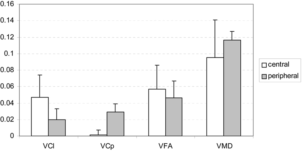
Fig. 9.
The comparisons of VFA, VMD, VCl and VCp between central (combing Genu, Splenium and PLIC) and peripheral (combining FC, CR and SLF) white matters. The VFA was significantly higher in central region (p=0.045), while VMD was significantly higher in peripheral white matter (p=0.0012). The VCl in the central was significantly higher than both the VCl (p<10−6) and VCp (p<10−4) in the peripheral. Contrarily, VCp was significantly higher in the peripheral than in the central (p<10−6).

