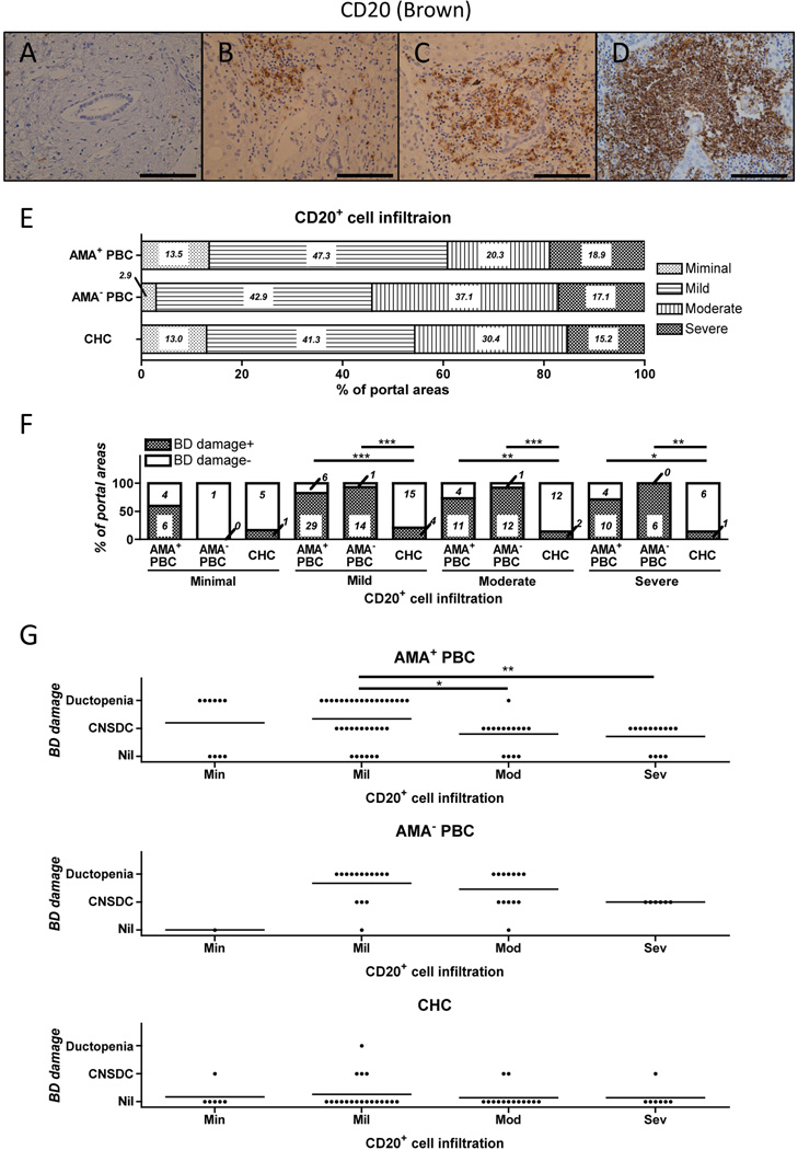
Figure 2.
A–D. Immunohistological evaluation of the degree of CD20+ cell infiltrates (stained brown) was conducted and profiles scored as minimal, mild, moderate and severe are shown (A to D). The frequency of each degree of CD20+ cellular infiltration was evaluated by the study of 74, 35 and 46 portal areas of liver tissues from AMA+ and AMA− PBC and CHC patients, respectively. E. CD20+ cellular infiltration was similar in the portal areas of PBC and CHC. F. Bile duct (BD) damage was more frequent in the mildly to severely CD20+ cellular infiltrated portal areas of liver tissues from PBC than CHC. Bile duct damage was more severe in liver tissues from PBC patients, but was not proportional to the degree of CD20+ cellular infiltration (G). (Scale bars indicate 100 µm in A–D. *: p < 0.05, **: p < 0.01 in Mann-Whitney Test.)

