Abstract
We constructed a reverse genetics system for avian paramyxovirus serotype 7 (APMV-7) to investigate the role of the fusion F glycoprotein in tissue tropism and virulence. The AMPV-7 F protein has a single basic residue arginine (R) at position −1 in the F cleavage site sequence and also is unusual in having alanine at position +2 (LPSSR↓FA) (underlining indicates the basic amino acids at the F protein cleavage site, and the arrow indicates the site of cleavage.). APMV-7 does not form syncytia or plaques in cell culture, but its replication in vitro does not depend on, and is not increased by, added protease. Two mutants were successfully recovered in which the cleavage site was modified to mimic sites that are found in virulent Newcastle disease virus isolates and to contain 4 or 5 basic residues as well as isoleucine in the +2 position: (RRQKR↓FI) or (RRKKR↓FI), named Fcs-4B or Fcs-5B, respectively. In cell culture, one of the mutants, Fcs-5B, formed protease-independent syncytia and grew to 10-fold-higher titers compared to the parent and Fcs-4B viruses. This indicated the importance of the single additional basic residue (K) at position −3. Syncytium formation and virus yield of the Fcs-5B virus was impaired by the furin inhibitor decanoyl-RVKR-CMK, whereas parental APMV-7 was not affected. APMV-7 is avirulent in chickens and is limited in tropism to the upper respiratory tract of 1-day-old and 2-week-old chickens, and these characteristics were unchanged for the two mutant viruses. Thus, the acquisition of furin cleavability by APMV-7 resulted in syncytium formation and increased virus yield in vitro but did not alter virus yield, tropism, or virulence in chickens.
INTRODUCTION
The family Paramyxoviridae comprises diverse viruses that have been isolated from mammalian, avian, reptilian, and fish species around the world (33). These are large pleomorphic, enveloped viruses containing a nonsegmented, single-stranded, negative-sense RNA genome. The paramyxoviruses that have been isolated to date from avian species mostly fall into the group that is called the avian paramyxoviruses (APMVs) and constitute the genus Avulavirus; the only other avian member of Paramyxoviridae is avian metapneumovirus, which is more distantly related and is classified in the genus Pneumovirus. APMVs have been divided into nine serotypes (APMV-1 to -9) based on hemagglutination inhibition (HI) and neuraminidase inhibition (NI) assays (1), and there is recent evidence for a 10th serotype (19). APMV-1, which consists of all Newcastle disease virus (NDV) strains, has been extensively characterized, because virulent NDV strains cause severe disease in chickens and are the most important cause of infectious disease in poultry (2). Complete genome sequences and reverse genetic systems are available for several NDV strains (6, 8, 14, 18, 23, 28, 30). The natural hosts and disease potential of the other APMV serotypes are mostly unknown. As an initial step toward characterizing the other APMV serotypes, complete genome sequences of one or more representative strains of APMV serotypes 2 to 9 have been determined (15, 24, 26, 34, 35, 37, 41, 42). Recently, reverse genetic systems for APMV-2 and -3 have been reported (16, 38).
APMV-7 was first isolated from a hunter-killed dove in 1975 in Tennessee and was designated a new serotype based on HI and NI assays (3). This isolate, called APMV-7/dove/Tennessee/4/75, is the prototype for APMV-7. Subsequently, APMV-7 was identified from natural outbreaks of respiratory tract disease in commercial turkey breeder flocks in Ohio in 1997, has been isolated from ostriches (40), and has been shown to be prevalent in pigeons, turkeys, and chickens (2). Warke et al. examined commercial chicken flocks in the United States by HI assay and found that 27% of serum samples were positive (HI ≥ 64) for APMV-7 (39). APMV-7 was also examined in isolates collected from dying birds in the United Kingdom and from Mallard ducks in New Zealand in which 47% of 315 sera were positive (HI ≥ 64) for antibody to APMV-7 (36). APMV-7 has not been causally associated with severe disease in avian species. Nevertheless, experimental infection of APMV-7 in turkeys caused respiratory disease and reduced egg production (32). In experimental infections of embryonated chicken eggs, the APMV-7 prototype strain dove/Tennessee/4/75 was not lethal, suggesting that it is avirulent in chickens (41).
The genome of APMV-7 strain dove/Tennessee/4/75 is 15,480 nucleotides (nt) long and contains six nonoverlapping genes in the order 3′-N-P/V/W-M-F-HN-L-5′, which encode a nucleocapsid protein (N), a phosphoprotein (P), a matrix protein (M), a fusion protein (F), a hemagglutinin-neuraminidase (HN), and a large polymerase protein (L), as well as two additional putative proteins, V and W, that are produced by RNA editing of the P gene. The 3′-leader and 5′-trailer sequences of the genome are 55 and 127 nt, respectively. The intergenic sequences are 11 to 70 nt in length. The organization of the APMV-7 genome is broadly similar to those of the other APMV serotypes except APMV-6, which contains an additional small hydrophobic protein (SH) gene between the F and HN genes (42).
The envelope of APMVs contains two glycoproteins, HN and F (except for that of APMV-6, which contains a third glycoprotein, SH). The HN protein is responsible for attachment to the host cell, and the F protein mediates fusion of the viral envelope with the cell membrane. Among the APMVs, the activities of these proteins have been studied in detail for NDV (17). The F protein is synthesized as an inactive precursor (F0) that is cleaved by host cell proteases into two biologically active F1 and F2 subunits that remain linked by a disulfide bond. Cleavage of the F protein is a prerequisite for virus entry and cell-to-cell fusion. The sequence of the F protein cleavage site is a well-characterized determinant of NDV pathogenicity in chickens (11, 22, 27, 28). The F protein of mesogenic and velogenic strains of NDV typically contains a polybasic cleavage site [(R/K)RQ(R/K)R↓F)] that contains the preferred recognition site for furin [RX(K/R)R↓], which is an intracellular protease present in a wide range of cells and tissues (basic residues are underlined, and the arrow indicates the site of cleavage). Consequently, the F protein of these strains can be cleaved in most tissues, making it possible for virulent strains to spread systemically. In contrast, avirulent NDV strains typically have basic residues at the −1 and −4 positions in the cleavage site [(G/E)(K/R)Q(G/E)R↓L)] and depend on a secreted protease (or, in cell culture, added trypsin or chicken egg allantoic fluid) for cleavage. This limits the replication of avirulent strains to the respiratory and enteric tracts, where the secreted protease is found. In addition, the residue at the +1 position that immediately follows the cleavage site can affect the efficiency of cleavage: phenylalanine and leucine are typically found in velogenic and lentogenic strains (4), respectively, and the latter has been associated with reduced cleavability of the APMV-1 F protein (21).
The putative F cleavage site of APMV-7 (LPSSR↓FA) has a single basic residue (underlined) at the −1 position. The F1 subunit of APMV-7 begins with a phenylalanine residue, as is characteristic of virulent NDV strains. It is also unusual in having alanine at position +2 rather than isoleucine, as seen in most virulent NDV strains, although it was not known whether this assignment affects cleavage. APMV-7 replicates in a wide range of cells without the addition of exogenous protease, and the inclusion of protease does not improve its efficiency of replication (41). This is incongruent with the lack of a preferred furin motif. Interestingly, APMV-7 produces single-cell infection and does not cause syncytium formation, a hallmark of paramyxovirus cytopathic effect (CPE). Also, APMV-7 is highly attenuated in chickens, which is incongruent with its independence from exogenous protease. Thus, questions remained about the cleavability of the F protein of APMV-7 and its role in infectivity and pathogenicity.
To investigate the role of the F protein cleavage site in replication and pathogenicity of APMV-7, we have developed a reverse genetics system. The APMV-7 cDNA clone was used to generate two APMV-7 mutants whose F protein cleavage sites were derived from virulent NDV isolates and contained 4 or 5 basic residues as well as isoleucine at the +2 position. Both F protein cleavage site mutants replicated efficiently and maintained the mutations after propagation in embryonated chicken eggs. However, only the mutant virus containing the cleavage site with five basic residues (RRKKR↓FI) exhibited protease independence, syncytium and plaque formation, and increased replication in vitro; in contrast, the mutant virus with four basic residues (RRQKR↓FI) did not form syncytia or plaques. However, the mutations did not change the avirulent nature of APMV-7, as determined by mean death time (MDT) in 9-day-old embryonated chicken eggs, by intracerebral pathogenicity index (ICPI) in 1-day-old chicks, and by natural infection in 2-week-old chickens. These results suggest that the F protein cleavage site sequence is not a major determinant for pathogenicity and virulence of APMV-7 in chickens.
MATERIALS AND METHODS
Viruses and cells.
APMV-7/dove/Tennessee/4/75 was obtained from the National Veterinary Services Laboratory, Ames, IA. The virus was propagated in the allantoic cavity of 9-day-old specific-pathogen-free (SPF) embryonated chicken eggs. Modified vaccinia virus strain Ankara expressing the T7 RNA polymerase (MVA/T7) was kindly provided by Bernard Moss (NIAID, NIH) and was propagated in primary chicken embryo fibroblast (CEF) cells. The chicken embryo fibroblast DF-1 cell line, human epidermoid carcinoma HEp-2 cell line, and Africa green monkey kidney Vero cell line were grown in Dulbecco's modified Eagle medium (DMEM) containing 10% fetal bovine serum (FBS) and maintained in DMEM with 5% FBS. In experiments that required supplementation of exogenous protease for cleavage of the F protein, either acetyl trypsin (Invitrogen) (1 μg/ml) or 5% chicken egg allantoic fluid was used.
Construction of plasmids expressing the full-length antigenome and support proteins.
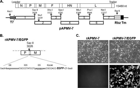
A full-length cDNA of the APMV-7 genome was constructed in plasmid pBR322/dr (5, 38). The full-length cDNA clone (pAPMV-7) expressing the complete 15,480-nt-long antigenome of APMV-7 was constructed as seven fragments that were generated by reverse transcription-PCR (RT-PCR) of RNA from APMV-7-infected embryonated chicken eggs. To facilitate construction, three restriction enzyme sites, SacII, SpeI, and MluI, were created downstream of the untranslated region (UTR) of the P, F, and HN genes, respectively. A unique NotI site was created in the L gene. These fragments were sequentially cloned into the pBR322/dr plasmid between the T7 promoter and the HDV antigenome ribozyme sequence (Fig. 1A). In the full-length cDNA, the two unique enzyme sites StuI and XhoI in the M and F genes made it possible to readily substitute a mutated F cleavage site fragment. The four F protein cleavage site mutants (Fig. 2A) were generated by overlapping PCR. The mutated fragments were digested with StuI and XhoI enzymes and were used to replace the corresponding fragments in the full-length cDNA of pAPMV-7. The full-length cDNAs of all fusion protein cleavage site mutants were sequenced in the M and F genes by the use of an ABI 3130xl genetic analyzer (Applied Biosystems). Support plasmids pAPMV-7 N, pAPMV-7-P, and pAPMV-7-L were constructed to individually express the N, P, and L proteins, respectively. cDNAs bearing the open reading frames (ORFs) of the N, P, and L genes (positions 141 to 1532, 1789 to 2973, and 8449 to 15132 in the complete genomic sequence, respectively) were cloned under the control of the T7 RNA polymerase promoter in pTM1 vector.
Fig 1.
Schematic diagram of the full-length cDNA encoding the APMV-7 antigenomic RNA. (A) The seven cDNA fragments were inserted sequentially in pBR322/dr vector, flanked on the upstream side by a T7 RNA polymerase promoter sequence and on the downstream side by the hepatitis delta virus ribozyme sequence (Rbz) followed by a T7 terminator sequence (Tm). The unique restriction enzymes used in the assembly and their nucleotide positions in the genome are shown: the SacII, SpeI, MluI, and NotI sites were created by nucleotide substitutions in nontranslated regions, while the other sites occurred naturally. (B) Diagram of the insertion of the EGFP open reading frame (ORF) into the APMV-7 cDNA construct at the SacII site present in the downstream nontranslated region of the P gene. The DNA insert bearing the EGFP ORF contained on its upstream end a gene end (GE) signal identical to that of the P gene, an intergenic sequence (IGS) identical to that of the F and HN genes, and a gene start (GS) signal identical to that of the N gene, followed by the EGFP ORF preceded by a sequence (Kozak) designed for efficient translation. (C) Expression of the EFGP protein in Vero cells from recombinant APMV-7. The cells were infected with either rAPMV-7 or rAPMV-7/EGFP at an MOI of 0.1. The EGFP expression was observed 24 h postinfection by light microscopy in the upper panels and fluorescence microscopy in the lower panels.
Fig 2.
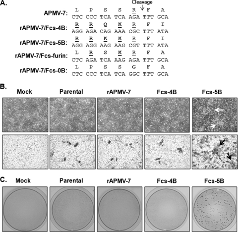
APMV-7 F protein cleavage site mutants. (A) Sequences of the cleavage sites of wt APMV-7 (top line) and four cleavage site mutants. Only wt APMV-7 and the Fcs-4B and -5B mutants could be recovered in infectious virus. The basic residues are underlined, and the mutated residues are shown in bold. (B) Syncytium formation. Vero cells were infected with parental biological APMV-7 and the indicated recombinant viruses at an MOI of 0.01 and incubated for 5 days in serum-free medium. The upper row of panels shows the cultures visualized by light microscopy. The lower row of panels shows the cultures following fixation with methanol and immunoperoxidase staining using a rabbit antiserum raised against the APMV-7 N protein. The arrows indicate syncytia. (C) Plaque formation. Vero cells were infected with the indicated viruses and incubated under conditions of methylcellulose overlay in serum-free media. On day 7, the plaques were fixed and immunostained as described for panel B.
Rescue of recombinant viruses.
Infectious virus was recovered from the cDNAs as previously described (14). Briefly, HEp-2 cells were grown overnight to 90% to 95% confluence in six-well culture plates and were cotransfected with 2 μg of the respective full-length cDNA plasmid, 1 μg of pAPMV-7 N, 0.5 μg of pAPMV-7-P, and 0.5 μg of pAPMV-7-L by using 5 μl of Lipofectamine 2000 (Invitrogen). Along with the transfection mixture, 1 focus-forming unit per cell of recombinant MVA/T7 was added. The transfection mixture was replaced after 20 h with DMEM containing 2% FBS. Two days after transfection, the HEp-2 cells were scraped into the medium and frozen and thawed three times, and the resulting supernatant was inoculated into the allantoic cavities of 9-day-old embryonated, SPF chicken eggs. The allantoic fluid was harvested 3 days postinoculation (dpi) and tested for hemagglutinin (HA) activity. Allantoic fluid with a positive HA titer was used for the isolation of the viral RNA followed by sequence confirmation of the F protein cleavage site. The recovered parental and mutant viruses were subjected to five passages in 9-day-old embryonated chicken eggs, and their stability was verified by sequencing the F cleavage site of each mutant virus.
Growth characteristics of parental and F protein cleavage site mutant viruses.
The growth characteristics of the parental and mutant viruses were evaluated in chicken embryos and in two cell lines, Vero and DF1, with and without 5% allantoic fluid supplementation in the medium. The SPF embryonated chicken eggs were inoculated with the viruses and incubated at 37°C. The allantoic fluids were harvested daily for titration. The ability of the mutant viruses to produce plaques was tested in Vero and DF1 cells under conditions that included a 0.8% methylcellulose overlay. The plaques were immunostained using a rabbit polyclonal antiserum raised against APMV-7 N protein (10) at 6 or 7 dpi, depending on the onset of plaques, followed by incubation with secondary antibody and detection by substrate AEC plus chromogen (Dako). The ability of mutant viruses to produce syncytia was tested in Vero cells. The growth kinetics of the parental, wild-type (wt), and mutant viruses was determined in Vero cells. Briefly, Vero cells grown in six-well plates were infected with each virus at a multiplicity of infection (MOI) of 1 or 0.01. At 24, 48, 72, 96, and 120 h postinfection, 200 μl of culture supernatants was collected and stored at −70°C for virus titration, and an equal volume of fresh medium was reintroduced. Virus titers of the samples were determined by serial endpoint assays on Vero cells in 96-well plates, with duplicate wells per virus per dilution. The infected cells were stained by an immunoperoxidase method using the polyclonal antiserum against APMV-7 N protein. The virus titers (50% endpoint tissue culture infectious dose [TCID50]/ml) were calculated using the method of Reed and Muench (29).
Inhibition of syncytium and plaque formation.
The Vero cells in 6-well plates were infected with parental and mutant viruses at an MOI of 0.01 or 1. After 1 h of virus adsorption, the cells were washed with PBS three times and the medium was replaced with serum-free DMEM or serum-free DMEM containing 0.8% methylcellulose and a furin inhibitor, decanoyl-RVKR-CMK (decanoyl-RVKR-chloromethylketone) (Calbiochem) (0, 1, 5, or 10 μM), and incubated at 37°C. At 4 dpi, the cells were processed by the following methods: (i) cells were immunostained with anti-APMV-7 N polyclonal antiserum for visualization of the syncytium and plaque formation as described above, and (ii) cells were lysed using sodium dodecyl sulfate-polyacrylamide gel electrophoresis (SDS-PAGE) sample buffer for Western blot analysis with anti-APMV-7 N and F rabbit polyclonal antiserums.
Pathogenicity tests.
The pathogenicity of the mutant viruses was determined by the mean death time (MDT) test in 9-day-old embryonated chicken eggs and the intracerebral pathogenicity index (ICPI) test in 1-day-old chicks (25). Briefly, for the MDT test, a series of 10-fold dilutions (10−6 to 10−9) of fresh allantoic fluid from infected eggs was made in sterile phosphate-buffered saline (PBS), 0.1 ml of each dilution was inoculated into the allantoic cavities of five 9-day-old embryonated chicken eggs per dilution, and the eggs were incubated at 37°C. Each egg was examined three times daily for 7 days, and the time of embryo death, if any, was recorded. The minimum lethal dose (MLD) is the highest virus dilution that causes all embryos inoculated with that dilution to die. The MDT is the mean time in hours required for the MLD to kill all of the inoculated embryos. The MDT has been used to classify APMV-1 strains into the following categories: velogenic strains (MDT less than 60 h), mesogenic strains (60 to 90 h), and lentogenic strains (more than 90 h).
For the ICPI test, 0.05 ml of a 1:10 dilution of fresh infective allantoic fluid for each virus was inoculated into groups of 10 1-day-old SPF chicks via the intracerebral route. The birds were observed for clinical signs and mortality once every 8 h for a period of 10 days. At each observation, the birds were scored as follows: 0 if normal, 1 if sick, and 2 if dead. The ICPI is the mean score per bird for all observations over the 10-day period. Highly virulent velogenic viruses give values approaching 2, and avirulent or lentogenic strains give values close to 0.
Effect of the F protein cleavage site mutant viruses on replication in 1-day- and 2-week-old chickens.
The effect of the F protein cleavage site mutations on viral replication and pathogenicity was determined by experimentally infecting 1-day- and 2-week-old SPF chickens. Briefly, groups of six 1-day- and 2-week-old SPF chickens were inoculated with 0.2 ml of 28 HA units of each virus by the oculonasal route. The birds were observed daily and scored for any clinical signs for 14 dpi. Three birds from each group were euthanized at 2, 4, and 6 dpi, and oral and cloacal swabs were taken. In addition, samples of the following tissues were collected for virus isolation: brain, lung, trachea, spleen, kidney, and intestine. The oral and cloacal swabs were collected in 1 ml of PBS containing antibiotics (2,000 units of penicillin G/ml, 200 μg of gentamicin sulfate/ml, and 4 μg of amphotericin B/ml; Sigma Chemical Co., St. Louis, MO). The swab-containing tubes were centrifuged at 1,000 ×g for 20 min, and the supernatants were removed for virus isolation. Virus isolation was performed by inoculating the supernatant into the allantoic cavities of 9-day-old embryonated chicken eggs, and the allantoic fluid was tested at 3 dpi for HA activity. The virus titers in the tissue samples were determined by the following method. Briefly, the tissue samples were homogenized, and the supernatant was serially diluted and used to infect Vero cells, with duplicate wells per dilution. Infected wells were identified by immunostaining, and the TCID50 (per milliliter) was calculated.
RESULTS
Development of an APMV-7 reverse genetics system.
A cDNA clone expressing the antigenome of APMV-7 was constructed from seven cDNA segments that were synthesized by RT-PCR from virion-derived genomic RNA (Fig. 1A). The cDNA segments were cloned in a sequential manner into low-copy-number pBR322/dr plasmid between a T7 promoter and the hepatitis delta virus ribozyme sequence. The resulting APMV-7 cDNA was a faithful copy of the published APMV-7 antigenome consensus sequence (40) except for 8 silent nucleotide changes that were introduced to create four new unique restriction enzyme sites (SacII, SpeI, MluI, and NotI) used in the construction (Fig. 1A). This construct contains a T7 promoter that initiates a transcript with three extra G residues at its 5′ end, which increases the efficiency of T7 RNA polymerase transcription and does not interfere with virus recovery (11, 16). Three support plasmids expressing the N, P, and L proteins of APMV-7 were also constructed under the control of the T7 promoter in the pTM1 vector.
A cDNA containing the open reading frame (ORF) encoding enhanced green fluorescent protein (EFGP) was inserted into the APMV-7 cDNA clone at the SacII site in the downstream nontranslated region of the P gene. The cDNA was designed so that, following insertion, the EGFP ORF was flanked by conserved gene end (GE) and gene start (GS) sequences of APMV-7 such that EGFP would be expressed as a separate mRNA by the APMV-7 polymerase. The translational start codon of EGFP ORF was placed in a sequence context favorable for efficient translation (13). Two extra nucleotides were inserted in the downstream of EGFP gene to keep the genome length of APMV-7 as an even multiple of six, which is important for efficient genome replication (12) (Fig. 1B).
The recombinant wt APMV-7 parent (rAPMV-7) virus and the rAPMV-7/EGFP virus expressing GFP were readily recovered by transfection of the respective antigenome plasmids into HEp-2 cells together with plasmids encoding the N, P, and L proteins, which are necessary for viral RNA replication and transcription. T7 RNA polymerase was supplied by infection with MVA/T7. The supernatants from the transfected HEp-2 monolayers were inoculated into the allantoic cavities of 9-day-old embryonated chicken eggs. Allantoic fluid was harvested 3 days after infection and tested for HA activity. Allantoic fluid with a positive HA titer was used as a preliminary viral stock. Both the rAPMV-7 parent and rAPMV-7/GFP derivative readily replicated in Vero cells (as described below). Neither recombinant virus required the addition of exogenous protease during transfection and recovery, which was as expected, since replication of the biological APMV-7 parent is unaffected by added protease. To examine EGFP expression, Vero cells were infected with rAPMV-7/EGFP at an MOI of 0.1. Examination of the cell monolayers by fluorescence microscopy 24 h later revealed abundant expression of EGFP (Fig. 1C).
Creation of F protein cleavage site mutant viruses.
We then made four derivatives of the wt AMPV-7 antigenomic cDNA containing mutations in the F protein cleavage site (Fig. 2A). The wt APMV-7F protein cleavage site (LPSSR↓FA) contains a single basic amino acid (underlined). In two mutants, called Fcs-4B and Fcs-5B, respectively, this site was placed with two different naturally occurring virulent NDV F protein cleavage sites that contain the following four or five basic amino acid sequences: RRQKR↓FI (the mutated residues are shown in bold), which is present in most velogenic NDV strains, and RRKKR↓FI, which is present in some velogenic strains such as Nigeria/95. In addition, in both of these mutants, the residue in the +2 position was changed from alanine to isoleucine so as to have the assignment that is present in most velogenic NDV strains. A third F cleavage site mutant, Fcs-furin (LRSKR↓FA), was made that had three basic residues that match the minimal furin cleavage site (RX[K/R]R↓): this mutant retained alanine at position +2. A fourth mutant, Fcs-0B (LPSSG↓FA), was made that lacks any basic residues in the F cleavage site (LPSSG↓FA; the arginine at position −1 was replaced by glycine [shown in bold]). These mutants were constructed by overlapping PCR and substituted into the wt full-length cDNA clone by the use of the StuI (nt position 3439) and XhoI (nt position 5322) sites.
The four F cleavage site mutants were recovered by transfection into HEp-2 cells and subsequent inoculation of the cell culture supernatants into embryonated chicken eggs, as described above. The Fcs-4B and -5B mutants were readily recovered. Part of the harvested allantoic fluid was processed to isolate viral genomic RNA, which was subjected to RT-PCR and partial sequence analysis of the F gene to confirm the sequence of the F protein cleavage site. As with the wt and GFP-expressing viruses, neither of these F protein cleavage site mutants required the addition of exogenous protease during transfection and recovery (i.e., in HEp-2 cells). To evaluate genetic stability, the Fcs-4B and -5B viruses were subjected to five passages in 9-day-old embryonated chicken eggs, and the sequence of the F gene was confirmed, showing that the introduced mutations were maintained without any adventitious mutations.
In contrast, neither the Fcs-furin nor the Fcs-0B mutants could be recovered in five different attempts whereas the positive-control rAPMV-7 was readily recovered. The inability to recover the Fcs-furin mutant was unexpected, since its cleavage site (LRSKR↓FA) contains the preferred furin cleavage site (RX[K/R]R↓). Presumably, some aspect of this sequence, possibly the unusual alanine residue at the +2 position, makes it incompatible with furin cleavage. The inability to recover the Fcs-0B (LPSSG↓FA) mutant indicates that the arginine residue in the −1 position is essential for cleavage and that cleavage is essential for virus viability. In contrast, the basic residue in Nipah virus is not needed at that position for cleavage based on the plasmid-expressed viral proteins in cells, because it is cleaved by a different protease (20).
Syncytium formation, infection, and growth characteristics of parental and F protein cleavage site mutant viruses.
All recombinant viruses replicated well in Vero cells, and supplementation of the growth medium with exogenous proteases (1 unit/ml furin, 1.0 μg/ml trypsin, or 10% allantoic fluid) did not enhance the growth of any of the viruses. The wt recombinant virus resembled its biological parent in growth characteristics, causing single-cell infection in Vero (Fig. 2B) and DF1 (not shown) cell lines rather than forming syncytia. Also, neither the biological nor recombinant wt APMV-7 produced plaques under conditions of methylcellulose overlay in the presence or absence of exogenous proteases (Fig. 2C). The F protein cleavage site mutant virus Fcs-4B also did not produce syncytia or plaques and thus resembled its parental virus in Vero and DF1 cells. In contrast, The F protein cleavage site mutant virus Fcs-5B gained the ability to produce syncytia and plaques under conditions of methylcellulose overlay in the Vero cells (Fig. 2B and C) but not in DF1 cells (data not shown). This indicated that there was a surprising large difference in the biological properties associated with the single amino acid residue difference between the Fcs-4B and -5B mutants, namely, the presence at the −3 position of glutamine in the former virus and arginine in the latter.
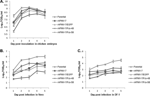
The replication kinetics of parental and F protein cleavage site mutant viruses were compared in chicken embryos and cell lines. The various viruses grew to titers of 108 to 109 TCID50/ml in embryonated chicken embryos (Fig. 3A). The F cleavage site mutant virus Fcs-5B grew to a titer that was 0.5 log higher than that of the parental virus, whereas the Fcs-4B virus grew to a titer that was 1 log lower (Fig. 3A). To examine replication in cell lines, monolayer cultures of Vero (Fig. 3B) and DF1 (Fig. 3C) cells were infected with the viruses at an MOI of 1, the cells were washed 1 h later, and samples from the medium overlay were collected at 24-h intervals and quantified in Vero cells by limiting dilution. This analysis showed that Fcs-5B grew to a 10-fold-higher titer than its biological and recombinant parents and the other mutant viruses, whereas the mutant virus Fcs-4B replicated poorly compared to other viruses. The yields of all viruses were higher in Vero cells than in DF1 cells (Fig. 3B and C).
Fig 3.
Replication kinetics of parental biological APMV-7, wt rAMPV-7, and the indicated recombinant mutant viruses in embryonated chicken eggs (A) and in Vero (B) and DF-1 (C) cells. (A) Nine-day-old SPF embryonated chicken eggs were inoculated with 103 TCID50 of the indicated viruses. Allantoic fluids were harvested daily for virus titration. (B and C) Vero (B) and DF-1 (C) cells were infected in duplicate with the indicated viruses at an MOI of 1, and samples were collected from the culture supernatant at 24-h intervals until 120 h postinfection. Virus titers of the samples were determined by serial endpoint dilution, with infected wells detected by immunoperoxidase staining using anti-APMV-7 N protein rabbit serum.
Effect of a furin inhibitor on syncytium and plaque formation and viral replication.
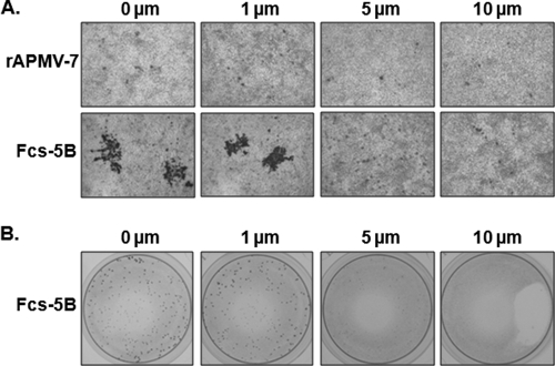
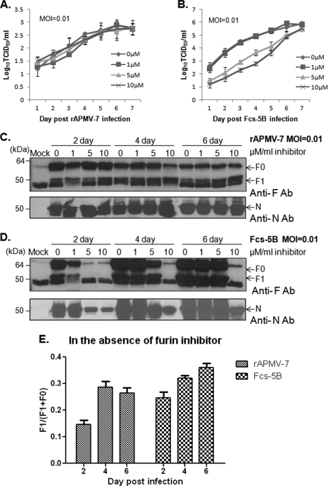
As noted, the precursor F protein (F0) of virulent NDV strains is cleaved by ubiquitous intracellular proteases such as furin, whereas avirulent strains are cleaved by a secreted, extracellular protease. The proteases involved in cleaving the other APMV serotypes are unknown but were assumed to be similar. To examine whether furin is responsible for cleavage of the F protein of wt AMPV-7 and the mutant Fcs-5B, Vero cells were infected with either virus at an MOI of 0.01 and incubated with serum-free medium containing a range of concentrations of decanoyl-RVKR-CMK, a furin inhibitor, with and without an overlay of methylcellulose (Fig. 4A and B, respectively). The cells were fixed 7 days postinoculation, and syncytia (Fig. 4A) and plaques (Fig. 4B) were visualized by immunostaining with an anti-APMV-7 N polyclonal antibody. Wt rAPMV-7 formed characteristic single-cell infections without protease in the absence of inhibitor, and this was not affected by the various concentrations of inhibitor (Fig. 4A). The Fcs-5B mutant formed large syncytia in the absence of inhibitor and at the lower concentration of 1 μM, but syncytium formation was strongly reduced at inhibitor concentrations of 5 and 10 μM, resulting in single-cell infections (Fig. 4A). Similarly, Fcs-5B induced plaque formation in the absence of inhibitor and at the lower concentration of 1 μM, but plaque formation was strongly inhibited at inhibitor concentrations of 5 and 10 μM (Fig. 4B). These results confirmed that furin-mediated cleavage of the Fcs-5B F protein enabled virus-induced formation of syncytia and plaques. In contrast, furin did not appear to be necessary for the single-cell infection characteristic of wt rAPMV-7 and also was not necessary for single-cell infection by the Fcs-5B mutant.
Fig 4.
Effects of a furin inhibitor on the formation of syncytia and plaques by wt rAPMV-7 and the Fcs-5B mutant. (A) Syncytium formation. Vero cells were infected with wt rAPMV-7 and the Fcs-5B mutant at an MOI of 0.01. The cultures were incubated with serum-free media containing furin inhibitor at 0, 1, 5, and 10 μM, as indicated. The infected cells were fixed with methanol at 4 dpi and visualized by immunoperoxidase staining with anti-APMV-7 N protein rabbit serum. (B) Plaque formation. Vero cells were infected with the Fcs-5B mutant at an MOI of 0.01 and incubated under conditions of methylcellulose overlay in serum-free media containing furin inhibitor at 0, 1, 5, and 10 μM, as indicated. At 7 dpi, the cells were fixed and stained as described for panel A.
In addition, we examined whether the furin inhibitor affected the yield of the viruses in cell culture. Vero cells were infected with the recombinant wt and Fcs-5B viruses at an MOI of 0.01 and maintained in the presence of the furin inhibitor. The growth of wt rAPMV-7 was not affected by multiple-step cycle replication at an MOI of 0.01 (Fig. 5A). However, the replication of the Fcs-5B virus was significantly reduced at 5 and 10 μM concentrations (Fig. 5B). Fcs-5B with and without furin inhibitor achieved similar titers by 7 dpi, which might reflect accumulation of furin and incomplete inhibition.
Fig 5.
Effects of a furin inhibitor on replication of wtrAPMV-7 and the Fcs-5B mutant. Vero cells in 6-well plates were infected with wt rAPMV-7 (A and C) or the Fcs-5B mutant (B and D) at an MOI of 0.01. The cells were incubated with serum-free media containing furin inhibitor at 0, 1, 5, and 10 μM, as indicated. (A and B) Virus replication. Samples of the culture medium supernatants were collected at 24-h intervals, and virus titers were determined by limiting dilution assay. (C and D) Western blot analysis. The cells in additional plates that had been infected and treated in parallel were harvested and lysed at 2, 4, and 6 dpi. Western blot analysis was performed by using rabbit antiserum that had been raised against a synthetic peptide representing the cytoplasmic tail of the APMV-7 F protein (upper panel in C and D) or using anti-N protein rabbit serum (lower panel in C and D). The precursor (F0) and cleaved subunit (F1) of the F protein are indicated. (E) Efficiency of cleavage of the F proteins of wt rAPMV-7 and the Fcs-5B mutant in the absence of the furin inhibitor. The relative levels of the F0 and F1 proteins in the absence of inhibitor in the Western blot images in panels C and D were measured by Bio-Rad Gel Image analysis, and the efficiency of cleavage was determined by dividing the amount of F1 by the amount of F1 plus F0.
The effect of the furin inhibitor on F protein cleavage was examined by Western blot analysis of cell lysates with a rabbit antiserum against a synthetic peptide representing the 24 residues (amino acids 516 to 539) in the cytoplasmic tail of APMV-7 F protein. This peptide was synthesized by GenScript. This rabbit antiserum, produced as described previously (10), reacts with both the F0 precursor and the F1 subunit but not with the F2 subunit. In cells infected with wt rAPMV-7 and grown in the absence of the inhibitor, there was accumulation of F0 protein precursor and, surprisingly, accumulation of the F1 cleavage product (Fig. 5C). The efficiency of cleavage of F0 was estimated by dividing the amount of F1 (quantified from the Western blot images) by the combined amounts of F1 plus F0. This provided a value of approximately 15% to 29% for the efficiency of cleavage (Fig. 5E). The addition of inhibitor had little effect on the magnitude of wt F protein expression or cleavage. The lack of effect of this inhibitor is consistent with the possibility that the cleavage of wt F protein is mediated by nonfurin host cell proteases. We have also analyzed the F protein in purified virions from egg allantoic fluids by Western blotting. We detected a single band of F1 protein that produced similar intensities for wt and mutant viruses (data not shown), indicating that the levels of F protein cleavage were equal for wt and mutant viruses.
In cells infected with the Fcs-5B mutant in the absence of inhibitor, both F0 and F1 were readily detected (Fig. 5D). The efficiency of cleavage was estimated to be 25% to 36% and thus was somewhat higher than for wt APMV-7 (Fig. 5E). When infected cells were incubated in the presence of the furin inhibitor, two effects were observed. First, there was an overall reduction in F protein expression (Fig. 5D). This was paralleled by a reduction in N protein expression, indicating that the reduction in the overall expression of these proteins was probably an indirect effect of reduced virus replication or spread. Second, cleavage of the F0 precursor appeared to be inhibited at the higher inhibitor concentrations. Specifically, the F1 subunit was not detected on day 2 with inhibitor concentrations of 5 and 10 μM or on days 4 and 6 with an inhibitor concentration of 10 μM. Similarly, the expression of N protein also was corresponding less that of the F protein, indicating that the viral protein expression of this mutant was decreased by the presence of the furin inhibitor. These results support the idea that furin plays an important role in cleavage of the F0 protein of the Fcs-5B mutant, but not of the F0 protein of the wt, and that this cleavage was important for multiple-step cycle replication of the Fcs-5B mutant but a similar though somewhat less efficient cleavage of wt F0 by a different enzyme is not sufficient for cleavage.
Pathogenicity in chicken embryos and 1-day-old chickens.
The pathogenicity of biological wt APMV-7, recombinant wt rAPMV-7, and the two F protein cleavage site mutant viruses was evaluated by two standard assays, namely, the mean death time (MDT) test in 9-day-old embryonated chicken eggs and the intracerebral pathogenicity index (ICPI) test in 1-day-old chicks. None of the viruses caused the death of any chicken embryos within the standard 7-day (168-h) time limit for the assay; thus, the MDT for all of the viruses was scored as >168 h. None of the viruses caused disease or death following intracerebral inoculation of 1-day-old chicks; therefore, those viruses had ICPI values of zero. Both tests suggested that APMV-7 is highly attenuated in eggs and young chicks and that the cleavage site of the F protein and the ability to form syncytia did not detectably affect the pathogenicity of APMV-7.
Pathogenicity in 1-day- and 2-week-old chickens inoculated by the natural oculonasal route.
The effect of the F protein cleavage site on viral pathogenesis was further studied by experimentally infecting 1-day- and 2-week-old SPF chickens with parental biological wt APMV-7 and each of the recombinant viruses. The chickens were infected with a high dose (107 TCID50) of infectious fresh allantoic fluids by the oculonasal route, mimicking a natural infection. The birds were observed daily for 10 days postinfection. Three birds from each group were sacrificed on days 2, 4, and 6 postinfection. Tissue samples were taken from the brain, lung, trachea, spleen, kidney, and colon. Upon sacrifice, oral and cloacal swabs were taken and analyzed for viral shedding.
There were no apparent clinical signs of disease throughout the study period in any of the virus-inoculated groups of either the 1-day- or 2-week-old chickens. The only tissue samples that contained infectious virus were from the trachea of inoculated 1-day-old chickens sacrificed at 2 dpi. In these samples, there was no significant difference in titers among the different viruses, although the Fcs-5B titer was the highest. No infectivity was found in the brain, lung, trachea, spleen, kidney, and colon tissue from the 2-week-old chickens collected on 2, 4, and 6 days postinoculation. Viral shedding was detected in oral swabs from both 1-day- and 2-week-old chickens but only on day 2 (Fig. 6). No oral shedding was detected on any other day. No cloacal viral shedding was detected on any day (data not shown). There were no significant differences in the amounts of viral shedding between the parental and F protein cleavage site mutant viruses. These results showed no consistent pattern with regard to the F protein cleavage site. Tissues of the inoculated chickens were also investigated by immunohistochemistry (data not shown), confirming the presence of viral antigen in the trachea of 1-day-old chickens on day 2 postinoculation. Viral antigen could not be unequivocally detected in any other tissue samples. All the infected birds were seropositive at 14 dpi, with a mean HI titer of 27. Although all viruses replicated with low systemic spread and viral shedding, all viruses were able to induce that high level of humoral immune response in chickens.
Fig 6.
Virus titration from APMV-7-infected chickens at 2 day postinfection. Virus titers from the indicated tissues harvested on day 2 from 1-day-old and from 2-week-old chickens infected with the viruses are shown. Chickens were inoculated by the oculonasal route. Each group is represented by 3 birds for each day. Titers are shown as mean TCID50 per gram for tissues or TCID50 per milliliter for swabs.
DISCUSSION
For paramyxoviruses in general, cleavage of the F0 protein precursor into the F1 and F2 subunits is a requirement for fusion activity. Virulent strains of NDV (APMV-1) have multibasic cleavage sites that are recognized and cleaved by furin, a ubiquitous intracellular protease whose preferred cleavage sequence is as follows: RX(R/K)R↓. These viruses are able to replicate in most cell types and cause systemic infection. Avirulent NDV strains have one or occasionally two unpaired basic amino acids and thus lack the preferred furin motif. These F proteins are thought to be cleaved by trypsin-like extracellular proteases. Hence, these viruses are restricted mostly to the respiratory and gastrointestinal tracts where these extracellular proteases are present, and they require supplementation with exogenous protease for growth in vitro. It has been shown that the F protein cleavage site is a major determinant of NDV virulence in chickens (27, 28). In contrast, the contribution of the F protein cleavage site to the growth properties and pathogenicity of the remaining APMV serotypes 2 to 9 is largely unknown.
The viruses of APMV-2 to -9 have been isolated from a variety of avian species, and the natural hosts of these viruses are not clearly defined. The sequences of the F protein cleavage site differ greatly among the APMV serotypes. APMV-7 causes mild or inapparent respiratory illness in turkeys and chickens and thus appears to be avirulent. In seeming agreement with this phenotype, the F protein cleavage sequence of APMV-7 (LPSSR↓FA) contains only a single basic residue and thus does not contain the furin motif. However, APMV-7 contains phenylalanine in the +1 position, which is typically found in velogenic strains of NDV and may aid cleavage (21). Furthermore, we previously showed that replication of APMV-7 in cell culture does not require, and is not enhanced by, exogenous protease (41). Therefore, there were incongruities in the biological properties of the APMV-7 F protein, and the contribution of the F protein cleavage site sequence to the pathogenicity of APMV-7 was unknown.
In the present study, we created a reverse genetic system for APMV-7 and used this system to evaluate the role of the F protein cleavage site in replication, formation of syncytia and plaques, F protein cleavage, and pathogenicity. Consistent with previous studies performed with NDV, the three viral ribonucleoprotein (RNP)-related proteins N, P, and L were necessary and sufficient as viral support proteins to mediate recovery. It was of interest to investigate whether any or all of the N, P, and L support plasmids were functional between APMV-7 and NDV. We therefore performed recovery experiments using antigenomic cDNAs of APMV-7 and NDV/LaSota with various combinations of the support plasmids of APMV-7 and NDV. Recombinant viruses were rescued only with the full set of homologous N, P, and L proteins (data not shown). This result demonstrated a virus serotype-specific interaction between viral support proteins and their genome. This was not completely unexpected, since the amino acid sequence identities of the N, P, and L proteins of APMV-7 and NDV are very low at 41%, 21%, and 38%, respectively. We also showed that APMV-7 readily accepted and expressed EGFP from a transcription cassette added between the P and M genes, with little effect on replication in vitro or in vivo. Like biological wt APMV-7, the recombinant wt APMV-7 did not require added protease for replication in cell culture, and replication was not enhanced by added protease.
We used the reverse genetic system to construct four recombinant APMV-7 F protein cleavage site mutants. Two of these mutants, Fcs-4B and -5B (RRQKR↓FI and RRKKR↓FI, respectively), contained two different cleavage sites from naturally occurring virulent strains of NDV that differed by a single basic residue in the −3 position, and both mutants also contained isoleucine in the +2 position rather than the alanine found in wt APMV-7 (LPSSR↓FA). The Fcs-4B and -5B mutants, with cleavage sites from virulent NDV, were readily recovered. For both Fcs-4B and -5B, replication in cell culture did not require, and was not enhanced by, added protease. However, the Fcs-furin (LRSKR↓FA) and Fcs-0B (LPSSG↓FA) mutants could not be recovered. The inability to recover the Fcs-furin mutant was surprising, since it contained the preferred furin cleavage site and differed from wt APMV-7 at two amino acid positions, −4 and −2. This indicated that, in addition to the furin motif, cleavage site residues are critical for cleavage and/or activity. The inability to recover the Fcs-0B mutant indicated that the presence of a basic residue at position −1 is essential for cleavage and/or activity.
Syncytium formation is a hallmark of infection with many of the paramyxoviruses, but APMV-7 is an exception in that it causes single-cell infections in cell culture even in the presence of added protease. It also does not form plaques. As seen with parental APMV-7, the wt rAPMV-7 virus caused single-cell infections and did not form syncytia or plaques. Although the mutant Fcs-4B contained a cleavage site with multiple basic residues and the preferred furin motif and was replication competent, it also did not form syncytia or plaques. Interestingly, however, mutant Fcs-5B produced both syncytia and plaques in Vero cells, although not in DF-1 cells. There is only one amino acid difference between Fcs-4B and -5B, namely, the presence of an additional basic residue at the −3 position in the latter virus. This suggests that the basic residue at position −3 is important in order for the virus to form syncytia and plaques in Vero cells.
Expression and cleavage of the F proteins of wt APMV-7 and the Fcs-5B mutant were evaluated directly by Western blot analysis of intracellular proteins from virus-infected Vero cells. This showed that the F protein of wt APMV-7 accumulated mostly as the F0 precursor and thus was not efficiently cleaved. Cleavage of the F protein of the Fcs-5B mutant was more efficient, although there was substantial accumulation of the F0 precursor. This increase in cleavage efficiency presumably accounts for the ability to form syncytia and plaques, more efficient spread, and virion production. It also is possible that the presence of the isoleucine in the +2 position in F cleavage site rendered the Fcs-5B F protein more fusogenic than the wt F protein with alanine in this position.
We also investigated the effects of a furin inhibitor on cell culture growth and F protein processing of wt APMV-7 and the Fcs-5B mutant. For wt APMV-7, the inhibitor had no apparent effect on infection and replication in cell culture or cleavage of the F protein. This was consistent with the idea that cleavage of wt APMV-7 is mediated by a nonfurin protease. There are multiple furin-like proteases, and some may cleave one of these better than another. It is also possible that this domain of the F protein is not properly presented for furin or a furin-like enzyme to cleave it. In contrast, for the Fcs-5B mutant, syncytium formation, plaque formation, replication, and F protein cleavage were impaired by the furin inhibitor. Thus, furin appears to play a role in cleavage of the Fcs-5B F protein, which thus differs from wt APMV-7 F protein.
The observation that the Fcs-5B mutant had gained the ability to form syncytia and plaques, and also replicated more efficiently than wt APMV-7 in cell culture, suggested that its tropism and virulence in vivo might be increased, based on the well-known example of NDV. Surprisingly, however, all of the recombinant viruses, including Fcs-5B, were found to have retained the avirulent phenotype of the APMV-7 parent. For all of the recombinant viruses, the MDT values were >168 h and the ICPI value was zero, the same as for wt APMV-7. Following oculonasal inoculation of 1-day- and 2-week-old chickens with a high titer of virus (107 TCID50), expression of all of the viruses, including the Fcs-5B mutant, was found to be highly restricted. Shedding was detected in both age groups, but the shedding was detected only in the oral swabs and only on day 2 postinoculation. Infectious virus was recovered from tissue samples only in the trachea, only in 1-day-old chicks, and only on day 2. These results indicate that the acquisition of furin cleavability did not alter the tropism or increase the virulence of APMV-7 in chickens, despite its ability to spread and produce more viruses. The slight increase in F protein cleavage by Fcs-5 might be enough to enable syncytium formation but might be inefficient enough to escape the immune system and cause disease in chickens. These results suggest that the F protein cleavage site is not a major determinant of virulence in APMV-7 and, in particular, is not responsible for the avirulent phenotype of wt APMV-7 in chickens.
We have shown previously that altering the F protein cleavage site of an avirulent strain of NDV to that of a neurovirulent strain did not convert the avirulent phenotype into a neurovirulent phenotype following oculonasal infection (27). Although the biological activities of the F protein and the growth characteristics of the virus in vivo improved for the mutant virus compared to the parental avirulent virus, the complete spectrum of the virulent phenotype was not obtained, suggesting that the F cleavage site is not the only significant determinant of virulence. Consistent with this, other NDV proteins, such as the HN, V, N, P, and L proteins, have been shown to contribute to the virulence phenotype along with the F protein (7, 9, 31). Therefore, it is likely that other APMV-7 proteins, in addition to the F protein, contribute to the avirulent phenotype of APMV-7, which requires further investigation. Recently, the role of the F protein cleavage site of APMV-2 was investigated (38). The replacement of the F protein cleavage site of wt APMV-2 (KPASR↓F) with that of any of the other APMV serotypes (APMV-1 to -9) regardless of whether it was multibasic or not was associated with the ability to form syncytia and plaques and with an increase in virus spread and production. Despite these enhancements, neither the APMV-2 nor the APMV-7 mutants exhibited increased virulence in chickens, again suggesting that enhanced F protein cleavage is not the only characteristic of a virulent phenotype. On the other hand, optimal cleavage may be necessary for acquisition of more virus characteristics. These features, in addition to the F protein cleavage site, are responsible for the avirulent phenotype of APMV-7. In addition, rAPMV-7 efficiently expressed a foreign gene (EGFP) with little effect on replication in vitro or in vivo, indicating that APMV-7 could be a potential vector for vaccine development.
ACKNOWLEDGMENTS
We thank Daniel Rockemann for his technical assistance. We thank Flavia Dias and Sachin Kumar for his help with handling of chickens during the animal experiments. The pTM-1 vector and MVA/T7 vaccinia virus recombinant were kindly provided by Bernard Moss, NIAID. This research was supported by NIAID contract no. N01A060009 (85% support) and NIAID, NIH Intramural Research Program (15% support).
The views expressed herein do not necessarily reflect the official policies of the Department of Health and Human Services; mention of trade names, commercial practices, or organizations does not imply endorsement by the U.S. government.
Footnotes
Published ahead of print 18 January 2012
REFERENCES
- 1. Alexander D. 2003. Paramyxoviridae, 11th ed Iowa State University Press, Ames, IA [Google Scholar]
- 2. Alexander D, Senne D. 2008. Newcastle disease and other avian paramyxovirus and pneumovirus infection, p 75–115 In Saif YM, Fadly AM, Glisson JR. (ed), Diseases of poultry. Iowa State University Press, Ames, IA [Google Scholar]
- 3. Alexander DJ, Hinshaw VS, Collins MS. 1981. Characterization of viruses from doves representing a new serotype of avian paramyxoviruses. Arch. Virol. 68:265–269 [DOI] [PubMed] [Google Scholar]
- 4. Collins MS, Bashiruddin JB, Alexander DJ. 1993. Deduced amino acid sequences at the fusion protein cleavage site of Newcastle disease viruses showing variation in antigenicity and pathogenicity. Arch. Virol. 128:363–370 [DOI] [PubMed] [Google Scholar]
- 5. Collins PL, et al. 1995. Production of infectious human respiratory syncytial virus from cloned cDNA confirms an essential role for the transcription elongation factor from the 5′ proximal open reading frame of the M2 mRNA in gene expression and provides a capability for vaccine development. Proc. Natl. Acad. Sci. U. S. A. 92:11563–11567 [DOI] [PMC free article] [PubMed] [Google Scholar]
- 6. de Leeuw OS, Koch G, Hartog L, Ravenshorst N, Peeters BP. 2005. Virulence of Newcastle disease virus is determined by the cleavage site of the fusion protein and by both the stem region and globular head of the haemagglutinin-neuraminidase protein. J. Gen. Virol. 86:1759–1769 [DOI] [PubMed] [Google Scholar]
- 7. Dortmans JC, Rottier PJ, Koch G, Peeters BP. 2010. The viral replication complex is associated with the virulence of Newcastle disease virus. J. Virol. 84:10113–10120 [DOI] [PMC free article] [PubMed] [Google Scholar]
- 8. Estevez C, King D, Seal B, Yu Q. 2007. Evaluation of Newcastle disease virus chimeras expressing the hemagglutinin-neuraminidase protein of velogenic strains in the context of a mesogenic recombinant virus backbone. Virus Res. 129:182–190 [DOI] [PubMed] [Google Scholar]
- 9. Huang Z, Krishnamurthy S, Panda A, Samal SK. 2003. Newcastle disease virus V protein is associated with viral pathogenesis and functions as an alpha interferon antagonist. J. Virol. 77:8676–8685 [DOI] [PMC free article] [PubMed] [Google Scholar]
- 10. Khattar SK, Kumar S, Xiao S, Collins PL, Samal SK. 2011. Experimental infection of mice with avian paramyxovirus serotypes 1 to 9. PLoS One 6:e16776. [DOI] [PMC free article] [PubMed] [Google Scholar] [Retracted]
- 11. Klenk HD, Garten W. 1994. Activation cleavage of viral spike proteins by host proteases, p 241–280 In Wimmer E. (ed), Cellular receptors for animal viruses. Cold Spring Harbor Laboratory, Cold Spring Harbor, NY [Google Scholar]
- 12. Kolakofsky D, et al. 1998. Paramyxovirus RNA synthesis and the requirement for hexamer genome length: the rule of six revisited. J. Virol. 72:891–899 [DOI] [PMC free article] [PubMed] [Google Scholar]
- 13. Kozak M. 1987. An analysis of 5′-noncoding sequences from 699 vertebrate messenger RNAs. Nucleic Acids Res. 15:8125–8148 [DOI] [PMC free article] [PubMed] [Google Scholar]
- 14. Krishnamurthy S, Huang Z, Samal SK. 2000. Recovery of a virulent strain of Newcastle disease virus from cloned cDNA: expression of a foreign gene results in growth retardation and attenuation. Virology 278:168–182 [DOI] [PubMed] [Google Scholar]
- 15. Kumar S, Nayak B, Collins PL, Samal SK. 2008. Complete genome sequence of avian paramyxovirus type 3 reveals an unusually long trailer region. Virus Res. 137:189–197 [DOI] [PMC free article] [PubMed] [Google Scholar]
- 16. Kumar S, Nayak B, Collins PL, Samal SK. 2011. Evaluation of the Newcastle disease virus F and HN proteins in protective immunity by using a recombinant avian paramyxovirus type 3 vector in chickens. J. Virol. 85:6521–6534 [DOI] [PMC free article] [PubMed] [Google Scholar] [Retracted]
- 17. Lamb R, Parks G. 2007. Paramyxoviridae: the viruses and their replication, p 1449–1496 In Knipe DM, Howley PM, Griffin DE, Lamb RA, Martin MA, Roizman B, Straus SE. (ed), Fields virology, 5th ed Lippincott Williams & Wilkins, Philadelphia, PA [Google Scholar]
- 18. Liu YL, et al. 2007. Generation of a velogenic Newcastle disease virus from cDNA and expression of the green fluorescent protein. Arch. Virol. 152:1241–1249 [DOI] [PubMed] [Google Scholar]
- 19. Miller PJ, et al. 2010. Evidence for a new avian paramyxovirus serotype 10 detected in rock hopper penguins from the Falkland Islands. J. Virol. 84:11496–11504 [DOI] [PMC free article] [PubMed] [Google Scholar]
- 20. Moll M, Diederich S, Klenk HD, Czub M, Maisner A. 2004. Ubiquitous activation of the Nipah virus fusion protein does not require a basic amino acid at the cleavage site. J. Virol. 78:9705–9712 [DOI] [PMC free article] [PubMed] [Google Scholar]
- 21. Morrison T, McQuain C, Sergel T, McGinnes L, Reitter J. 1993. The role of the amino terminus of F1 of the Newcastle disease virus fusion protein in cleavage and fusion. Virology 193:997–1000 [DOI] [PubMed] [Google Scholar]
- 22. Nagai Y, Klenk HD, Rott R. 1976. Proteolytic cleavage of the viral glycoproteins and its significance for the virulence of Newcastle disease virus. Virology 72:494–508 [DOI] [PubMed] [Google Scholar]
- 23. Nakaya T, et al. 2001. Recombinant Newcastle disease virus as a vaccine vector. J. Virol. 75:11868–11873 [DOI] [PMC free article] [PubMed] [Google Scholar]
- 24. Nayak B, Kumar S, Collins PL, Samal SK. 2008. Molecular characterization and complete genome sequence of avian paramyxovirus type 4 prototype strain duck/Hong Kong/D3/75. Virol. J. 5:124. [DOI] [PMC free article] [PubMed] [Google Scholar]
- 25. Office International des Epizooties 2009. Newcastle disease, p. 576–589 Terrestrial manual. World Organisation for Animal Health, Paris, France: http://www.oie.int/fileadmin/Home/eng/Health_standards/tahm/2.03.14_NEWCASTLE_DIS.pdf [Google Scholar]
- 26. Paldurai A, Subbiah M, Kumar S, Collins PL, Samal SK. 2009. Complete genome sequences of avian paramyxovirus type 8 strains goose/Delaware/1053/76 and pintail/Wakuya/20/78. Virus Res. 142:144–153 [DOI] [PMC free article] [PubMed] [Google Scholar]
- 27. Panda A, Huang Z, Elankumaran S, Rockemann DD, Samal SK. 2004. Role of fusion protein cleavage site in the virulence of Newcastle disease virus. Microb. Pathog. 36:1–10 [DOI] [PMC free article] [PubMed] [Google Scholar]
- 28. Peeters BP, de Leeuw OS, Koch G, Gielkens AL. 1999. Rescue of Newcastle disease virus from cloned cDNA: evidence that cleavability of the fusion protein is a major determinant for virulence. J. Virol. 73:5001–5009 [DOI] [PMC free article] [PubMed] [Google Scholar]
- 29. Reed LJ, Muench H. 1938. A simple method of estimation of 50% end points. Am. J. Hyg. 27:493–497 [Google Scholar]
- 30. Römer-Oberdörfer A, Mundt E, Mebatsion T, Buchholz UJ, Mettenleiter TC. 1999. Generation of recombinant lentogenic Newcastle disease virus from cDNA. J. Gen. Virol. 80(Pt. 11):2987–2995 [DOI] [PubMed] [Google Scholar]
- 31. Rout SN, Samal SK. 2008. The large polymerase protein is associated with the virulence of Newcastle disease virus. J. Virol. 82:7828–7836 [DOI] [PMC free article] [PubMed] [Google Scholar]
- 32. Saif YM, et al. 1997. Natural and experimental infection of turkeys with avian paramyxovirus-7. Avian Dis. 41:326–329 [PubMed] [Google Scholar]
- 33. Samal SK. (ed). 2011. The biology of paramyxoviruses, p 69–114 Caister Academic Press, Norfolk, United Kingdom [Google Scholar]
- 34. Samuel AS, Kumar S, Madhuri S, Collins PL, Samal SK. 2009. Complete sequence of the genome of avian paramyxovirus type 9 and comparison with other paramyxoviruses. Virus Res. 142:10–18 [DOI] [PMC free article] [PubMed] [Google Scholar]
- 35. Samuel AS, Paldurai A, Kumar S, Collins PL, Samal SK. 2010. Complete genome sequence of avian paramyxovirus (APMV) serotype 5 completes the analysis of nine APMV serotypes and reveals the longest APMV genome. PLoS One 5:e9269. [DOI] [PMC free article] [PubMed] [Google Scholar]
- 36. Stanislawek WL, et al. 2002. Avian paramyxoviruses and influenza viruses isolated from mallard ducks (Anas platyrhynchos) in New Zealand. Arch. Virol. 147:1287–1302 [DOI] [PubMed] [Google Scholar]
- 37. Subbiah M, Xiao S, Collins PL, Samal SK. 2008. Complete sequence of the genome of avian paramyxovirus type 2 (strain Yucaipa) and comparison with other paramyxoviruses. Virus Res. 137:40–48 [DOI] [PMC free article] [PubMed] [Google Scholar]
- 38. Subbiah M, Khattar SK, Collins PL, Samal SK. 2011. Mutations in the fusion protein cleavage site of avian paramyxovirus serotype 2 increase cleavability and syncytium formation but do not increase viral virulence in chickens. J. Virol. 85:5394–5405 [DOI] [PMC free article] [PubMed] [Google Scholar]
- 39. Warke A, Appleby L, Mundt E. 2008. Prevalence of antibodies to different avian paramyxoviruses in commercial poultry in the United States. Avian Dis. 52:694–697 [DOI] [PubMed] [Google Scholar]
- 40. Woolcock PR, Moore JD, McFarland MD, Panigrahy B. 1996. Isolation of paramyxovirus serotype 7 from ostriches (Struthio camelus). Avian Dis. 40:945–949 [PubMed] [Google Scholar]
- 41. Xiao S, et al. 2009. Complete genome sequence of avian paramyxovirus type7 (strain Tennessee) and comparison with other paramyxoviruses. Virus Res. 145:80–91 [DOI] [PMC free article] [PubMed] [Google Scholar]
- 42. Xiao S, et al. 2010. Complete genome sequences of avian paramyxovirus serotype 6 prototype strain Hong Kong and a recent novel strain from Italy: evidence for the existence of subgroups within the serotype. Virus Res. 150:61–72 [DOI] [PMC free article] [PubMed] [Google Scholar]


