Abstract
Molecular clone technology has proven to be a powerful tool for investigating the life cycle of flaviviruses, their interactions with the host, and vaccine development. Despite the demonstrated utility of existing molecular clone strategies, the feasibility of employing these existing approaches in large-scale mutagenesis studies is limited by the technical challenges of manipulating relatively large molecular clone plasmids that can be quite unstable when propagated in bacteria. We have developed a novel strategy that provides an extremely rapid approach for the introduction of mutations into the structural genes of West Nile virus (WNV). The backbone of this technology is a truncated form of the genome into which DNA fragments harboring the structural genes are ligated and transfected directly into mammalian cells, bypassing entirely the requirement for cloning in bacteria. The transfection of cells with this system results in the rapid release of WNV that achieves a high titer (∼107 infectious units/ml in 48 h). The suitability of this approach for large-scale mutagenesis efforts was established in two ways. First, we constructed and characterized a library of variants encoding single defined amino acid substitutions at the 92 residues of the “pr” portion of the precursor-to-membrane (prM) protein. Analysis of a subset of these variants identified a mutation that conferred resistance to neutralization by an envelope protein-specific antibody. Second, we employed this approach to accelerate the identification of mutations that allow escape from neutralizing antibodies. Populations of WNV encoding random changes in the E protein were produced in the presence of a potent monoclonal antibody, E16. Viruses resistant to neutralization were identified in a single passage. Together, we have developed a simple and rapid approach to produce infectious WNV that accelerates the process of manipulating the genome to study the structure and function of the structural genes of this important human pathogen.
INTRODUCTION
Flaviviruses are a group of ∼73 positive-stranded RNA viruses that cause significant morbidity and mortality worldwide (39). Several flaviviruses are considered emerging or reemerging pathogens due to recent changes in virus distribution and epidemiology (41). West Nile virus (WNV) is a mosquito-borne neurotropic flavivirus that was introduced into North America in 1999 and quickly spread across the United States and into parts of Canada, the Caribbean, and South America (61). WNV is now an endemic pathogen responsible for ∼1,000 severe illnesses in the United States each year (www.cdc.gov). While most WNV infections are subclinical, overt manifestations of infection range from a mild febrile illness to severe neuroinvasive disease characterized by encephalitis, meningitis, and poliomyelitis (64). The most severe disease is experienced by the elderly and immunocompromised (2, 25, 70). To date, there are no licensed vaccines or therapeutics available for use in humans.
Flaviviruses are small spherical virus particles composed of the capsid (C), precursor-to-membrane (prM), and envelope (E) proteins, a lipid membrane, and the ∼11-kb viral genomic RNA (46). The translation of the viral RNA yields a single polyprotein that is processed by host and viral proteases into the three structural proteins (C, prM, and E) and seven nonstructural proteins (39). Flanking the open reading frame are highly structured untranslated regions (UTRs) that control viral replication and translation (72). The E protein is composed of three domains connected by flexible linkers and is attached to the viral membrane by a flexible helical stem (reviewed in references 22 and 46). All three domains are recognized by antibodies (Abs) capable of neutralizing virus infectivity albeit with widely varying efficiencies (reviewed in reference 60). The most potent WNV-specific neutralizing Abs bind an epitope on the lateral ridge of domain III (DIII-LR) on the E protein (E-DIII-LR) (3, 51). prM is a small (∼20-kDa) glycoprotein that serves to facilitate E protein folding and prevent adventitious fusion during virus trafficking and release (15, 21, 23, 38, 76). Recent studies suggested that antibodies to prM may be common in vivo, although they are characterized by limited neutralizing activity (5, 6, 11, 45, 71, 73).
Flaviviruses assemble at membranes derived from the endoplasmic reticulum and bud into the lumen as immature virions. Immature virus particles incorporate 60 heterotrimeric spikes of E and prM arranged with icosahedral symmetry (79, 80). Virion maturation occurs during transit through the Golgi network and is driven by the cleavage of prM by a furin-like serine protease (67). The “pr” portion of prM is detached from the virion upon release from the cell, leaving behind the M peptide in the viral membrane (38, 76). While genetic studies indicate that virion maturation is a required step in the virus life cycle (15), this process appears to be inefficient. Viruses that incorporate uncleaved prM proteins may be infectious (10, 20, 59); the extent of cleavage required for the infectious virion is not known. The E proteins of mature virions lie flat against the viral membrane and are arranged with T=3 pseudoicosahedral symmetry (35). Flavivirus-infected cells release a heterogeneous mixture of mature, immature, and partially mature viruses that incorporate various amounts of uncleaved prM (27).
A principal target of the humoral response is the viral glycoproteins incorporated into virions (60). The structure and heterogeneity of the flavivirus virion add considerably to the antigenic complexity of flaviviruses (14). The neutralization activity of anti-flavivirus Abs is governed by the functional affinity of the antibody for viral antigens and the number of epitopes displayed on the virus particle (55). Recent studies suggested that epitope accessibility is a complex and dynamic factor that limits neutralization. Steric constraints arising from the dense arrangement of E proteins on a mature virion limit the exposure of many of the epitopes recognized by antibodies, including those commonly elicited by natural infection (28, 49, 53, 69). Changes in the arrangement of E proteins that occur during virion maturation significantly modulate neutralization by many classes of antibodies (8, 47). In addition, the dynamic motion of E proteins on virions that occurs at equilibrium also modulates neutralizing activity through changes in epitope accessibility (13, 40). Because even high-affinity antibodies may be incapable of neutralizing WNV due to an inability to bind poorly accessible epitopes with a stoichiometry that exceeds the requirements for neutralization (55), identifying where antibodies bind the virus particle is a critical aspect of understanding the humoral response.
Molecular clone technology provides a powerful tool for studying many aspects of the virus life cycle and pathogenesis. Molecular clones of flaviviruses take advantage of the fact that the positive-stranded RNA genome is infectious once introduced into cells (reviewed in reference 63). In the majority of these constructs, the viral genome is cloned downstream of a bacteriophage promoter to allow the production of RNA in vitro, which can be purified and transfected into eukaryotic cells. Developed first for yellow fever virus (58), this strategy has since been adapted for use in several flavivirus systems, including WNV (4, 63, 65, 74). The production of viral RNA with high specific infectivity using these methods is challenging; this may reflect difficulties in producing and purifying full-length copies of a highly structured ∼11-kb RNA molecule, inefficient cap addition in vitro, and errors introduced by SP6 or T7 polymerases during synthesis (63). As an alternative, Khromykh and colleagues developed methods for the production of the nonpathogenic Kunjin strain of WNV by the transfection of cells with a plasmid harboring the viral cDNA under the transcriptional control of a eukaryotic promoter, which has since been adapted for use with a second strain of WNV (30, 54). This “DNA-launched” approach enables the production of viral RNA in cells simply by transfecting the molecular clone plasmid. While this strategy confers some logistical advantages over infectious clones that require the production of RNA in vitro, DNA-launched constructs are of limited utility for the study of RNA replication in transfected cells due to the continuous production of the viral genome from transfected plasmids. Nonetheless, both strategies may be hampered by the marked instability of full-length flavivirus constructs when propagated in bacteria. To increase stability, many molecular clone systems are composed of multiple plasmids that must be assembled prior to RNA synthesis (63). In this study, we describe the development of a novel molecular clone system for the production and manipulation of infectious WNV that enables the functional analysis of the antigenic surface of the virus on an unprecedented scale.
MATERIALS AND METHODS
Cell culture.
HEK-293T and BHK-21 cells were cultured in Dulbecco's modified Eagle's medium containing 10% fetal bovine serum (FBS) and 1% penicillin-streptomycin (PS). Raji-DC-SIGNR cells were propagated in RPMI containing 10% FBS and 1% PS. All tissue culture products were purchased from Invitrogen.
Plasmid construction.
pWNV-backbone was constructed by modifying a two-plasmid molecular clone of the lineage I NY99 strain of WNV, generously provided by Richard Kinney (CDC, Fort Collins, CO) (4). Plasmid WN-AB was modified by introducing the cytomegalovirus (CMV) promoter upstream of the 5′-terminal nucleotide of the viral genome by overlap-extension PCR (OE-PCR), followed by introduction into unique MluI and BglII sites in pWN-AB (to generate pWNV-CMV-AB). Plasmid WN-CG was modified by introducing nucleotides encoding the ribozyme of hepatitis delta virus (HDV) and the polyadenylation sequence of simian virus 40 (SV40) at the 3′ terminus of the genome by OE-PCR (to generate pWNV-CG-HDV); the template for the ribozyme and the polyadenylation sequence was a previously described subgenomic WNV lineage II replicon (56). Elements of these two plasmids were combined by OE-PCR to create a DNA fragment containing the CMV promoter, the 5′ UTR and the first 92 nucleotides of C from pWNV-CMV-AB, a BssHII site, a portion of NS1 (nucleotides 2494 to 2514 of WNV NY99 [GenBank accession number DQ211652.1]), a BamHI site, and nucleotides 2521 to 3628 of WNV NY99. This fragment was inserted into MluI and SphI sites present in pWNV-CG-HDV.
pWNV-complement was generated by using pWN-AB and pWN-CG as templates for OE-PCR; the resulting DNA fragment encoded nucleotides 195 to 2514 of WNV NY99, flanked by unique BssHII and BamHI sites. Altogether, this fragment encodes the entire sequence deleted from pWNV-backbone. This fragment was introduced into the entry vector pDonr221 by Gateway-mediated cloning according to the manufacturer's instructions (Invitrogen). To increase the utility of pWNV-complement as a template for PCR mutagenesis, two variants were constructed, which encode stop codons that render them defective. pWNV-complement-5′ was constructed by cleaving pWNV-complement with MfeI (which cleaves the structural gene fragment in capsid [at nucleotide 270 of WNV NY99]), followed by blunting with Klenow fragment (New England BioLabs) and intramolecular religation. The resulting construct encodes a stop codon in the capsid gene; downstream sequences of this insertion are no longer in frame. pWNV-complement-3′ was constructed by the cleavage of pWNV-complement with PshAI and AleI (removing nucleotides 1920 to 1933 of WNV NY99) (New England BioLabs), followed by intramolecular religation. This construct also encodes frameshifting to introduce a stop codon in the E gene; downstream flavivirus sequences in this construct are no longer in frame.
pWNV-GFP-backbone was designed by using a configuration described previously (66). A large DNA fragment encoding the 5′ portion of this construct was created by using sequential OE-PCRs. First, a DNA segment encoding the CMV promoter, the 5′ UTR, the first 60 nucleotides of capsid (C60), the green fluorescent protein (GFP) gene, and the 2A protein of foot-and-mouth disease virus (FMDV) was generated by using pWNV-backbone and the previously described subgenomic WNV lineage II replicon pWNII-rep-GFP as templates (56). This fragment was then fused to a second PCR product encoding the first 92 nucleotides of C, a BssHII site, a portion of NS1 (nucleotides 2494 to 2514 of WNV NY99), a BamHI site, and nucleotides 2521 to 3628 of WNV NY99. The resulting product was cloned into pWNV-backbone using unique SnaBI and SphI sites to generate pWNV-GFP-backbone V1.
Two additional versions of pWNV-GFP-backbone were also constructed to after the cyclization sequence (CS) encoded by the C gene positioned downstream of the GFP reporter gene. First, four synonymous mutations were introduced into this cyclization sequence (GCT GTC AAT ATG CTA to GCA GTG AAC ATG TTA [mutations are underlined]) by OE-PCR; the resulting fragment was cloned into pWNV-GFP-backbone V1 using SnaBI and BamHI sites to generate pWNV-GFP-backbone V2. Alternatively, a construct in which the entire downstream C60 sequence was randomized using synonymous mutations was constructed. This was accomplished by synthesizing a fragment encoding the CMV promoter, the 5′ UTR, the upstream unmodified C60 sequence, GFP, the FMDV 2A protein, randomized downstream C60, a portion of C (nucleotides 157 to 188 of WNV NY99), a BssHII site, a portion of NS1 (nucleotides 2494 to 2514 of WNV NY99), and the unique BamHI site. The fragment was synthesized by OriGene Technologies, Inc. The synthetic fragment was introduced into pWNV-GFP-backbone V1 using unique SnaBI and BamHI sites to create pWNV-GFP-backbone V3. Unless indicated otherwise, all experiments with GFP-expressing viruses presented in this paper were performed by using the third generation of this construct (referred to in the text as pWNV-GFP-backbone).
Overlap-extension PCR to introduce variations into pWNV-complement.
OE-PCR was performed by adapting methods described previously by Ho et al. (24). Briefly, each OE-PCR process involved three different reactions. Mutations were introduced into the prME genes of WNV in OE-PCRs using plasmid pWNV-complement (∼50 ng) or two derivatives that encode stop codons and frameshifts at the 5′ and 3′ ends, respectively. The use of these defective constructs eliminates the possibility that the wild-type (WT) WNV structural gene sequence will contribute to the production of infectious virus when ligated into pWNV-backbone. Mutations were introduced into two distinct PCR products using partially overlapping “inner primers.” A “left” PCR was performed by using a common forward “outer” primer (5′-GGA CTG AAG CGC GCT ATG TTG AGC CTG ATC GAC-3′) and an antisense inner primer; the use of pWNV-complement-3′ as a template in this reaction eliminated the presence of DNA encoding a WT structural gene sequence from the PCR. A “right” PCR included a sense inner primer and a common “outer” reverse primer (5′-CAC TCC GGA TCC ACA TCT CAG CTC TTG CCG GCT GAT GTC TAT GGC ACA CCC-3′); the use of pWNV-complement-5′ in this reaction eliminated the WT structural gene sequence from this PCR. Inner primers were designed to be roughly 30 nucleotides in length and encoded either a desired mutation or a degenerate sequence (NNN) at the 5′ end. These two first-round products were gel purified and used together as a template for a second PCR employing the forward and reverse outer primers detailed above (which encode BssHII and BamHI recognition sequences, respectively). PCRs were run with the following parameters: (i) 94°C for 2 min; (ii) 5 cycles of 94°C for 30 s, 52°C for 30 s, and 68°C for 2 min 30 s; and (iii) 30 cycles of 94°C for 30 s, 56°C for 30 s, 68°C for 2 min 30 s, and (iv) 68°C for 10 min.
Virus production.
Virus was produced by using the pWNV-backbone system by ligating a fragment of DNA encoding the structural genes into unique BssHII and BamHI restriction sites introduced into the truncated genome, followed by transfection directly into mammalian cells. DNA fragments encoding the structural genes were amplified by PCR and cut with BssHII and BamHI or were cut from plasmid pWNV-complement using the same enzymes. Ligation reactions were performed by incubating the backbone (0.2 μg) and the structural gene fragment (1 μg) with 400 units of T4 DNA ligase (New England BioLabs) in a final volume of 20 μl for 2 h at room temperature or at 16°C overnight. The entire unpurified ligation reaction mixture was then transfected into HEK-293T cells in a six-well plate using Lipofectamine LTX (Invitrogen) according to the manufacturer's instructions. Transfected cells were cultured at 37°C in 7% CO2. At the indicated times, virus-containing supernatants were harvested, filtered through a 0.22-μm filter (Millipore), and frozen at −80°C until use.
Viral RNA analysis.
WNV-containing supernatants were treated with 100 units of recombinant DNase I (Roche), followed by RNA isolation using the QiaAmp viral RNA kit according to the manufacturer's instructions, including an additional on-column treatment with DNase (Qiagen). The amplification of the structural gene cassette from viral genomic RNA was accomplished by using the Superscript III one-step reverse transcription (RT)-PCR system (Invitrogen). PCR products were sequenced directly (analysis of prM variants) or after cloning into the pDonr221 vector (prM K9A, N15A, G42A, and R85A mutants and for analysis of variation at position T332 on E proteins). Quantitative real-time RT-PCR (qRT-PCR) was performed as described previously (17).
Determination of virus titer.
The infectious titer of WNV stocks was determined by infecting Raji-DC-SIGNR cells with serial 2-fold dilutions of virus-containing supernatants. For virus stocks produced by using pWNV-backbone, infection was scored at 16 h postinfection as a function of E protein expression. Briefly, infected cells were fixed in 2% paraformaldehyde for 1 h, permeabilized with 0.1% saponin, and stained with 1 μg/ml of an Alexa-647 conjugate of E protein-reactive monoclonal antibody (MAb) 4G2 at 4°C for 30 min. Infected cells were enumerated by using flow cytometry. For virus stocks produced by using pWNV-GFP-backbone, infection (GFP expression) was monitored at 16 h postinfection by flow cytometry. The titer of each virus was calculated by using linear portions of the virus-dose infectivity curve using the following formula: infectious units (IU)/ml = (percent infected cells × number of cells in the experiment)/(volume of virus-containing supernatant).
Neutralization assays.
The antibody-mediated neutralization of GFP-expressing variants of WNV was measured as described previously for WNV reporter virus particles (RVPs) (57). Briefly, stocks of WNV of a determined titer were diluted to a level sufficient to infect 3 to 5% of Raji-DC-SIGNR cells and to ensure an antibody excess at informative portions of the antibody dose-response profile. Diluted virus was mixed with serial 4-fold dilutions of E protein-specific MAb E16, E53, or E121 and incubated for 1 h at room temperature to allow for steady-state binding. Virus-antibody complexes were then added to 5 × 10⁴ cells in a total volume of 300 μl and incubated for 16 h at 37°C, after which infectivity was assessed by flow cytometry.
Western blot analysis.
Culture media from WT- or N15A mutant-infected BHK-21 cells were collected at 72 h postinfection and filtered through 0.22-μm filters (Millipore). Viruses were overlaid onto a 20% sucrose cushion and subjected to ultracentrifugation at 35,000 rpm (210,000 × g) at 4°C for 4 h. Concentrated viruses were lysed with 200 μl of lysis buffer (1× phosphate-buffered saline [PBS], 150 mM NaCl, 50 mM Tris-HCl, 2 mM EDTA, 1% Triton-X, 1× protease inhibitor cocktail [Roche]) and analyzed by using SDS-PAGE and Western blotting for E, prM, and M protein levels. The E protein was detected by the panspecific flavivirus MAb 4G2 at 1 μg/ml, whereas the prM and M proteins were detected by an Ab recognizing residues 8 to 27 of the WNV M protein at 1 μg/ml (Imgenex).
Nucleotide sequence accession numbers.
The annotated sequences of plasmids pWNV-backbone and pWNV-GFP backbone V3 have been deposited in GenBank under accession numbers JQ582804 and JQ582805, respectively.
RESULTS
Modification of a WNV molecular clone system to simplify the production of fully infectious virions.
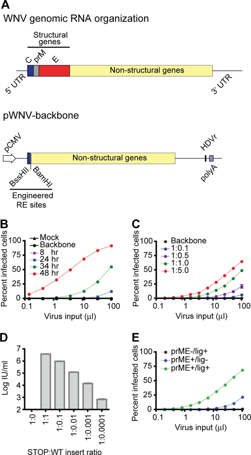
Here, we describe a novel approach that markedly simplifies the production of infectious WNV from an otherwise unstable genome. We modified a previously described two-plasmid molecular clone system of the NY99 strain of WNV (4) to construct a single plasmid encoding a truncated form of the viral genome under the transcriptional control of the CMV promoter (pWNV-backbone) (Fig. 1A); the assembly of a single plasmid encoding the full-length genome in this configuration was not possible due to significant instability in bacteria. The deleted portion of the viral genome in this construct (nucleotides 195 to 2514 of WNV NY99) encoded the entire sequences of prM and E as well as small portions of C and nonstructural protein 1 (NS1). Flanking the deleted structural gene segment are unique restriction endonuclease recognition sequences (BssHII and BamHI) introduced into the vector using synonymous mutations. The ribozyme from hepatitis delta virus (HDV) and the poly(A) signal from SV40 were introduced adjacent to the 3′ end of the genome to ensure the correct processing of the 3′ terminus and to protect the RNA from degradation. The RNA encoded by pWNV-backbone is incapable of replicating in transfected cells because the signal sequence at the amino terminus of NS1 required to direct the correct topology of downstream nonstructural proteins is within the deleted segment (16).
Fig 1.
A rapid and simple method for the production of infectious WNV. (A) Schematics of the pWNV-backbone construct. Plasmid pWNV-backbone was constructed by a modification of a two-plasmid infectious clone system of WNV NY99 (4). A truncated form of the viral genome was placed under the transcriptional control of the CMV immediate-early promoter/enhancer (pCMV). In this construct, the majority of the structural genes have been deleted, along with a small portion of the amino terminus of NS1 (nucleotides 195 to 2514 of NY99). This deleted region was flanked by unique BssHII and BamHI restriction sites introduced into the vector via synonymous mutations. The ribozyme from HDV and the poly(A) sequence signal from SV40 were placed downstream of the viral 3′ UTR to ensure the correct processing of the 3′ terminus of the genome and to protect the RNA from degradation. (B) Kinetics of WNV production using the pWNV-backbone system. A DNA fragment carrying WT structural genes was generated by PCR, ligated into pWNV-backbone, and transfected into HEK-293T cells. Virus-containing supernatants were harvested at the indicated times posttransfection. Transfections of ligation reaction mixtures lacking the structural gene fragment (backbone) or with no DNA (mock) were included as controls. The virus titer was determined by infecting Raji cells expressing the attachment factor DC-SIGNR with serial 2-fold dilutions of supernatant. Infection was scored as a function of the percentage of cells expressing viral E proteins. Data are representative of four independent experiments. (C) Effect of varying the ratio of backbone to insert (by mass) in the ligation reaction mixture. The virus titer was determined as described above. Data are representative of two independent experiments. (D) The sensitivity of the ligation-transfection approach for capturing functional structural gene fragments was evaluated by varying the ratio of the WT structural gene sequence to a nonviable mutant in the ligation reaction mixture. The ratios of the two fragments in the ligation reaction mixture are indicated, where STOP refers to the sequence obtained from pWNV-complement-5′. Virus-containing supernatants were obtained at 120 h posttransfection, and titers were determined as described above (limit of detection of 102 IU/ml). The data are representative of two independent experiments; error bars represent titer results from three dilutions of virus. Similar results were obtained when plasmids harboring WT structural gene sequences and a variant shown previously to confer sensitivity to a neutralizing antibody (57) were mixed; virus with a resistant phenotype was isolated in the presence of MAb E16 even when only 1 in 10,000 structural gene molecules encoded a resistant genotype (data not shown). (E) The production of virus using the pWNV-backbone system is not strictly ligase dependent. Ligation reaction mixtures were assembled in the presence or absence of ligase enzyme, incubated at 16°C overnight, and transfected into cells. Virus was harvested, and titers were determined at 48 h posttransfection. Data are representative of two independent experiments.
To produce infectious virus using this plasmid, a sequence carrying the deleted structural gene segment was generated by PCR and digested with restriction endonucleases that recognize BssHII and BamHI sites incorporated into the primers used for amplification. Alternatively, this fragment was obtained by the digestion of a helper plasmid (pWNV-complement) using the same enzymes. In either case, the structural gene segment was ligated into BssHII- and BamHI-digested pWNV-backbone. Next, the unpurified ligation reaction mixture was transfected directly into HEK-293T cells. Importantly, this step bypassed the requirements for both the propagation of the full-length flavivirus genome in bacteria (which is associated with significant plasmid instability) and the production and purification of viral RNA in vitro. Analysis of transfected-cell supernatants revealed a rapid release of infectious virus, detectable as early as 24 h posttransfection, which steadily increased over the course of 2 days (8.5 × 10⁶ ± 0.5 × 10⁶ infectious units [IU]/ml; n = 4 at 48 h posttransfection) (Fig. 1B). Altering the ratio of the backbone to the structural gene segment in the ligation reaction mixture demonstrated that WNV production is possible over a broad stoichiometric range (Fig. 1C), indicating that the quantity (or quality) of the insert is an important yet not critical parameter for virus recovery. In fact, this approach is sensitive to the presence of a small number of structural gene sequences against a background of nonfunctional inserts. The transfection of mixtures of WT pWNV-complement with a nonfunctional variant encoding a stop codon demonstrated the ability to produce virus when only 1 in 104 inserts in the ligation mixture harbored an intact structural gene (Fig. 1D). While the omission of DNA ligase from ligation reaction mixtures significantly reduced the efficiency of virus production (∼23-fold; n = 2), it is not absolutely dependent on ligase activity in vitro, presumably due to the activity of endogenous cellular ligases on transfected DNA (Fig. 1E) (62).
Construction of GFP-expressing WNV using a backbone construct.
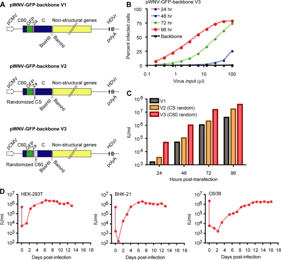
To simplify the detection and quantification of WNV-infected cells, we next modified pWNV-backbone to express GFP using a previously described configuration (43, 66) (Fig. 2A). Briefly, the gene encoding GFP was introduced into pWNV-backbone between the 5′ UTR and the C gene. This reporter gene was flanked on the 5′ end by the first 60 nucleotides of the C gene (C60) that contains RNA structures required for efficient cyclization and replication of the viral genome (30, 72). The sequence encoding the foot-and-mouth disease virus (FMDV) 2A protein was added downstream of GFP to liberate the full-length C protein from the reporter gene by translational “skipping” (12). The resulting construct is referred to as pWNV-GFP-backbone (version 1 [V1]). Because the duplication of the C60 sequence may interfere with the correct circularization of the viral genome, we constructed two additional versions of the construct containing silent mutations in the downstream C sequence. In the second version (V2), four synonymous mutations were introduced into the cyclization sequence (CS) of the downstream C gene. In version 3 (V3), the first 20 codons of the downstream C sequence were randomized by using synonymous mutations. All three constructs supported the production of infectious virions when ligated with a DNA fragment harboring the structural genes and transfected into HEK-293T cells (Fig. 2B and C). The most rapid production and highest yield of virus (1.5 × 10⁷ ± 0.2 × 10⁷ IU/ml; n = 2 at 72 h posttransfection) were achieved with pWNV-GFP-backbone V3 (Fig. 2C); this variant was used in all subsequent experiments. Notably, the kinetics of virus production using all three variants of pWNV-GFP-backbone was delayed by roughly 24 h compared to experiments with the wild-type backbone described in the legend of Fig. 1. To characterize the ability of GFP-expressing WNV to replicate in cells, one-step growth curve experiments were performed with the HEK-293T, BHK-21, and C6/36 (insect) cell lines (Fig. 2D). Virus replication and spread were observed in all three cell lines. The maximal viral yield was achieved at 3 to 4 days postinfection in mammalian cell cultures, whereas it took more than 1 week in insect C6/36 cells. Serial propagation studies of Vero cells confirmed that flaviviruses encoding GFP in this configuration were stable for at least five passages, as previously reported (66; data not shown).
Fig 2.
Production of infectious WNV carrying a GFP reporter gene. (A) Schematics of the pWNV-GFP-backbone constructs. A variant of pWNV-backbone was constructed by introducing the GFP reporter gene between the 5′ UTR and the C gene using a strategy described previously by Shustov and colleagues (66). The first 60 nucleotides of capsid (C60) were duplicated and introduced at the amino terminus of GFP to ensure the proper cyclization and replication of pWNV-GFP-backbone. Three versions of pWNV-GFP-backbone were constructed. These three versions share the same upstream C60 at the amino terminus of GFP but are different in the downstream genomic capsid. In version 1 (V1), both upstream and downstream C60 sequences are identical. In version 2 (V2), synonymous mutations were introduced into the four amino acids comprising the cyclization sequence (CS) of the downstream capsid. In version 3 (V3), the first 60 nucleotides of the downstream capsid (including the CS) were scrambled using synonymous mutations. (B) Kinetics of WNV production using V3 of the WNV-GFP-backbone system. WNV-GFP viruses were produced as described in the legend of Fig. 1. Virus-containing supernatants were harvested at the indicated times, and titers were determined by using Raji-DC-SIGNR cells. Infection was scored as a function of GFP expression at 16 h postinfection. Data are representative of two independent experiments. (C) Comparison of the kinetics of WNV production using the three versions of the WNV-GFP-backbone system. The infectious titer of virus in supernatants harvested at the indicated hours posttransfection was calculated by using the linear portion of the virus dose-infectivity curve. Data are representative of two separate experiments. (D) Growth kinetics of WNV production using WNV-GFP-backbone V3. A virus stock produced in HEK-293T cells was used to inoculate HEK-293T, BHK-21, and C6/36 cells at a multiplicity of infection (MOI) of 0.5. Viruses were collected from infected cells at the indicated time points, and titers were determined on Raji-DC-SIGNR cells. Similar results were obtained from independent experiments performed at MOIs of 3 and 0.05.
Rapid construction of a library of variants in the prM protein.
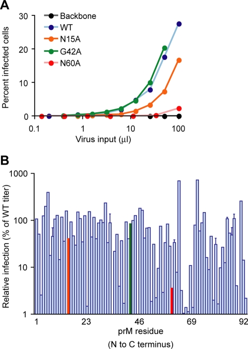
The ability to produce WNV using a strategy that does not involve conventional cloning procedures suggested that this approach would be an efficient and rapid method for introducing mutations into the structural genes of the virus. As a proof of principle, we constructed a panel of 92 viruses encoding single alanine or glycine substitutions into each position of the “pr” portion of prM. Mutations were introduced into the structural gene segment by OE-PCR, ligated into pWNV-GFP-backbone, and transfected directly into HEK-293T cells. The relative infectivity of the virus released from transfected cells was assessed on day 3 by infecting Raji-DC-SIGNR cells (10) with serial 2-fold dilutions of virus-containing supernatants for a set of mutants. The titer of each variant was calculated by using linear portions of the virus-dose infectivity curve and is shown relative to the titer of virus produced in parallel cultures using the WT prM sequence (Fig. 3A and B). Analysis of the sequence of viral RNA in the transfection supernatants of each infectious clone verified the presence of the desired mutation in prM. Infectious virus incorporating the engineered mutation was recovered in the majority of cases (89 of 92); when harvested at 72 h posttransfection, ∼20% of the mutants displayed a greater-than-20-fold reduction in titer relative to that of the WT.
Fig 3.
Rapid mutagenesis of the prM gene of WNV using pWNV-GFP-backbone. A panel of viruses encoding single-amino-acid substitutions in the 92 amino acids of the “pr” portion of the prM protein was constructed. PCR fragments encoding an alanine mutation at each position were generated by OE-PCR; alanine residues of prM were replaced by glycine substitutions. Each PCR fragment was ligated into pWNV-GFP-backbone V3 and transfected into HEK-293T cells. Virus-containing supernatants were harvested at 72 h posttransfection; the titer was determined as described in the legend of Fig. 2. (A) Production of the WT and representative prM variants N15A, G42A, and N60A. Data from representative prM variants are shown and color coded to correspond to the data presented in the bottom panel. (B) The infectious titer of each prM variant was determined from linear portions of the virus dose-infectivity curve. Infectivity is expressed as the titer of each variant relative to those determined in paired experiments performed with the WT virus (×100%). In cases where independent preparations of virus were produced, error bars represent standard errors.
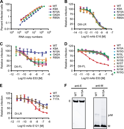
The efficiency of virion maturation has been shown to markedly influence the sensitivity of WNV to neutralization by MAbs specific for several structurally distinct epitopes and polyclonal antibodies elicited by two candidate WNV vaccines (47). For example, MAb E53 is an E-DII-fusion loop (E-DII-FL)-reactive antibody that has a limited capacity to neutralize mature virions; previous structural studies demonstrated that this MAb binds E proteins only in the conformation associated with the prM protein present on immature and partially mature virions (8). To explore the feasibility of using a library of mutants to study residues in prM that modulate neutralization by antibodies that bind the E protein, four variants were selected for further study, including the asparagine residue critical for glycosylation (N15A) and a residue that is part of one of the two overlapping cleavage recognition motifs for the cellular furin protease (R85A). Alanine substitution variants at K9, N15, G42, and R85 were produced by using pWNV-GFP-backbone and mutated forms of pWNV-complement constructed by using structural gene sequence obtained from viruses produced as part of the studies described in the legend of Fig. 3. Virus-containing supernatants were collected at 72 h posttransfection, and titers were subsequently determined on Raji-DC-SIGNR cells. The viral RNA content of each virus stock was measured by qRT-PCR (Fig. 4A). These studies indicated that differences in specific infectivity between WT and mutant viruses were less than 2-fold.
Fig 4.
Neutralization of prM mutants by monoclonal antibodies. PCR fragments containing K9A, N15A, G42A, and R85A mutations or the WT sequence were generated by using viral sequences cloned during the experiments described in the legend of Fig. 3 and ligated into pWNV-GFP-backbone to produce WNV-GFP viruses. (A) Specific infectivity of the WT virus and mutant viruses encoding K9A, N15A, G42A, or R85A prM mutations. Virus was harvested from culture supernatants at 72 h posttransfection, and titers were determined as described above. Infectivity is expressed as a function of the viral RNA content of DNase-treated virus stocks. (B and C) Determinations of the sensitivities of the WT and the K9A, N15A, G42A, and R85A mutants to neutralization by MAbs E16 (B) and E53 (C) were performed as described previously (57). Assays were performed with two independent virus stocks, and data are representative of a total of three independent experiments. (D) Sensitivity of the WT or the N15 variants to neutralization by MAb E53. Viruses were generated and titers were determined as described above for panel A. Sensitivity to MAb E53 neutralization was determined as described above. (E) Sensitivities of the WT and the N15A and T17A mutants to neutralization by MAb E121. Viruses were generated, titers were determined, and sensitivity to MAb E121 neutralization was determined as described above. Data shown are representative of two independent experiments. DIII-LR, E protein domain III lateral ridge; DII-FL, E protein domain II fusion loop; DI-LR, E protein domain I lateral ridge. (F) Western blot analysis for detection of WNV E, prM, and M proteins. WT and N15A viruses were harvested from infected BHK-21 cells, and the levels of WNV E, prM, and M proteins were determined by Western blotting using anti-E (MAb 4G2) and anti-M antibodies.
We next examined the sensitivity of each variant to neutralization by MAbs shown previously to be sensitive to the maturation state of WNV. As a control, we used the well-characterized WNV-specific MAb E16, which recognizes E-DIII-LR; neutralization by this antibody is not sensitive to the maturation state of the virus particle (50, 51). All variants were equally sensitive to neutralization by E16 (Fig. 4B). In contrast, a significant difference was observed for the sensitivity of the N15A variant of WNV to neutralization by MAbs E53 (Fig. 4C) and E121 (data not shown). Both of these antibodies were shown previously to be sensitive to the maturation state of the virus particle (47). Similar results were obtained with other substitutions at N15, or with a T17A variant (which is also part of the N-linked glycosylation signal), indicating that the change in sensitivity was likely due to the absence of the carbohydrate rather than a change in the amino acid itself (Fig. 4D and E). Furthermore, a biochemical analysis of the efficiency of prM cleavage by the N15A variant revealed that it was similar to the WT virus (Fig. 4F), suggesting that the carbohydrate on prM is not critical for the efficient cleavage of prM, consistent with data from a prior study of Japanese encephalitis virus (34). The mechanism of resistance of the N15A variant to neutralization by maturation-state-sensitive antibodies is currently being investigated.
Directed evolution of WNV.
Despite the relatively low fidelity of the RNA polymerases used by viruses to replicate their genomes (7), virus adaptation experiments can be limited by the variant frequency and size of the virus population achievable in vitro, particularly when multiple mutations are required for adaptation to a particular selective pressure (75). Because our approach for producing WNV does not include a cloning step that may bottleneck genetic diversity, we explored the possibility of using this molecular clone system to facilitate virus adaptation studies by significantly increasing the genetic complexity of virus populations. The feasibility of using this system in selection experiments was evaluated in the context of identifying mutations that confer escape from antibody-mediated neutralization. Biochemical mapping and structural studies both demonstrated that interactions between MAb E16 and its epitope are mediated primarily by four residues on the E protein (S306, K307, T330, and T332) (50, 51). Mutations at T332 were shown previously to result in escape from neutralization by E16 and other DIII-LR-specific antibodies in vitro and in vivo (3, 57, 78).
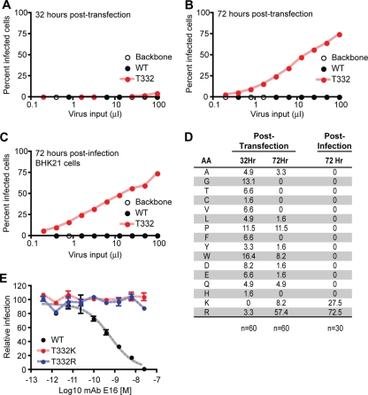
To generate a population of viruses that differ randomly at the T332 residue contacted by E16, we amplified the WNV structural genes using OE-PCR and primers with degenerate sequences at the T332 codon. This pool of PCR fragments was digested with restriction enzymes, ligated into the pWNV-backbone construct, and transfected into HEK-293T cells in the presence of a relatively high concentration of E16 (5 μg/ml; the 50% effective concentration [EC50] of this antibody is ∼0.01 μg/ml). Virus-containing supernatants were harvested at 32 h posttransfection, and titers were determined on Raji-DC-SIGNR cells in the presence of MAb E16 (Fig. 5A). Infectious virus was detected in the T332 population at very low levels (∼6.0 × 10⁴ IU/ml); no infectious virus was observed for cultures transfected with the WT sequence. Analysis of the sequences of viruses released from transfected cells at this time point revealed extensive variation at T332; 15 different amino acid substitutions were represented in a survey of 60 cloned sequences (Fig. 5D). Analysis at 72 h posttransfection, however, revealed the growth of virus resistant to neutralization by E16 (Fig. 5B) and a marked change in the proportion of amino acid variants in transfected-cell supernatants (Fig. 5D). Notably, the frequencies of codons for arginine (R) and lysine (K) residues increased remarkably during this sampling interval (3.3% to 57.4% and undetected to 8.2%, respectively). A single passage of the virus onto BHK-21 cells in the presence of E16 yielded an even more homogeneous resistant population composed solely of the lysine and arginine variants (Fig. 5C and D). While the T332K substitution was shown previously to reduce binding and neutralization by E16 (51, 57), a T332R variant has not been described. To confirm that both substitutions were sufficient to confer a resistant phenotype, structural genes encoding these variants were reintroduced into pWNV-GFP-backbone and used to produce virus. Titers of virus-containing supernatants were then determined on Raji-DC-SIGNR cells, and these viruses were tested for sensitivity to neutralization by E16 (Fig. 5E). Both variants displayed high-level resistance to E16.
Fig 5.
Directed evolution of WNV: rapid escape from a neutralizing antibody. A PCR fragment containing random nucleotides at all three positions of codon 332 of the E protein was generated by overlap-extension PCR using degenerate primers and ligated into pWNV-GFP-backbone. Ligation reaction mixtures were transfected into HEK-293T cells in the presence of a high concentration of E16 (5 μg/ml). PCR amplicons encoding the WT sequence were processed in parallel. Transfections of ligation reaction mixtures lacking the structural gene fragment (Backbone) were included as a negative control. (A and B) Virus-containing supernatants were harvested from transfected HEK-293T cells at 32 h (A) and 72 h (B) posttransfection, and titers were determined in the presence of 5 μg/ml of E16 using Raji-DC-SIGNR cells as described above. (C) A single passage of virus harvested from transfected cells at 72 h posttransfection on BHK-21 cells was performed in the presence of E16 (5 μg/ml). The titer of virus released from BHK-21 cells at 72 h postinfection was determined, and the virus was sequenced as described above. (D) The proportion of T332 codons encoding a particular amino acid (AA) was monitored by sequence analysis of viral RNA. RNA was isolated from culture supernatants and used to amplify the structural gene segment. PCR products were cloned into the pDonr221 entry vector by using Gateway cloning (Invitrogen). The percentages of clones that encoded a particular amino acid are presented for viruses harvested from transfected cells (n = 60 for viruses in panels A and B) and infected cells (n = 30 for viruses in panel C). (E) Sensitivity of the T332K and T332R variants to neutralization by MAb E16. The prME fragments encoding the T332K or T332R variant were cut from the constructed clones in panel C and ligated into plasmid pWNV-GFP-backbone to generate viruses. Titers of virus-containing supernatants were then determined on Raji-DC-SIGNR cells, and the virus was tested for sensitivity to neutralization by E16 as described previously (57).
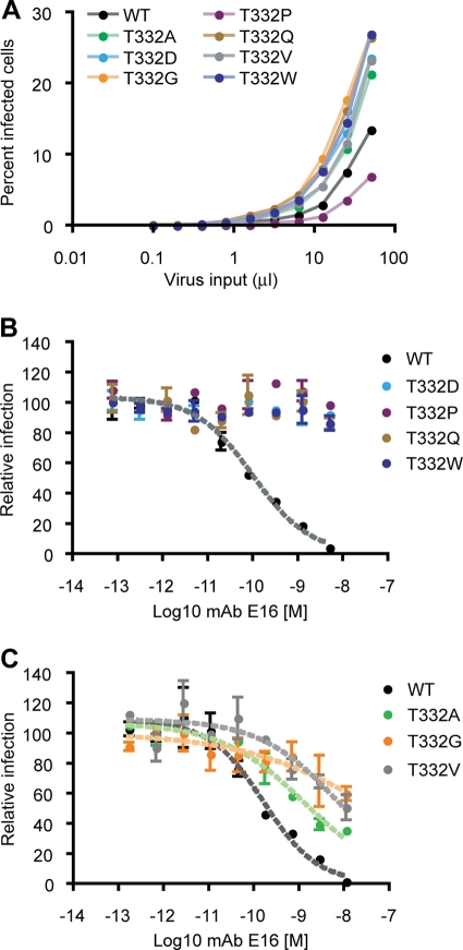
The selection experiment described above identified two variants that allowed for escape from neutralization. However, it is important to note that antibody pressure was not the only selective force in the experiment; our approach identified the most fit variants in the presence of antibody. To investigate the dynamics of our selection experiment in greater detail, we investigated the sensitivities of other variants detected in the supernatants of HEK-293T cells to neutralization by E16 at 32 h posttransfection (Fig. 5D). Viruses encoding the T332A, T332D, T332G, T332P, T332Q, T332V, and T332W substitutions were produced by using the ligation-transfection strategy described above, harvested at 72 h posttransfection, and titered on Raji-DC-SIGNR cells (Fig. 6A). Neutralization studies with E16 revealed two patterns. Four variants (D, P, Q, and W) were completely resistant to neutralization (Fig. 6B), as observed with the T332K and T332R viruses identified by selection (Fig. 5E). The T332A, T332G, and T332V variants were markedly less sensitive to neutralization by E16 (characterized by 3.7-, 5.6-, and 5.5-fold reductions in neutralization, respectively) (Fig. 6C). In agreement with our findings, a previous yeast display study revealed a 15 to 19% decrease in the binding of E16 to the T322A and T332V variants, respectively (51). The fact that many of the variants produced from transfected cells were resistant to E16 yet not maintained after a single passage suggests that factors beyond antibody binding contributed to the selection of the most fit variant during our antibody selection studies. Altogether, these studies confirm the feasibility of using this molecular clone technology to increase the genetic heterogeneity of WNV populations and rapidly identify variants with desirable phenotypes under selection.
Fig 6.
Neutralization of envelope T332 mutants by monoclonal antibody E16. (A) The production of E protein T332 variants that were not selected for by E16 in experiments described in the legend of Fig. 5 was accomplished by using WNV-GFP-backbone as described in the legend to Fig. 2. (B and C) The sensitivity of each variant to neutralization by E16 was established as previously described (57). Data are representative of two independent experiments.
DISCUSSION
In this study, we describe a novel molecular clone system for the production of WNV that simplifies the methodology required to produce infectious virus and allows for the introduction of genetic variation into the structural genes on an unprecedented scale. An innovative aspect of this approach is that it does not require conventional cloning procedures, which can be time-consuming and challenging due to the instability of many existing flavivirus molecular clone plasmids. Instead, a sequence carrying the structural genes, which can be manipulated in a defined or random fashion using PCR, is ligated into a truncated stable molecular clone construct and transfected directly into cells. This translates into a significantly reduced time required for mutagenesis. In our experience, this has enabled the construction and partial characterization of new WNV variants at a rate of ∼24 full-length viruses/week/investigator.
The applications of this approach for studying the biology of flaviviruses are numerous. The ability to create large panels of mutants will enhance the resolution of structure-function studies by allowing the exploration of the effect of all possible amino acid substitutions at a given residue rather than a limited number of selected mutations. Alternatively, the ability to study a larger fraction of the genetic space of the structural genes of WNV allows a more unbiased approach for an understanding of structure-function relationships. In this study, we demonstrate both principles by rapidly generating a panel of variants at each position of the prM protein (Fig. 3), followed by the more intensive mutagenesis of a residue of interest (Fig. 5), as discussed further below.
The utility of the molecular clone strategy described here extends beyond logistical advantages for virus production and mutagenesis. The determination of the molecular basis of adaptation to selection is a powerful approach used extensively in virology. While the selection of variants at a single position in the genome of an RNA virus is trivial in vitro due to the low fidelity of the RNA-dependent RNA polymerase, the selection of variants under conditions that require multiple substitutions is improbable. In the absence of recombination, the frequency with which variants encoding two substitutions arise can be estimated as the product of the variant frequency of each single change; for a flavivirus, this is roughly 1/1010 (26). The ability to produce virus with extensive genetic diversity, either at defined positions or randomly throughout the structural genes, will increase the complexity of selection schemes that can be investigated or used to create viruses with desirable phenotypes. Furthermore, it provides an interesting opportunity to study the dynamics and evolution of a large population of viral sequences under selection (36).
The ability to create large panels of mutants will enhance the resolution of structure-function studies by allowing the exploration of the effect of all possible amino acid substitutions at a given residue rather than a limited number of selected mutations. We note that while this technology streamlines the molecular biology of handling WNV molecular clones and virus production, the requirements to characterize the resulting viruses remain the same. Not all mutations will be stable or will have predictable phenotypes; the most insightful results will be obtained from fully sequenced, biologically cloned virus stocks regardless of the method of production.
Analysis of the functional properties of anti-flavivirus antibodies.
The functional properties of antibodies that bind flaviviruses are determined by a combination of factors, including the affinity and isotype of the antibody molecule as well as where it binds the virus particle (reviewed in reference 14). This complexity is best studied in the context of authentic virions. Mutagenesis is an established approach for studying antibody reactivity, which may be accelerated by this molecular clone technology (6, 9, 18, 19, 37, 42, 51, 68, 77). Panels of structural protein variants of WNV can be constructed by using the ligation-transfection method described here and screened for sensitivity to antibody-mediated neutralization. The ease of construction of large numbers of mutants increases the chance of identifying interesting phenotypes from variants that might not be included in more limited mutagenesis studies performed by using conventional cloning methods.
In this study, 86 alanine and 6 glycine mutations were introduced into the “pr” portion of the WNV protein, and a subset of these variants was screened for sensitivity to neutralization by E protein-reactive antibodies (Fig. 4). Mutations that prevent the glycosylation of WNV prM on residue 15 significantly reduced the sensitivity of viruses to neutralization by antibodies shown previously to be sensitive to the maturation state of the virion (47). Of significant interest, biochemical studies of the cleavage efficiency of N15A virions produced in HEK-293T cells did not reveal an increased efficiency of virion maturation (prM cleavage) that could explain the resistance of these virions to maturation-sensitive antibodies. In agreement with these studies, the ablation of the N-linked sugar on Japanese encephalitis virus prM does not significantly change the maturation of this virus (34). The molecular basis for the resistance of variants lacking the glycosylation site in prM to neutralization requires further investigation. One interesting possibility is that the removal of the carbohydrate on the prM protein changes the distribution of uncleaved prM on infectious virus particles. The stoichiometric requirements for the cleavage of prM on infectious “partially mature” virions are unknown. While uncleaved prM may be distributed widely among wild-type virions released from cells, as suggested by a recent study of dengue virus (27), the cleavage of prM on the N15 variant may occur more or less in an all-or-none pattern, resulting in a population with fewer partially mature virions and a greater proportion of fully mature (neutralization-resistant) or immature (noninfectious) virus particles.
Several approaches have been used to investigate the complexity of the humoral response to flavivirus infection. The use of recombinant proteins to enumerate and characterize the repertoire of antibodies elicited by infection in vivo has been informative. For example, previous studies of flaviviruses demonstrated that antibodies that bind the highly conserved fusion loop at the tip of E-DII constitute a significant fraction of antibodies elicited by infection (9, 52). However, these approaches do not directly provide insight into specificities that contribute to the functional complexity of the humoral response, such as virus neutralization. Furthermore, analyses of antibody specificity using recombinant proteins have limitations that arise from the complexity of the virus particle. Epitopes that are readily bound by antibody molecules on recombinant proteins may not be accessible for antibody binding on the virus particle (69); epitope accessibility may be modulated further by the maturation state of the virus particle (8, 47) and the dynamic motion of E proteins incorporated into the virions (13, 40). Conversely, composite epitopes composed of more than one E protein subunit of the icosahedral virion may not be present on recombinant proteins (29). To study the complexity of the interaction of antibodies, we have employed pseudoinfectious reporter virus particles (RVPs) that incorporate the structural genes of several flaviviruses (1, 44, 47, 56, 57). RVPs are produced by the genetic complementation of a subgenomic replicon, are capable of only a single round of infection, and allow virus entry to be scored as a function of reporter gene expression (33). One desirable feature of this approach is that it allows the study of variants of the structural genes simply by changing the plasmids used during complementation (48). However, the scope of these efforts is limited by the number of structural gene plasmids that can be constructed. Furthermore, because RVPs cannot support multiple rounds of replication, insight into flavivirus biology arising from the selection of desirable phenotypes is not possible.
Additional applications in virology.
The concept of transfecting eukaryotic cells with ligated fragments of viral genomes is not limited to studies of the structural genes of flaviviruses. pWNV-backbone was designed with a large deletion encompassing the structural genes due to the potential contribution of E gene sequences to plasmid stability and our interests in the biology of the E protein. However, ligations involving three or more pieces may be possible using molecular clones like pWNV-backbone bearing deletions in other regions. In cases where relatively stable “DNA-launched” molecular clones exist, rapid and large-scale mutagenesis of any region of the genome is possible by ligating mutated fragments into existing restriction endonuclease recognition sites (30, 54). For flaviviruses, the spatial and temporal coupling between RNA replication and packaging (32) and a failure of many of the nonstructural proteins to function in trans (31) increase the likelihood that the genotype of the RNA packaged into a virion corresponds to the phenotype of the structural proteins that comprise the virion. DNA-launched molecular clones of other positive-stranded viruses have been developed; it is likely that this approach will be of considerable value in the study of other RNA viruses.
ACKNOWLEDGMENTS
This work was funded by the intramural research program of the National Institute of Allergy and Infectious Diseases, NIH.
We thank Michael Diamond, Thomas Kristie, and Heather Hickman for critically reading the manuscript and for helpful discussions.
We declare no conflicts of interest.
Footnotes
Published ahead of print 18 January 2012
REFERENCES
- 1. Ansarah-Sobrinho C, Nelson S, Jost CA, Whitehead SS, Pierson TC. 2008. Temperature-dependent production of pseudoinfectious dengue reporter virus particles by complementation. Virology 381:67–74 [DOI] [PMC free article] [PubMed] [Google Scholar]
- 2. Asnis DS, Conetta R, Teixeira AA, Waldman G, Sampson BA. 2000. The West Nile virus outbreak of 1999 in New York: the Flushing Hospital experience. Clin. Infect. Dis. 30:413–418 [DOI] [PubMed] [Google Scholar]
- 3. Beasley DW, Barrett AD. 2002. Identification of neutralizing epitopes within structural domain III of the West Nile virus envelope protein. J. Virol. 76:13097–13100 [DOI] [PMC free article] [PubMed] [Google Scholar]
- 4. Beasley DW, et al. 2005. Envelope protein glycosylation status influences mouse neuroinvasion phenotype of genetic lineage 1 West Nile virus strains. J. Virol. 79:8339–8347 [DOI] [PMC free article] [PubMed] [Google Scholar]
- 5. Beltramello M, et al. 2010. The human immune response to dengue virus is dominated by highly cross-reactive antibodies endowed with neutralizing and enhancing activity. Cell Host Microbe 8:271–283 [DOI] [PMC free article] [PubMed] [Google Scholar]
- 6. Calvert AE, et al. 2011. Human monoclonal antibodies to West Nile virus identify epitopes on the prM protein. Virology 410:30–37 [DOI] [PubMed] [Google Scholar]
- 7. Castro C, Arnold JJ, Cameron CE. 2005. Incorporation fidelity of the viral RNA-dependent RNA polymerase: a kinetic, thermodynamic and structural perspective. Virus Res. 107:141–149 [DOI] [PMC free article] [PubMed] [Google Scholar]
- 8. Cherrier MV, et al. 2009. Structural basis for the preferential recognition of immature flaviviruses by a fusion-loop antibody. EMBO J. 28:3269–3276 [DOI] [PMC free article] [PubMed] [Google Scholar]
- 9. Crill WD, Trainor NB, Chang GJ. 2007. A detailed mutagenesis study of flavivirus cross-reactive epitopes using West Nile virus-like particles. J. Gen. Virol. 88:1169–1174 [DOI] [PubMed] [Google Scholar]
- 10. Davis CW, et al. 2006. West Nile virus discriminates between DC-SIGN and DC-SIGNR for cellular attachment and infection. J. Virol. 80:1290–1301 [DOI] [PMC free article] [PubMed] [Google Scholar]
- 11. Dejnirattisai W, et al. 2010. Cross-reacting antibodies enhance dengue virus infection in humans. Science 328:745–748 [DOI] [PMC free article] [PubMed] [Google Scholar]
- 12. Donnelly ML, et al. 2001. Analysis of the aphthovirus 2A/2B polyprotein ‘cleavage’ mechanism indicates not a proteolytic reaction, but a novel translational effect: a putative ribosomal ‘skip’. J. Gen. Virol. 82:1013–1025 [DOI] [PubMed] [Google Scholar]
- 13. Dowd KA, Jost CA, Durbin AP, Whitehead SS, Pierson TC. 2011. A dynamic landscape for antibody binding modulates antibody-mediated neutralization of West Nile virus. PLoS Pathog. 7:e1002111. [DOI] [PMC free article] [PubMed] [Google Scholar]
- 14. Dowd KA, Pierson TC. 2011. Antibody-mediated neutralization of flaviviruses: a reductionist view. Virology 411:306–315 [DOI] [PMC free article] [PubMed] [Google Scholar]
- 15. Elshuber S, Allison SL, Heinz FX, Mandl CW. 2003. Cleavage of protein prM is necessary for infection of BHK-21 cells by tick-borne encephalitis virus. J. Gen. Virol. 84:183–191 [DOI] [PubMed] [Google Scholar]
- 16. Falgout B, Chanock R, Lai CJ. 1989. Proper processing of dengue virus nonstructural glycoprotein NS1 requires the N-terminal hydrophobic signal sequence and the downstream nonstructural protein NS2a. J. Virol. 63:1852–1860 [DOI] [PMC free article] [PubMed] [Google Scholar]
- 17. Geiss BJ, Pierson TC, Diamond MS. 2005. Actively replicating West Nile virus is resistant to cytoplasmic delivery of siRNA. Virol. J. 2:53. [DOI] [PMC free article] [PubMed] [Google Scholar]
- 18. Gromowski GD, Barrett AD. 2007. Characterization of an antigenic site that contains a dominant, type-specific neutralization determinant on the envelope protein domain III (ED3) of dengue 2 virus. Virology 366:349–360 [DOI] [PubMed] [Google Scholar]
- 19. Gromowski GD, Barrett ND, Barrett AD. 2008. Characterization of dengue virus complex-specific neutralizing epitopes on envelope protein domain III of dengue 2 virus. J. Virol. 82:8828–8837 [DOI] [PMC free article] [PubMed] [Google Scholar]
- 20. Guirakhoo F, Bolin RA, Roehrig JT. 1992. The Murray Valley encephalitis virus prM protein confers acid resistance to virus particles and alters the expression of epitopes within the R2 domain of E glycoprotein. Virology 191:921–931 [DOI] [PMC free article] [PubMed] [Google Scholar]
- 21. Guirakhoo F, Heinz FX, Mandl CW, Holzmann H, Kunz C. 1991. Fusion activity of flaviviruses: comparison of mature and immature (prM-containing) tick-borne encephalitis virions. J. Gen. Virol. 72(Pt 6):1323–1329 [DOI] [PubMed] [Google Scholar]
- 22. Harrison SC. 2008. Viral membrane fusion. Nat. Struct. Mol. Biol. 15:690–698 [DOI] [PMC free article] [PubMed] [Google Scholar]
- 23. Heinz FX, et al. 1994. Structural changes and functional control of the tick-borne encephalitis virus glycoprotein E by the heterodimeric association with protein prM. Virology 198:109–117 [DOI] [PubMed] [Google Scholar]
- 24. Ho SN, Hunt HD, Horton RM, Pullen JK, Pease LR. 1989. Site-directed mutagenesis by overlap extension using the polymerase chain reaction. Gene 77:51–59 [DOI] [PubMed] [Google Scholar]
- 25. Hubalek Z, Halouzka J. 1999. West Nile fever—a reemerging mosquito-borne viral disease in Europe. Emerg. Infect. Dis. 5:643–650 [DOI] [PMC free article] [PubMed] [Google Scholar]
- 26. Jin Z, Deval J, Johnson KA, Swinney DC. 2011. Characterization of the elongation complex of dengue virus RNA polymerase: assembly, kinetics of nucleotide incorporation, and fidelity. J. Biol. Chem. 286:2067–2077 [DOI] [PMC free article] [PubMed] [Google Scholar]
- 27. Junjhon J, et al. 2010. Influence of pr-M cleavage on the heterogeneity of extracellular dengue virus particles. J. Virol. 84:8353–8358 [DOI] [PMC free article] [PubMed] [Google Scholar]
- 28. Kaufmann B, et al. 2006. West Nile virus in complex with the Fab fragment of a neutralizing monoclonal antibody. Proc. Natl. Acad. Sci. U. S. A. 103:12400–12404 [DOI] [PMC free article] [PubMed] [Google Scholar]
- 29. Kaufmann B, et al. 2010. Neutralization of West Nile virus by cross-linking of its surface proteins with Fab fragments of the human monoclonal antibody CR4354. Proc. Natl. Acad. Sci. U. S. A. 107:18950–18955 [DOI] [PMC free article] [PubMed] [Google Scholar]
- 30. Khromykh AA, Meka H, Guyatt KJ, Westaway EG. 2001. Essential role of cyclization sequences in flavivirus RNA replication. J. Virol. 75:6719–6728 [DOI] [PMC free article] [PubMed] [Google Scholar]
- 31. Khromykh AA, Sedlak PL, Westaway EG. 2000. cis- and trans-acting elements in flavivirus RNA replication. J. Virol. 74:3253–3263 [DOI] [PMC free article] [PubMed] [Google Scholar]
- 32. Khromykh AA, Varnavski AN, Sedlak PL, Westaway EG. 2001. Coupling between replication and packaging of flavivirus RNA: evidence derived from the use of DNA-based full-length cDNA clones of Kunjin virus. J. Virol. 75:4633–4640 [DOI] [PMC free article] [PubMed] [Google Scholar]
- 33. Khromykh AA, Varnavski AN, Westaway EG. 1998. Encapsidation of the flavivirus Kunjin replicon RNA by using a complementation system providing Kunjin virus structural proteins in trans. J. Virol. 72:5967–5977 [DOI] [PMC free article] [PubMed] [Google Scholar]
- 34. Kim JM, et al. 2008. A single N-linked glycosylation site in the Japanese encephalitis virus prM protein is critical for cell type-specific prM protein biogenesis, virus particle release, and pathogenicity in mice. J. Virol. 82:7846–7862 [DOI] [PMC free article] [PubMed] [Google Scholar]
- 35. Kuhn RJ, et al. 2002. Structure of dengue virus: implications for flavivirus organization, maturation, and fusion. Cell 108:717–725 [DOI] [PMC free article] [PubMed] [Google Scholar]
- 36. Lauring AS, Andino R. 2010. Quasispecies theory and the behavior of RNA viruses. PLoS Pathog. 6:e1001005. [DOI] [PMC free article] [PubMed] [Google Scholar]
- 37. Li L, Barrett AD, Beasley DW. 2005. Differential expression of domain III neutralizing epitopes on the envelope proteins of West Nile virus strains. Virology 335:99–105 [DOI] [PubMed] [Google Scholar]
- 38. Li L, et al. 2008. The flavivirus precursor membrane-envelope protein complex: structure and maturation. Science 319:1830–1834 [DOI] [PubMed] [Google Scholar]
- 39. Lindenbach BD, Thiel HJ, Rice CM. 2007. Flaviviridae: the viruses and their replication, p 1101–1152 In Knipe DM, et al. (ed), Fields virology, 5th ed, vol 1 Lippincott Williams & Wilkins, Philadelphia, PA [Google Scholar]
- 40. Lok SM, et al. 2008. Binding of a neutralizing antibody to dengue virus alters the arrangement of surface glycoproteins. Nat. Struct. Mol. Biol. 15:312–317 [DOI] [PubMed] [Google Scholar]
- 41. Mackenzie JS, Gubler DJ, Petersen LR. 2004. Emerging flaviviruses: the spread and resurgence of Japanese encephalitis, West Nile and dengue viruses. Nat. Med. 10:S98–S109 [DOI] [PubMed] [Google Scholar]
- 42. Matsui K, et al. 2009. Characterization of dengue complex-reactive epitopes on dengue 3 virus envelope protein domain III. Virology 384:16–20 [DOI] [PubMed] [Google Scholar]
- 43. McGee CE, et al. 2009. Infection, dissemination, and transmission of a West Nile virus green fluorescent protein infectious clone by Culex pipiens quinquefasciatus mosquitoes. Vector Borne Zoonotic Dis. 10:267–274 [DOI] [PMC free article] [PubMed] [Google Scholar]
- 44. Mehlhop E, et al. 2009. Complement protein C1q reduces the stoichiometric threshold for antibody-mediated neutralization of West Nile virus. Cell Host Microbe 6:381–391 [DOI] [PMC free article] [PubMed] [Google Scholar]
- 45. Montgomery SP, et al. 2005. Rhabdomyolysis in patients with West Nile encephalitis and meningitis. Vector Borne Zoonotic Dis. 5:252–257 [DOI] [PubMed] [Google Scholar]
- 46. Mukhopadhyay S, Kuhn RJ, Rossmann MG. 2005. A structural perspective of the flavivirus life cycle. Nat. Rev. Microbiol. 3:13–22 [DOI] [PubMed] [Google Scholar]
- 47. Nelson S, et al. 2008. Maturation of West Nile virus modulates sensitivity to antibody-mediated neutralization. PLoS Pathog. 4:e1000060. [DOI] [PMC free article] [PubMed] [Google Scholar]
- 48. Nelson S, Poddar S, Lin TY, Pierson TC. 2009. Protonation of individual histidine residues is not required for the pH-dependent entry of West Nile virus: evaluation of the “histidine switch” hypothesis. J. Virol. 83:12631–12635 [DOI] [PMC free article] [PubMed] [Google Scholar]
- 49. Nybakken GE, Nelson CA, Chen BR, Diamond MS, Fremont DH. 2006. Crystal structure of the West Nile virus envelope glycoprotein. J. Virol. 80:11467–11474 [DOI] [PMC free article] [PubMed] [Google Scholar]
- 50. Nybakken GE, et al. 2005. Structural basis of West Nile virus neutralization by a therapeutic antibody. Nature 437:764–769 [DOI] [PMC free article] [PubMed] [Google Scholar]
- 51. Oliphant T, et al. 2005. Development of a humanized monoclonal antibody with therapeutic potential against West Nile virus. Nat. Med. 11:522–530 [DOI] [PMC free article] [PubMed] [Google Scholar]
- 52. Oliphant T, et al. 2007. Induction of epitope-specific neutralizing antibodies against West Nile virus. J. Virol. 81:11828–11839 [DOI] [PMC free article] [PubMed] [Google Scholar]
- 53. Oliphant T, et al. 2006. Antibody recognition and neutralization determinants on domains I and II of West Nile virus envelope protein. J. Virol. 80:12149–12159 [DOI] [PMC free article] [PubMed] [Google Scholar]
- 54. Pierson TC, et al. 2005. An infectious West Nile virus that expresses a GFP reporter gene. Virology 334:28–40 [DOI] [PubMed] [Google Scholar]
- 55. Pierson TC, Fremont DH, Kuhn RJ, Diamond MS. 2008. Structural insights into the mechanisms of antibody-mediated neutralization of flavivirus infection: implications for vaccine development. Cell Host Microbe 4:229–238 [DOI] [PMC free article] [PubMed] [Google Scholar]
- 56. Pierson TC, et al. 2006. A rapid and quantitative assay for measuring antibody-mediated neutralization of West Nile virus infection. Virology 346:53–65 [DOI] [PubMed] [Google Scholar]
- 57. Pierson TC, et al. 2007. The stoichiometry of antibody-mediated neutralization and enhancement of West Nile virus infection. Cell Host Microbe 1:135–145 [DOI] [PMC free article] [PubMed] [Google Scholar]
- 58. Rice CM, Grakoui A, Galler R, Chambers TJ. 1989. Transcription of infectious yellow fever RNA from full-length cDNA templates produced by in vitro ligation. New Biol. 1:285–296 [PubMed] [Google Scholar]
- 59. Rodenhuis-Zybert IA, et al. 2010. Immature dengue virus: a veiled pathogen? PLoS Pathog. 6:e1000718. [DOI] [PMC free article] [PubMed] [Google Scholar]
- 60. Roehrig JT. 2003. Antigenic structure of flavivirus proteins. Adv. Virus Res. 59:141–175 [DOI] [PubMed] [Google Scholar]
- 61. Rossi SL, Ross TM, Evans JD. 2010. West Nile virus. Clin. Lab. Med. 30:47–65 [DOI] [PMC free article] [PubMed] [Google Scholar]
- 62. Roth DB, Wilson JH. 1985. Relative rates of homologous and nonhomologous recombination in transfected DNA. Proc. Natl. Acad. Sci. U. S. A. 82:3355–3359 [DOI] [PMC free article] [PubMed] [Google Scholar]
- 63. Ruggli N, Rice CM. 1999. Functional cDNA clones of the Flaviviridae: strategies and applications. Adv. Virus Res. 53:183–207 [DOI] [PubMed] [Google Scholar]
- 64. Sejvar JJ. 2007. The long-term outcomes of human West Nile virus infection. Clin. Infect. Dis. 44:1617–1624 [DOI] [PubMed] [Google Scholar]
- 65. Shi PY, Tilgner M, Lo MK, Kent KA, Bernard KA. 2002. Infectious cDNA clone of the epidemic West Nile virus from New York City. J. Virol. 76:5847–5856 [DOI] [PMC free article] [PubMed] [Google Scholar]
- 66. Shustov AV, Mason PW, Frolov I. 2007. Production of pseudoinfectious yellow fever virus with a two-component genome. J. Virol. 81:11737–11748 [DOI] [PMC free article] [PubMed] [Google Scholar]
- 67. Stadler K, Allison SL, Schalich J, Heinz FX. 1997. Proteolytic activation of tick-borne encephalitis virus by furin. J. Virol. 71:8475–8481 [DOI] [PMC free article] [PubMed] [Google Scholar]
- 68. Stiasny K, Brandler S, Kossl C, Heinz FX. 2007. Probing the flavivirus membrane fusion mechanism by using monoclonal antibodies. J. Virol. 81:11526–11531 [DOI] [PMC free article] [PubMed] [Google Scholar]
- 69. Stiasny K, Kiermayr S, Holzmann H, Heinz FX. 2006. Cryptic properties of a cluster of dominant flavivirus cross-reactive antigenic sites. J. Virol. 80:9557–9568 [DOI] [PMC free article] [PubMed] [Google Scholar]
- 70. Tsai TF, Popovici F, Cernescu C, Campbell GL, Nedelcu NI. 1998. West Nile encephalitis epidemic in southeastern Romania. Lancet 352:767–771 [DOI] [PubMed] [Google Scholar]
- 71. Vazquez S, et al. 2002. Immune response to synthetic peptides of dengue prM protein. Vaccine 20:1823–1830 [DOI] [PubMed] [Google Scholar]
- 72. Villordo SM, Gamarnik AV. 2009. Genome cyclization as strategy for flavivirus RNA replication. Virus Res. 139:230–239 [DOI] [PMC free article] [PubMed] [Google Scholar]
- 73. Wong SH, Smith DW, Fallon MJ, Kermode AG. 2005. Murray valley encephalitis mimicking herpes simplex encephalitis. J. Clin. Neurosci. 12:822–824 [DOI] [PubMed] [Google Scholar]
- 74. Yamshchikov VF, Wengler G, Perelygin AA, Brinton MA, Compans RW. 2001. An infectious clone of the West Nile flavivirus. Virology 281:294–304 [DOI] [PubMed] [Google Scholar]
- 75. Yewdell JW, Webster RG, Gerhard WU. 1979. Antigenic variation in three distinct determinants of an influenza type A haemagglutinin molecule. Nature 279:246–248 [DOI] [PubMed] [Google Scholar]
- 76. Yu IM, et al. 2008. Structure of the immature dengue virus at low pH primes proteolytic maturation. Science 319:1834–1837 [DOI] [PubMed] [Google Scholar]
- 77. Zhang S, et al. 2010. Role of BC loop residues in structure, function and antigenicity of the West Nile virus envelope protein receptor-binding domain III. Virology 403:85–91 [DOI] [PMC free article] [PubMed] [Google Scholar]
- 78. Zhang S, et al. 2009. Development of resistance to passive therapy with a potently neutralizing humanized monoclonal antibody against West Nile virus. J. Infect. Dis. 200:202–205 [DOI] [PMC free article] [PubMed] [Google Scholar]
- 79. Zhang Y, et al. 2003. Structures of immature flavivirus particles. EMBO J. 22:2604–2613 [DOI] [PMC free article] [PubMed] [Google Scholar]
- 80. Zhang Y, Kaufmann B, Chipman PR, Kuhn RJ, Rossmann MG. 2007. Structure of immature West Nile virus. J. Virol. 81:6141–6145 [DOI] [PMC free article] [PubMed] [Google Scholar]


