Abstract
Background
The first positive patient with influenza A (H1N1) was recorded in March 2009 and the pandemic continued with new outbreaks throughout 2010. This study’s objective was to quantify the total cost of inpatient care and identify factors associated with the increased cost of the 2009–2010 influenza A pandemic in comparison with nonviral respiratory infection.
Methods
In total, 133 positive and 103 negative H1N1 patients were included from three tertiary care hospitals during the two waves of H1N1 in 2009 and 2010. The health costs for protective equipment and pharmaceuticals and hospitalization (medications, laboratory, and diagnostic tests) were compared between H1N1 positive and negative patients.
Results
The objective of the study was to quantify the means of daily and total costs of inpatient care. Overall, cost was higher for H1N1 positive (€61,0117.72) than for H1N1-negative patients (€464,923.59). This was mainly due to the protection measures used and the prolonged hospitalization in intensive care units. In H1N1-negative patients, main contributors to cost included additional diagnostic tests due to concern regarding respiratory capacity and laboratory values, as well as additional radiologic and microbial culture tests. The mean duration of hospitalization was 841 days for H1N1 positive and 829 days for negative patients.
Conclusion
Cost was higher in H1N1 patients, mainly due to the protection measures used and the increased duration of hospitalization in intensive care units. An automated system to monitor patients would be desirable to reduce cost in H1N1 influenza.
Keywords: cost effect, H1N1, health care resource utilization, respiratory infection
Introduction
Since May 2009, the pandemic influenza A (H1N1) virus has been spreading throughout the world and has reached pandemic proportions.1–5 The World Health Organization promptly advised northern countries to prepare for a second wave of pandemic.6 To date, this second wave has been documented in most countries worldwide.7,8 Importantly, not only are influenza viruses highly contagious, but they can also mutate, developing resistance to standard treatment.9–11 The incidence, clinical characteristics, and factors affecting patient outcome of the first wave have already been described.12–16
According to UK data, 2% of influenza patients require hospitalization and 10%–25% treatment in an intensive care unit (ICU).15,17–19 The average patient age with health complications due to influenza A infection is lower than seasonal influenza.20 Although the number of H1N1-positive patients was lower in 2010, hospitalization rates and the proportion of hospitalized cases admitted to ICU were higher than the numbers and rates of 2009. The reasons for these differences are not yet completely understood. What is clear, however, is that hospitalization, including precautionary measures and treatment, may increase health cost.
The purpose of this multicenter retrospective study was to quantify the means of daily and total costs of inpatient care and identify any major factors associated with increased cost of the 2009–2010 H1N1 virus infection versus nonviral respiratory infection.
Patients and methods
Study design
This was a respective study of the records of patients admitted during 2009 and 2010 to three tertiary hospitals who were either H1N1 positive or H1N1 negative but with respiratory infection. During these 2 years, patients were admitted through the emergency room with suspicion of H1N1 virus. Data relating to laboratory findings, medications, and protection measures of admitted patients were analyzed. The present analysis comprised only patients for whom full cost data were evaluable. In total, 236 patients were enrolled. These included 133 (77 male and 56 female) H1N1-positive patients and 103 (36 males and 67 females) H1N1-negative patients.
Control measures and personal protective equipment
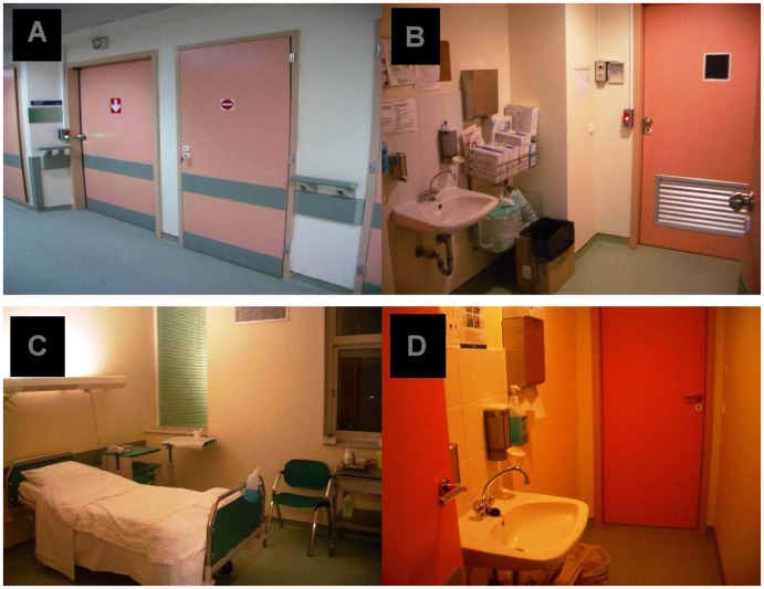
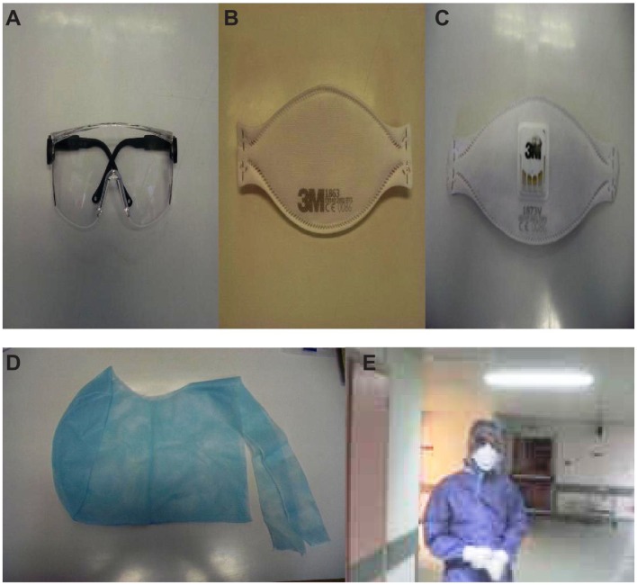
H1N1-positive patients were hospitalized in units with negative pressure especially designed for isolating patients with airborne viral infections8,9,12,13 (Figures 1 and 2). For health care personnel in close contact (defined as working within 6 feet of the patient or entering into a small enclosed airspace shared with the patient) with suspected or confirmed 2009 H1N1 influenza patients, standard precautions included the use of nonsterile gloves for any contact with potentially infectious material, followed by hand hygiene immediately after glove removal, and the use of gowns along with eye protection for any activity that might generate splashes of respiratory secretions or other infectious material.
Figure 1.
(A) Entrance (↓) and exit (−), (B) entrance, (C) negative pressure room, (D) exit.
Note: Photos by Paul Zarogoulidis, Unit of Infectious Diseases, University General Hospital of Alexandroupolis, Thrace, Greece.
Figure 2.
Protection measures: (A) protective glasses, (B) 3M™ protective mask without micro filter, (C) 3M™ protective mask with micro filter, (D) protective cup, (E) protective clothing.
Note: Photos by Paul Zarogoulidis, Unit of Infectious Diseases, University General Hospital of Alexandroupolis, Thrace, Greece.
Study sample
All patients with flu-like symptoms (ie, sore throat, cough, rhinorrhea, or nasal congestion) and fever >37.5°C were admitted to the units of infectious diseases and had pharyngeal or nasopharyngeal swabs taken. Swabs were tested with real-time reverse-transcriptase-polymerase chain reaction. 21–24 Data on underlying diseases were also recorded; diseases included chronic obstructive pulmonary disease (COPD), cancer, asthma, coronary heart disease, and diabetes mellitus. The criteria for discharge were absence of hypoxemia, normal chest X-ray, and a temperature <37°C for 1 day without antipyretic treatment. Upon discharge, they did not exit the negative pressure chambers to enter the general medical wards, but continued their hospitalization until hospital discharge. This was due to public prejudice about H1N1, as evidenced further by the fact that some patients with H1N1 even left the hospital because they did not want to become stigmatized. Patients negative for H1N1 were admitted to the general medical wards and were discharged from there. A second reverse-transcriptase-polymerase chain reaction test for H1N1 was not performed (Table 1).
Table 1.
Patients’ characteristics and clinical data
| With/Without | SD | P | ||
|---|---|---|---|---|
|
|
||||
| H1N1 positive | H1N1 negative | |||
| Age (years) | 38.65 | 57.90 | 16.974/23.645 | <0.001 |
| Chest X-ray with findings upon admission | 42 (31.6%)/91 (68.4%) | 103 (100%)/0 (0%) | <0.001 | |
| Days under oseltamivir | 5.4 | 42.00 | 2.542/0.00 | <0.001 |
| Days of hospitalization | 6.32 | 8.05 | 3.336/4.418 | NS |
| Patients with obesity | 68 (51.1%)/65 (48.9%) | 54 (52.4%)/49 (47.6%) | NS | |
| PO2 upon admission (mmHg) | 74.83 | 65.55 | 13.888/14.156 | NS |
| SPO2 upon admission (mmHg) | 94.62 | 93.06 | 12.308/16.64 | 0.001 |
| Sex (male/female) | 77/56 (57.9%/42.1%) | 57/46 (55.34%/44.66%) | NS | |
| Patients administered macrolides | 96 (72.2%)/37 (27.8%) | 60 (58.3%)/43 (41.7%) | 0.005 | |
| Patients administered quinolones | 53 (39.8%)/80 (60.2%) | 43 (41.7%)/60 (58.3%) | NS | |
| Patients changed/added quinolones from/to macrolides* | 16/96 | – | ||
| Respiratory distress upon admission | 20 (15%)/113 (85%) | 52 (50.5%)/51 (49.5%) | <0.001 | |
| Patient with respiratory disease background | 46 (34.5%)/87 (65.4%) | 33 (32%)/70 (67.9%) | NS | |
| Intensive care unit admission | 9 (6.7%) | 4 (3.8%) | NS | |
| Asthma | 22 (16.5%) | 9 (8.7%) | ||
| COPD | 21 (15.7%) | 24 (23.3%) | ||
| IPF | 3 (2.2%) | – | ||
| CHF | 18 (13.5%) | 44 (42.7%) | ||
| DM | 20 (15%) | 20 (19.4%) | ||
| Cancer | 10 (7.5%) | 6 (5.8%) | ||
Note:
Patients had to change or add their antibiotic treatment.
Abbreviations: CHF, coronary heart failure; COPD, chronic obstructive pulmonary disease; DM, diabetes mellitus; IPF, interstitial pulmonary fibrosis; SD, standard deviation.
Cost analysis
Contributors to health cost were precaution measures, pharmaceuticals administered, length of hospitalization (separately assessed in ICU and non-ICU), and diagnostic examinations (ie, laboratory, radiologic). Expenditure was calculated in Euros. The average prices of drugs were based on Greek retail prices. Hospitalization costs including nursing were calculated based on data provided by the hospital’s health economy personnel. The cost of diagnostics and drugs was the same in all hospitals, with some very minor differences in the costs of hospital-based care. This is due to the organization of the Greek health care system, which reimburses expenses via a national health insurance scheme in a uniform way.
Statistical analysis
Analysis was carried out with the use of the SPSS statistical software package (v 17.01; SPSS Inc, Chicago, IL). Continuous variables were presented as mean ± standard deviation or median (with interquartile range). For categorical variables, the percentages of patients in each category were calculated. Unpaired t-test was used in normally distributed quantitative variables to compare mean values. Chi square test was used to compare qualitative variables (frequencies in characteristics). A P-value of <0.05 was considered significant.
Results
Patients’ characteristics
In H1N1-positive patients, the mean age was 38.65 years, while it was 57.90 years in H1N1-negative patients. The frequency of obesity (ie, body mass index ≥30)25 was the same in the two groups (52.4% versus 51.1%). Among H1N1 patients, 46 (43.5%) had underlying respiratory disease (asthma, 22 [16.5%]; COPD, 21 [15.7%]; idiopathic pulmonary fibrosis, three [2.2%]), compared with 33 (32%) negative patients (asthma, 9 [8.7%]; COPD, 24 [23.3%]). In H1N1-positive patients, comorbidities were as follows: congestive heart failure, 18 (13.5%); diabetes mellitus, 20 (15%); and cancer, ten (7.5%). H1N1-negative patients’ comorbidities were: congestive heart failure, 44 (42.7%); diabetes mellitus, 20 (19.4%); and cancer, six (5.8%). Nine (6.8%) H1N1-positive patients and four (3.9%) H1N1-negative patients had to be admitted to the ICU (Tables 1 and 2).
Table 2.
Significant findings regarding influenza A (H1N1)-positive patients
| Most H1N1-positive patients did not have chest X-ray findings upon admission | P < 0.001 |
| Most H1N1-positive patients did not have chest X-ray findings upon discharge | P < 0.001 |
| Respiratory distress upon admission was less common in H1N1 positive than in H1N1-negative patients | P < 0.001 |
| H1N1-positive patients were mainly administered macrolides | P < 0.005 |
| H1N1-positive patients used mainly protection measures | P < 0.001 |
Pharmaceuticals
First, the cost of the oseltamivir 75 mg regimen was recorded. In H1N1-positive patients, €4655.32 was spent, in contrast to €1324.58 in H1N1-negative patients. The cost of macrolide treatment was €1927.36 for positive and €1588.17 for H1N1-negative patients. The cost of quinolone treatment was €2550.84 for H1N1-positive patients and €2404.24 for H1N1-negative patients. Aerosol and oxygen therapy were used freely, as judged by the clinicians. Aerosol therapy was provided with jet nebulizers and a combination of budesonide and ipratropium bromide was administered daily. Respiratory distress (PO2 < 60 mmHg) for H1N1 was noted in 20/133 (15%) positive and 52/103 (50.5%) negative patients, but this treatment was not included in the cost, since it is provided throughout hospitalization without any additional charge. Furthermore, €3427.20 was spent for aerosol therapy for H1N1-positive patients and €2833.6 for H1N1-negative patients (Table 3).
Table 3.
Cost analysis
| H1N1 (+) | H1N1 (−) | |
|---|---|---|
| Days under oseltamivir 75 mg | 724 days | 206 days |
| Total amount | €4655.32 | €1324.58 |
| Days of hospitalization* | 841 days | 829 days |
| Total amount | €354902 | €349838 |
| Protection measures | 524 days | 188 days |
| Total amount | €188640 | €67680 |
| ICU hospitalization*** | 73 days | 43 days |
| Total amount | €41610 | €24510 |
| Patients under macrolide treatment | 96 (72.2%) 608 days |
60 (58.3%) 501 days |
| Total amount | €1927.36 | €1588.17 |
| Patients under quinolone treatment | 53 (39.8%) 348 days |
43 (41.7%) 328 days |
| Total amount | €2550.84 | €2404.24 |
| Oxygen, cost/day | No additional cost | |
| Total amount | ||
| Aerosol therapy**** | 306 days | 253 days |
| Total amount | €3427.2 | €2833.6 |
| Patients diagnostic examinations | ||
| Chest X-ray | 464 X-rays | 532 X-rays |
| Total amount | €2320 | €2660 |
| Blood biochemistry** | During hospitalization | |
| Total amount | €9812 | €9671 |
| Arterial blood gas | 588 measurements | 503 measurements |
| Total amount | €2352 | €2012 |
| Sputum culture | 36 measurements | 67 measurements |
| Total amount | €216 | €402 |
| Total sum | €610,117.72 | €464,923.59 |
Notes:
In the days of ward (non-ICU) hospitalization cost the nursing care and nutrition is included;
In the blood biochemistry, complete blood count, glucose, electrolyte, renal, liver function test, C reactive protein, and procalcitonin are included. Reverse-transcriptase-polymerase chain reaction for the virus diagnosis not included;
Nursing care, materials used, and hospital day charge are included (pharmaceuticals not included);
Pharmaceuticals used were namely budesonide and ipratropium bromide for four times daily with nebulizer.
Abbreviations: H1N1, influenza A; ICU, intensive care unit.
Diagnostic examinations
Chest X-ray findings upon admission were implemented in 42 (31.6%) positive and 103 (100%) negative patients. Mean saturation (SpO2%) was 94.62% in positive and 93.06% in negative patients. Mean partial oxygenation was 74.83 mmHg for positive and 65.55 mmHg for negative patients. Respiratory distress (PO2 < 60 mmHg) was noted in 20 (15%) positive and 52 (50.5%) negative patients. The cost for arterial blood gas was €2352 for positive and €2012 for negative patients. The chest X-ray cost was €2320 for positive and €2660 for negative patients. Blood biochemistry cost, which included complete blood count, glucose, electrolyte, renal, liver function test, C-reactive protein, and procalcitonin, was €9812 for positive and €9671 for negative patients. The cost of sputum culture was €216 for positive and €402 for negative patients (Table 3).
Hospitalization cost
The cost of hospitalization was considered the same either in the negative pressure chambers or on the general medical wards. Nursing care and nutrition cost was the same. The major difference was the cost of protection measures used. For H1N1-positive patients, €188,640 was spent, in contrast with €67,680 for H1N1-negative patients. The overall cost of hospitalization was €354,902 for H1N1-positive patients and €349,838 for H1N1-negative patients. The ICU cost was €41,610 for positive and €24,510 for negative patients (Table 3).
Discussion
The major finding is that therapy for H1N1 incurred a higher cost compared with that for H1N1-negative infections. Of note, the cost was especially high during the first days of influenza, stabilizing at a lower level during the following days. The latter pattern is ascribable to the adoption of strict protection measures, according to the Hellenic Centre for Disease Control and Prevention guidelines.26 Indeed, while waiting for the swab results, all patients were treated with oseltamivir and medical staff had to keep using the aforementioned protection measures. These were pursued until the result was available, which was no sooner than 48 hours. These observations concur with current experience. Despite accurate diagnosis and prompt treatment, in one report numerous pathogens were wrongly diagnosed as influenza A (H1N1).27 From a health system point of view, this misdiagnosis of influenza A (H1N1)/2009 may have led to the underestimation of other serious conditions.28 For the same reason, a large number of patients is likely to have been unnecessarily treated with oseltamivir, resulting in not only unnecessary cost and patient exposure to the side effects of this agent but also overimplementation of infection control procedures in hospitals.29–31 Taken together, these findings underline and partly explain the high cost associated with H1N1.
In terms of protection measures, H1N1-positive patients incurred higher expenses. It should be emphasized that not only medical staff but also any visiting patients’ relatives employed costly protection measures. Therefore, frequent visits induced a higher cost due to the widespread use of protection measures. Additionally, the cost due to the extensive use of personal protective equipment is directly related to the bed design deficiencies observed in most negative-pressure chambers. In such chambers, patient medical monitoring is not automated, thus requiring regular visits by medical and nursing personnel, which, in turn, resulted in wide use of protective measures. Continuous monitoring and recording of patients’ vital signs is necessary to minimize the visits of medical staff and save cost on personal protective equipment.
In this study’s dataset, the cost of non-ICU ward care was approximately the same for both negative and positive patients. Conversely, the cost for hospitalization in the ICU was almost double for H1N1-positive patients, which was associated with more frequent virus-induced acute respiratory distress necessitating intubation and mechanical support. In general, though, admission to ICU led to a substantial increase in cost, due to the larger number of nursing staff and the use of special equipment.
Moreover, the cost associated with diagnostic examinations did not differ significantly between the two groups. This is largely due to the different clinical presentations of H1N1 and respiratory infection. Indeed, H1N1-positive patients had to undergo more regular blood-biochemistry examinations to monitor liver and renal function. Naturally, this increased cost, as reported in previous studies.15,19 H1N1-negative patients had to be regularly monitored for C-reactive protein and white blood cell count to assess clinical course.32–34 Arterial blood gas examination was performed more commonly in the H1N1-positive patients due to fear of acute respiratory distress development.15,19 In contrast, H1N1-negative patients had their usual check due to the hypoxia associated with radiographic opacities. Chest X-ray observation was more expensive for the H1N1-negative group because of the necessity for continuous clinical evaluation. This was due to the initial chest X-ray findings, since all H1N1-negative patients exhibited opacities upon admission X-rays. These findings were attributed to bacterial respiratory infection, based on clinical and laboratory findings. In addition, H1N1-negative patients had more frequent sputum cultures to determine the pathogen. This contrasts with H1N1-positive patients, in whom radiographic opacities were less common and clinical suspicion of bacterial co-infection upon admission was rarely a problem. As might be expected from previous studies,12,14–16 H1N1-positive patients were younger in comparison with H1N1-negative patients.
A further issue to be discussed is the pharmaceuticals used and how these affect cost. The H1N1 influenza virus is known to induce a “cytokine storm,”35–39 giving rise to the endeavor at patient prophylaxis. Macrolides are well-known for their anti-inflammatory and immunomodulatory actions, mostly attributable to the inhibition of intracellular hemagglutinin HA0 proteolysis.40–44 This observation agrees with the finding that only a small proportion of H1N1 patients receiving macrolides had to change antibiotic treatment. These agents could emerge as cost-effective when administered as first-line treatment, independently of their antibiotic property, due to their additional immunomodulatory ability to control influenza.
The present analysis may have a number of limitations. First, examinations of urine antigen for Legionella/Streptococcus and antibodies for Mycoplasma, Legionella, Rickettsia, and Streptococcus were not available for all patients and could therefore not be included. Moreover, diagnostic examinations at the emergency department were not described separately for the two groups, as the patients were admitted and the cost was incorporated in their overall hospitalization cost. Finally, the two groups differ in their absolute numbers and, therefore, fewer patients with underlying respiratory disease are included in the H1N1-negative patients. However, patients with co-morbidities are expected to be susceptible to H1N1, regardless of any bacterial coinfection, due to the immunomodulating and inflammatory properties of the virus.35–38,42
Conclusion
Treatment for H1N1 incurred higher cost compared with that for respiratory infection. The main reasons for this higher cost include widespread use of protection measures for patients and health care professionals, as well as prolonged ICU hospitalization. These results suggest that implementation of an automated system to monitor patients isolated in negative-pressure chambers would be desirable to reduce cost in H1N1.
Footnotes
Authors’ contributions
P Zarogoulidis conceived and wrote the first draft and contributed to data collection and interpretation. D Glaros, G Trakada, I Kouroumichakis, E Fouka, A Tsiostios, M Froudarakis, K Porpodis, I Kioumis, and D Papakosta contributed to writing the final draft. A Kallianos, P Steiropoulos, and I Kioumis contributed to data collection and interpretation. M Panopoulou was the microbiologist who provided the data regarding the microbiology exam cost. N Courcoutsakis evaluated the radiographic exams. T Kerenidi, TC Constantinidis, and E Nena performed statistical analysis. A Rapti, T Kontakiotis, K Zarogoulidis, and E Maltezos provided useful insights.
Disclosure
The authors declare no conflicts of interest in this work.
References
- 1.Centers for Disease Control and Prevention (CDC) Update: swine influenza A (H1N1) infections – California and Texas, April 2009. MMWR Morb Mortal Wkly Rep. 2009;58(16):435–437. [PubMed] [Google Scholar]
- 2.Centers for Disease Control and Prevention (CDC) Update: novel influenza A (H1N1) virus infection – Mexico, March–May, 2009. MMWR Morb Mortal Wkly Rep. 2009;58(21):585–589. [PMC free article] [PubMed] [Google Scholar]
- 3.Centers for Disease Control and Prevention (CDC) Update: novel influenza A (H1N1) virus infection – worldwide. MMWR Morb Mortal Wkly Rep. 2009;58(17):453–458. [PubMed] [Google Scholar]
- 4.World Health Organization (WHO) Global alert and response (GAR): pandemic (H1N1) 2009 – update 69 [webpage on the Internet] Geneva: WHO; 2009. [Accessed October 19, 2009]. Available at: http://www.who.int/csr/don/2009_10_09/en/ [Google Scholar]
- 5.Devaux I, Kreidl P, Penttinen P, Salminen M, Zucs P, Ammon A. ECDC influenza surveillance group; national coordinators for influenza surveillance. Initial surveillance of 2009 influenza A (H1N1) pandemic in the European union and European economic area, April–September 2009. Euro Surveill. 2010;15(49) doi: 10.2807/ese.15.49.19740-en. pii:19740. [DOI] [PubMed] [Google Scholar]
- 6.WHO. Global alert and response (GAR): preparing for the second wave; lessons from current outbreaks; pandemic (H1N1) 2009 briefing note 9 [webpage on the Internet] Geneva: WHO; 2009. [Accessed January 25, 2012]. Available from: http://www.who.int/csr/disease/swineflu/notes/h1n1_second_wave_20090828/en/index.html. [Google Scholar]
- 7.Barr IG, Cui L, Komadina N, et al. A new pandemic influenza A (H1N1) genetic variant predominated in the winter 2010 influenza season in Australia, New Zealand and Singapore. Euro Surveill. 2010;15(42) doi: 10.2807/ese.15.42.19692-en. pii:19692. [DOI] [PubMed] [Google Scholar]
- 8.Bandaranayake D, Jacobs M, Baker M, et al. The second wave of 2009 influenza A (H1N1) in New Zealand, January–October 2010. Euro Surveill. 2011;16(6) pii:19788. [PubMed] [Google Scholar]
- 9.Granados A, Goodman C, Eklund L. Pandemic influenza: using evidence on vaccines and antivirals for clinical decisions and policy making. Eur Respir J. 2006;27(4):661–663. doi: 10.1183/09031936.06.00017406. [DOI] [PubMed] [Google Scholar]
- 10.Lackenby A, Moran Gilad J, Pebody R, et al. Continued emergence and changing epidemiology of oseltamivir-resistant influenza A (H1N1)2009 virus, United Kingdom, Winter 2010/11. Euro Surveill. 2011;16(5) pii:19784. [PubMed] [Google Scholar]
- 11.Hurt AC, Deng YM, Ernest J, et al. Oseltamivir-resistant influenza viruses circulating during the first year of the influenza A (H1N1) 2009 pandemic in the Asia–Pacific region, March 2009 to March 2010. Euro Surveill. 2011;16(3) pii:19770. [PubMed] [Google Scholar]
- 12.Santa-Olalla Peralta P, Cortes-García M, Vicente-Herrero M, et al. Risk factors for disease severity among hospitalized patients with 2009 pandemic influenza A (H1N1) in Spain, April–December 2009. Euro Surveill. 2010;15(38) doi: 10.2807/ese.15.38.19667-en. pii:19667. [DOI] [PubMed] [Google Scholar]
- 13.Gubbels S, Perner A, Valentiner-Branth P, Molbak K. National surveillance of pandemic influenza A (H1N1) infection-related admissions to intensive care units during the 2009–2010 winter peak in Denmark: two complementary approaches. Euro Surveill. 2010;15(49) doi: 10.2807/ese.15.49.19743-en. pii:19743. [DOI] [PubMed] [Google Scholar]
- 14.Zarogoulidis P, Constantinidis T, Steiropoulos P, Papanas N, Zarogoulidis K, Maltezos E. Are there any differences in clinical and laboratory findings on admission between H1N1 positive and negative patients with flu-like symptoms? BMC Res Notes. 2011;1(1):141. doi: 10.1186/1756-0500-4-4. [DOI] [PMC free article] [PubMed] [Google Scholar]
- 15.Cao B, Li XW, Mao Y, et al. Clinical features of the initial cases of 2009 pandemic influenza A (H1N1) virus infection in China. N Engl J Med. 2009;361(26):2507–2517. doi: 10.1056/NEJMoa0906612. [DOI] [PubMed] [Google Scholar]
- 16.Zarogoulidis P, Kouliatsis G, Papanas N, et al. Long-term respiratory follow-up of H1N1 infection. Virol J. 2011;8:319. doi: 10.1186/1743-422X-8-319. [DOI] [PMC free article] [PubMed] [Google Scholar]
- 17.CDC. Outbreak of swine-origin influenza A (H1N1) virus infection – Mexico, March–April 2009. MMWR Morb Mortal Wkly Rep. 2009;58:467–70. [PubMed] [Google Scholar]
- 18.Mauad T, Hajjar LA, Callegari GD, et al. Lung pathology in fatal novel human influenza A (H1N1) infection. Am J Respir Crit Care Med. 2010;181:72–79. doi: 10.1164/rccm.200909-1420OC. [DOI] [PubMed] [Google Scholar]
- 19.Riquelme R, Riquelme M, Rioseco ML, et al. Characteristics of hospitalized patients with 2009 H1N1 influenza in Chile. Eur Respir J. 2010;36:864–869. doi: 10.1183/09031936.00180409. [DOI] [PubMed] [Google Scholar]
- 20.Riquelme R, Torres A, Rioseco ML, et al. Influenza pneumonia: a comparison between seasonal influenza virus and H1N1 pandemic. Eur Respir J. 2011;38(1):106–111. doi: 10.1183/09031936.00125910. [DOI] [PubMed] [Google Scholar]
- 21.WHO. CDC protocol of real-time RTPCR for swine influenza A (H1N1) Geneva: World Health Organization; 2009. [Accessed November 30, 2009]. Available from: http://www.who.int/csr/resources/publications/swineflu/CDCrealtimeRTPCRprotocol-20090428.pdf. [Google Scholar]
- 22.Newman AP, Reisdorf E, Beinemann J, et al. Human case of swine influenza (H1N1) triple reassortant virus infection, Wisconsin. Emerg Infect Dis. 2008;14:1470–1472. doi: 10.3201/eid1409.080305. [DOI] [PMC free article] [PubMed] [Google Scholar]
- 23.Taubenberger JK, Reid AH, Lourens RM, Wang R, Jin G, Fanning TG. Characterization of the 1918 influenza virus polymerase genes. Nature. 2005;437:889–893. doi: 10.1038/nature04230. [DOI] [PubMed] [Google Scholar]
- 24.CDC. Interim recommendations for clinical use of influenza diagnostic tests during the 2009–2010 influenza season. Atlanta, GA: CDC; 2009. [Accessed January 25, 2012]. Available from: http://www.cdc.gov/h1n1flu/guidance/diagnostic_tests.htm. [Google Scholar]
- 25.WHO. WHO global database on body mass index: BMI classification [webpage on the Internet] Geneva: WHO; 2006. [Accessed January 25, 2012]. [updated January 25, 2012]. Available from: http://apps.who.int/bmi/index.jsp?introPage=intro_3.html. [Google Scholar]
- 26.Lytras T, Theocharopoulos G, Tsiodras S, Mentis A, Panagiotopoulos T, Bonovas S Influenza Surveillance Report Group. Enhanced surveillance of influenza A(H1N1)v in Greece during the containment phase. Euro Surveill. 2009;14(29):19275. doi: 10.2807/ese.14.29.19275-en. [DOI] [PubMed] [Google Scholar]
- 27.Gunson RN, Carman WF. During the summer 2009 outbreak of “swine flu” in Scotland what respiratory pathogens were diagnosed as H1N1/2009? BMC Infect Dis. 2011;11:192. doi: 10.1186/1471-2334-11-192. [DOI] [PMC free article] [PubMed] [Google Scholar]
- 28.Egerer G, Goldschmidt H, Müller I, Karthaus M, Günther H, Ho AD. Ceftriaxone for the treatment of febrile episodes in non-neutropenic patients with hematooncological disease or HIV infection: comparison of outpatient and inpatient care. Chemotherapy. 2001;47(3):21–225. doi: 10.1159/000063225. [DOI] [PubMed] [Google Scholar]
- 29.Anderson RM. How well are we managing the influenza A/H1N1 pandemic in the UK? BMJ. 2009;339:b2897. doi: 10.1136/bmj.b2897. [DOI] [PubMed] [Google Scholar]
- 30.Jefferson T, Jones M, Doshi P, Del Mar C. Possible harms of oseltamivir – a call for urgent action. Lancet. 2009;374(9698):1312–1313. doi: 10.1016/S0140-6736(09)61804-3. [DOI] [PubMed] [Google Scholar]
- 31.Gerrard J, Keijzers G, Zhang P, Vossen C, Macbeth D. Clinical diagnostic criteria for isolating patients admitted to hospital with suspected pandemic influenza. Lancet. 2009;374(9702):1673. doi: 10.1016/S0140-6736(09)61983-8. [DOI] [PubMed] [Google Scholar]
- 32.Flood RG, Badik J, Aronoff SC. The utility of serum C – reactive protein in differentiating bacterial from nonbacterial pneumonia in children: a meta-analysis of 1230 children. Infect Dis J. 2008;27:95–99. doi: 10.1097/INF.0b013e318157aced. [DOI] [PubMed] [Google Scholar]
- 33.Ortega-Deballon P, Radais F, Facy O, et al. C-Reactive protein is an early predictor of septic complications after elective colorectal surgery. World J Surg. 2010;34(4):808–814. doi: 10.1007/s00268-009-0367-x. [DOI] [PMC free article] [PubMed] [Google Scholar]
- 34.George EL, Panos A. Does a high WBC count signal infection? Nursing. 2005;35(1):20–22. doi: 10.1097/00152193-200501000-00014. [DOI] [PubMed] [Google Scholar]
- 35.Belser JA, Zeng H, Katz JM, Tumpey TM. Infection with highly pathogenic H7 influenza viruses results in an attenuated proinflammatory cytokine and chemokine response early after infection. J Infect Dis. 2011;203(1):40–48. doi: 10.1093/infdis/jiq018. [DOI] [PMC free article] [PubMed] [Google Scholar]
- 36.Maines TR, Szretter KJ, Perrone L, et al. Pathogenesis of emerging avian influenza viruses in mammals and the host innate immune response. Immunol Rev. 2008;225:68–84. doi: 10.1111/j.1600-065X.2008.00690.x. [DOI] [PubMed] [Google Scholar]
- 37.Us D. Cytokine storm in avian influenza. Mikrobiyol Bul. 2008;42(2):365–380. [PubMed] [Google Scholar]
- 38.Woo PC, Tung ET, Chan KH, Lau CC, Lau SK, Yuen KY. Cytokine profiles induced by the novel swine-origin influenza A/H1N1 virus: implications for treatment strategies. J Infect Dis. 2010;201(3):346–353. doi: 10.1086/649785. [DOI] [PMC free article] [PubMed] [Google Scholar]
- 39.Lee SM, Gardy JL, Cheung CY, et al. Systems-level comparison of host-responses elicited by avian H5N1 and seasonal H1N1 influenza viruses in primary human macrophages. PLoS One. 2009;4(12):e8072. doi: 10.1371/journal.pone.0008072. [DOI] [PMC free article] [PubMed] [Google Scholar]
- 40.Leelarasamee A, Jongwutiwes U, Tantipong H, Puthavathana P, Siritantikorn S. Fulminating influenza pneumonia in the elderly: a case demonstration. J Med Assoc Thai. 2008;9:924–930. [PubMed] [Google Scholar]
- 41.Miyamoto D, Hasegawa S, Sriwilaijaroen N, et al. Clarithromycin inhibits progeny virus production from human influenza virus-infected host cells. Biol Pharm Bull. 2008;31:217–222. doi: 10.1248/bpb.31.217. [DOI] [PubMed] [Google Scholar]
- 42.Zhirnov O, Klenk HD. Human influenza A viruses are proteolytically activated and do not induce apoptosis in CACO-2 cells. Virology. 2003;313:198–212. doi: 10.1016/s0042-6822(03)00264-2. [DOI] [PubMed] [Google Scholar]
- 43.Shneĭder MA, Shtil’bans EB, Rachkovskaia LA, Poltorak VA. Antiviral action of carbonyl-conjugated pentaene macrolides. Antibiotiki. 1983;28:352–357. [PubMed] [Google Scholar]
- 44.Bermejo-Martin JF, Kelvin DJ, Eiros JM, Castrodeza J, Ortiz de Lejarazu R. Macrolides for the treatment of severe respiratory illness caused by novel H1N1 swine influenza viral strains. J Infect Dev Ctries. 2009;3(3):159–161. doi: 10.3855/jidc.18. [DOI] [PubMed] [Google Scholar]


