Abstract
Heterotopic ossification refers to formation of lamellar bone in soft tissues. The etiology is diverse and includes genetic, post-traumatic, and metabolic causes. Elicitation of bone morphogenic proteins are thought to play a key role in the pathogenic process. Initially, heterotopic ossification presents a clinical and radiographic challenge in that it can be mistaken for other more worrisome entities which present with calcified soft tissue masses. However, a spontaneous clinical resolution, temporal relationship to an inciting agent, and radiographic evolution to a peripherally-calcified lesion are all clues to the diagnosis. Here we present the clinical and radiographic features of heterotopic ossification as a result of an infiltrated peripheral IV.
Case Report
Summary
A newborn infant was hospitalized with complications related to a congenital heart disorder. On the day of birth, intravenous access was obtained through a peripheral vein in the dorsum of her foot. There was infiltration of this IV after one day, resulting in localized soft tissue swelling and erythema, and it was removed 2 days after placement. Two weeks later, a hard palpable soft tissue mass was noted at the same location in the dorsum of the foot. This was tender and continued to enlarge. Imaging studies of the foot were obtained 3 weeks after the initial IV placement.
Imaging Findings
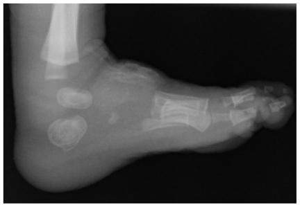
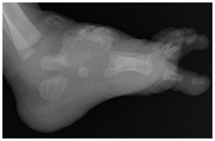
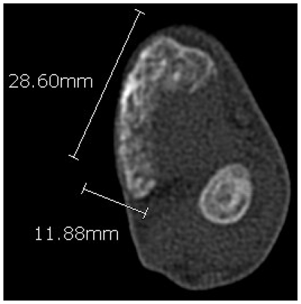
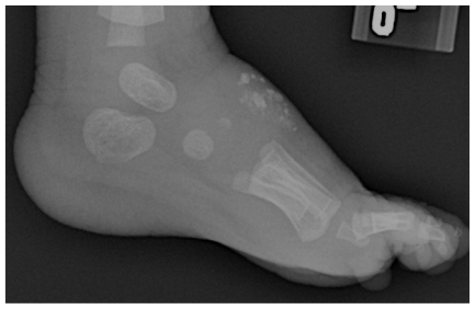
Radiographic studies of the foot were performed. An amorphous soft tissue mass with mineralization in the dorsum of the foot is seen on the initial radiograph (Fig. 1). The disease process does not appear to involve the underlying bones of the foot. After 1 month, repeat radiographs demonstrate evolution of the process, with enlargement of the mass and peripheral mineralization (Fig. 2). A CT scan of the foot confirms the mass to be mostly mature at the periphery, with less ossification at the center, and soft tissue separating the lesion from the adjacent cortex (Fig. 3). After 8 weeks of conservative treatment, repeat radiographs demonstrate interval shrinkage and dissolution of the mass (Fig. 4).
Figure 1.
Initial lateral radiograph of the foot demonstrating an amorphous mineralized mass in the dorsal soft tissues.
Figure 2.
Lateral radiograph obtained one month later demonstrates progression of the mineralization and size of the mass.
Figure 3.
Axial CT image of the foot demonstrating the large region of heterotopic ossification at the dorsum of the foot anterior to the talus. Mineralization is most notable at the periphery of the mass.
Figure 4.
Lateral radiograph showing interval improvement after 8 weeks of conservative management.
Discussion
Heterotopic ossification refers to the formation of lamellar bone within soft tissues, secondary to genetic, traumatic, or iatrogenic causes (1, 2). It can present as small, clinically-insignificant flecks of bone or large deposits of ossification. Its initial radiographic appearance can be confused with metastatic calcification in hypercalcemic disorders or more worrisome entities such as parosteal osteosarcoma or juxtacortical chondroma. The pathogenesis for heterotopic bone formation is not fully understood. Though there are inherited disorders such as fibrodysplasia ossificans progressiva and progressive osseous heteroplasia which are associated with advanced and often disfiguring manifestations of heterotopic ossification, these disorders are rare and tend to arise from spontaneous mutations (1, 2, 3). The disorder is seldom seen in infants, and heterotopic ossification in children most frequently presents secondary to inheritable causes (4, 5, 6). Neurogenic and post-traumatic forms of the disease are the most common clinically encountered in the non-pediatric age group, with bone morphogenetic proteins elicited from the site of soft tissue injury playing a key role in the reactive ossification process (1, 2). Treatment ranges from conservative therapy with spontaneous resolution to bisphosphonates and radiation for more advanced cases (2).
Post-traumatic heterotopic ossification, referred to by some authors as myositis ossificans traumatica, occurs in various clinical settings. Errant repair of muscle contusion injury in athletes is a well-documented entity that can result in various forms of heterotopic bone formation (7, 8, 9). Less common examples of post-traumatic ossification in other soft tissues have been described. Montoli et. al describe a case of large post-traumatic ossification within the Achilles tendon (10). A case of mesenteric ossification 2 weeks following blunt abdominal trauma and exploratory laparotomy has been described as well (11). A rare case of post-traumatic heterotopic ossification in the neck soft tissues of a 5-month old infant has been described (6). The time course of the disease is also variable; heterotopic ossification occurring several years after injury has been described as well, though rare (12).
In the case described above, there was clinically evident injury to the soft tissue secondary to an infiltrated IV in a newborn with progression over several weeks to development of an ossified mass. Radiographic features over the disease course were congruent with those previously-described for myositis ossificans traumatica, including the initial amorphous calcified soft tissue lesion which progressed to a mostly peripherally-ossified mass (2, 13). After 8 weeks of conservative therapy, clinical improvement and radiographic regression was noted.
Teaching Point
Heterotopic ossification can occur in a plethora of clinical settings and sometimes is confused for other entities such as metastatic calcification or osseous/soft-tissue tumors; the distinction can often be made based on the radiographic and clinical course. This case illustrates an unusual occurrence of post-traumatic heterotopic ossification in the setting of an infiltrated intravenous catheter.
Footnotes
Abbreviations: None
References
- 1.McCarthy EF, Sundaram M. Heterotopic Ossification: A Review. Skeletal Radiol. 2005;34(10):609–19. doi: 10.1007/s00256-005-0958-z. [DOI] [PubMed] [Google Scholar]
- 2.Bossche LV, Vanderstraeten G. Heterotopic Ossification: A Review. J Rehabil Med. 2005;37:129–136. doi: 10.1080/16501970510027628. [DOI] [PubMed] [Google Scholar]
- 3.Kaplan FS, Glaser DL, Hebela N, et al. Heterotopic Ossification. J Am Acad Orthop Surg. 2004;12(2):116–25. doi: 10.5435/00124635-200403000-00007. [DOI] [PubMed] [Google Scholar]
- 4.Kaplan FS, Shore EM, et al. Early Diagnosis of Fibrodysplasia Ossificans Progressiva. Pediatrics. 2008;121( 5):1295–1300. doi: 10.1542/peds.2007-1980. [DOI] [PMC free article] [PubMed] [Google Scholar]
- 5.Feldman GJ, Shore EM, et al. Over-expression of BMP4 and BMP5 in a Child with Axial Skeletal Malformations and Heterotopic Ossification: A New syndrome. Am J Med Genet A. 2007;143(7):699–706. doi: 10.1002/ajmg.a.31649. [DOI] [PubMed] [Google Scholar]
- 6.Toshitaka Y, Hideo E. Heterotopic Ossification in the Upper Cervical Spine in a 5-month-old Infant: A Case Report. J of Japanese Paediatric Orthopaedic Assoc. 2005;14(1):44–47. [Google Scholar]
- 7.Beiner JM, Jokl P. Muscle Contusion Injury and Myositis Ossificans Traumatica. Clin Orthop Relat Res. 2002;403(sup):S110–9. doi: 10.1097/00003086-200210001-00013. [DOI] [PubMed] [Google Scholar]
- 8.King JB. Post-traumatic Ectopic Calcification in the Muscles of Athletes: A Review. Br J Sports Med. 1993;32:287–290. doi: 10.1136/bjsm.32.4.287. [DOI] [PMC free article] [PubMed] [Google Scholar]
- 9.Arrington ED, Miller MD. Skeletal Muscle Injuries. Orthop Clin North Am. 1995;26:411–22. [PubMed] [Google Scholar]
- 10.Motoli C, Barbieri S, DePietri M, et al. Post-traumatic Ossification of the Achilles Tendon: Description of a Case. Chir Organi Mov. 2004;89(1):51–7. [PubMed] [Google Scholar]
- 11.Gouello J-P, Auvray L, Bouix A, et al. Early Post-traumatic Heterotopic Mesenteric Ossification: Report of a Case. Intensive Care Med. 2006;32(6):950–1. doi: 10.1007/s00134-006-0144-3. [DOI] [PubMed] [Google Scholar]
- 12.Todd W, Gianfortune PJ, Laughner T. Post-traumatic Myositis Ossificans Circumscripta: An Unusually Large Example. J Am Podiatr Med Assoc. 2007;97(3):229–33. doi: 10.7547/0970229. [DOI] [PubMed] [Google Scholar]
- 13.Parikh J, Hyare H, Saifuddin A. The Imaging Features of Post-Traumatic Myositis Ossificans with Emphasis on MRI. Clinical Rad. 2002;57:1058–66. doi: 10.1053/crad.2002.1120. [DOI] [PubMed] [Google Scholar]


