Abstract
Intracranial pneumatocele is a non-infected accumulation of air within the cranial cavity. We report a case of a 22-year-old male who sustained a fracture of anterior cranial fossa following a motor vehicle accident and the imaging findings showed a concave-convex epidural haematoma. Simple traumatic pneumocephalus usually does not require surgical treatment and non-operative management has been advocated for small, asymptomatic convexity extradural haematomas.
Keywords: Epidural haematoma, pneumocephalus, trauma, head injury
CASE REPORT
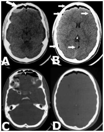
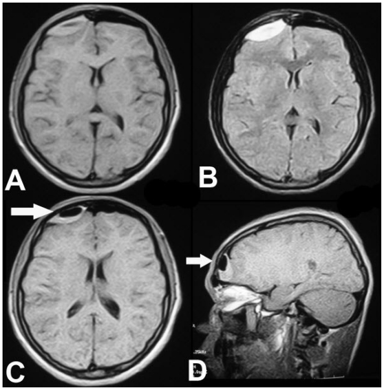
Intracranial pneumatocele is a noninfected accumulation of air within the cranial cavity (1) and this air accumulation is the result of a traumatic communication between the paranasal sinuses, cribriform plate or mastoid air cells, and the cranial fossa with an associated dural tear (2). A 22-year-old male sustained a fracture of anterior cranial fossa following a motor vehicle accident. He had transient loss of consciousness, nasal bleed and was complaining of mild headache. Axial CT scan showed a large bilateral frontal and also scattered specks of pneumatocele, bilateral anterior cranial fossa fractures and concavo-convex right frontal acute epidural haematoma (Fig. 1) There was localized but not significant mass effect over right frontal lobe and no surrounding edema. Further investigation with MRI suggested that the air was in the subdural space and confirmed the epidural nature of the haematoma (Fig. 2). The patient recovered with conservative management.
Figure 1.
22-year-old male with Mount Fuji sign. Axial noncontrast CT scan in bone window, showing bifrontal and also multiple specks (B-small arrows) of pneumocephalus, bilateral anterior skull base fracture and right frontal epidural haematoma with concavo-convex appearance (A-large arrow)
Figure 2.
22-year-old male with Mount Fuji sign. MRI T1 and FLAIR axial (A, B and C) and T1 sagittal (D) images showing the epidural nature of the haematoma and air collection in the subdural space (C and D) giving rise to concave-convex appearance to the haematoma.
DISCUSSION
The most common location of pneumocephalus is in the subarachnoid and subdural spaces and the fewer sites for air collection include the intraventricular, intracerebral, and extradural spaces (1). Because of the peculiar meningeal anatomy of the anterior cranial fossa (the dura being thin and closely applied to bone and the arachnoid adherent to the frontal lobe), fronto-ethmoidal meningeal lacerations frequently result in subdural air collection (2). This intracranial air collection sometime can result in tension pneumopcephalus with characteristic Mount Fuji sign The Mount Fuji sign is a finding observed in cases of tension pneumocephalus on CT scans of the brain, in which bilateral subdural hypo attenuating air collections causes compression and separation of the frontal lobes from the skull (4,5). The differential diagnosis of acute extra-axial haematoms includes epidural haematoma, subdural haematoma and subarachnoid hemorrhage (SAH). Anatomically the epidural hematomas is located between the inner table of the skull and the dura and the typical biconvex appearance is due to the fact that their outer border follows the inner table of the skull and their inner border is limited by locations at which the dura is firmly adherent to the skull (3,6). The subdural haematoma is located between the dura and the brain and their outer edge is convex, while their inner border is irregularly concave as these are not limited by the intracranial suture lines; an important feature that helps to differential these two lesions from each other (3,6). The SAH appears as diffuse thin hyperdensity on CT scan, scattered over the cortical surface (3,6). CT appearance of epidural haematoma is a characteristic homogeneous high density lens-shaped or bi-convex blood collection against the calvarium (3). In present case both the lesions existed simultaneously on right side and the indentation of the epidural haematoma by subdural air resulted in the concavo-convex appearance of the epidural haematoma (Fig. 1 and Fig. 2). Simple traumatic pneumocephalus usually does not require surgical treatment (7) and non-operative management has been advocated for epidural haematomas that are less than 1.5 cm in width, associated with minimal or no midline shift, and located in the convexities (8).
TEACHING POINT
Mount Fuji sign is seen in bilateral tension pneumocephalus causing compression and separation of the frontal lobes from the skull. Simple traumatic pneumocephalus usually does not require surgical treatment.
ABBREVIATIONS
- CT scan
Computerized tomography scan
- MRI
Magnetic resonance imaging
REFERENCES
- 1.Gordon IJ, Hardman DR. The traumatic pneumomyelogram: a previously undescribed entity. Neuroradiology. 1977;13:107–108. doi: 10.1007/BF00339843. [DOI] [PubMed] [Google Scholar]
- 2.Markham JW. Vinken PJ, Bruyn GW. Handbook of clinical neurology. Amsterdam, Oxford: North Holland Publishing Company; 1976. Pneumocephalus; pp. 201–213. [Google Scholar]
- 3.Donnelly LF. Computerized tomography (CT) in acute head trauma. AJR Am J Roentgenol. 2000;175(5):1370. doi: 10.2214/ajr.175.5.1751370. [DOI] [PubMed] [Google Scholar]
- 4.Ishiwata Y, Fujitsu K, Sekino T, et al. Subdural tension pneumocephalus following surgery for chronic subdural hematoma. J Neurosurg. 1988;68:58–61. doi: 10.3171/jns.1988.68.1.0058. [DOI] [PubMed] [Google Scholar]
- 5.Michel SJ. The Mount Fuji Sign. Radiology. 2004;232:449–450. doi: 10.1148/radiol.2322021556. [DOI] [PubMed] [Google Scholar]
- 6.Zee CS, Go JL. CT of head trauma. Neuroimaging Clin N Am. 1998;8(3):525–39. [PubMed] [Google Scholar]
- 7.Joshi SM, Demetriades A, Vasani SS, Ellamushi H, Yeh J. Tension pneumocephalus following head injury. Emerg. Med J. 2006;23:324. doi: 10.1136/emj.2005.028175. [DOI] [PMC free article] [PubMed] [Google Scholar]
- 8.Sagher O, Ribas GC, Jane JA. In discussion of: Hamilton M, Wallace C. Nonoperative management of acute epidural haematoma diagnosed by CT: the neuroradiologist’s role. AJNR Am J Neuroradiol. 1992;13:853–862. [PMC free article] [PubMed] [Google Scholar]


