Abstract
Arterial pseudoaneurysm formation of the genicular vessels following orthopaedic surgery to the knee is an extremely rare occurrence. Here we report the successful management of two cases as a complication of total knee arthroplasty and a tibial interlocking nail, utilising coil embolisation by interventional radiological techniques and negating the need for further surgery. To our knowledge this is one of the few reported cases of pseudoaneurysms of the descending genicular artery secondary to drain placement and only the second following tibial interlocking nail placement.
Keywords: Pseudoaneurysm, peripheral, coil, embolisation, orthopaedic, trauma
INTRODUCTION
Arterial pseudoaneurysm formation is a rare entity following elective and trauma orthopaedic surgery. A retrospective review by Wilson et al (1) discovered that of 4350 patients undergoing elective orthopaedic procedures, only 27 patients presented to the vascular surgery service with arterial injury following hip and knee arthoplasty and ankle reconstruction, of which only three had developed pseudoaneurysm formation. Pseudoaneurysms have been reported to occur following total knee arthroplasty (TKA) (2–4), open synovectomy (5), meniscectomy (6) and arthroscopy (7,8). They may occur in the popliteal artery (9–11), the superolateral (12–14), inferolateral (15) and inferomedial geniculate arteries (16,17). Here we report the successful management of two cases utilising coil embolisation by interventional radiology: the first after placement of a tibial interlocking nail and the second after the first stage of a revision knee replacement. Both patients subsequently avoided the need for further surgery.
Case report 1
A 64 year old man presented to the Accident & Emergency department with rigors and a painful left knee and ankle. A few years previously he had undergone a successful left knee replacement for tricompartmental osteoarthritis
Clinical examination revealed he was febrile with cellulitis surrounding the left ankle. A mild effusion of the left knee was present with an increase in the surrounding skin temperature. No range of movement of the left knee could be elicited due to pain. Laboratory tests revealed a leucocytosis and raised inflammatory markers. Aspiration revealed purulent fluid, from which, Group G Streptococci was cultured.
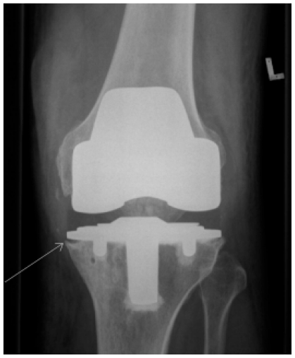
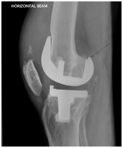
Antibiotics were commenced and he underwent arthroscopic washout and partial synovectomy. Unfortunately due to uncontrolled infection he returned to theatre for further washout and debridement. AP and lateral X-rays showed loosening of tibial and femoral components, soft tissue swelling and an effusion (Fig. 1,2), and the decision was made to remove the implant and insert a cement spacer. Extensive medial and lateral pseudomembranes were removed and the components were removed with difficulty due to problems with access secondary to a contracted patella tendon. Two drains were placed and exited superomedially. These were removed 48 hours post-operatively.
Figure 1.
Case Report 1 - AP X-ray of left knee demonstrating loosening medially of the tibial component with accompanying bony changes (white arrow), soft tissue swelling and an effusion.
Figure 2.
Case Report 1 - Lateral X-ray of left knee demonstrating loosening posteriorly of the femoral component (black arrow), soft tissue swelling and an effusion.
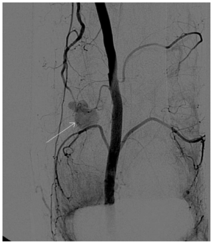
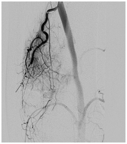
A few days after implant removal, an erythematous pulsatile swelling was noted at the old drain site, over the superomedial aspect of the left knee. Distal circulation and peripheral pulses were normal. The patient was investigated by digital subtraction angiography which confirmed a pseudoaneurysm arising from the descending genicular artery (Fig. 3). Embolisation was successfully performed by coil occlusion of the feeding artery proximal and distal to the neck of the pseudo-aneurysm using a total of 4 fibred microcoils (three 2 × 20 mm and one 3 × 30 mm ) (Fig. 4).
Figure 3.
Case Report 1 - Digital Subtraction Angiography of the left superficial femoral and popliteal artery demonstrating the pseudoaneurysm (white arrow) arising from the descending genicular artery.
Figure 4.
Case Report 1 - Digital Subtraction Angiography after selective catheterisation demonstrating embolisation with coils in-situ (black arrow) and no flow into the pseudoaneurysm.
The patient was discharged a further two weeks later on antibiotics. The second stage of the knee replacement was performed a few months after the first stage and at one year follow up, there was no sign of recurrence of infection or the pseudoaneurysm.
Case report 2
A 47 year old man presented to the Accident & Emergency department with a closed spiral fracture of the right distal third of the tibia following a fall. Neurovascular status of the limb was intact and the following day he underwent closed intramedullary nailing of the tibia with two static locking screws proximally in the mediolateral plane.
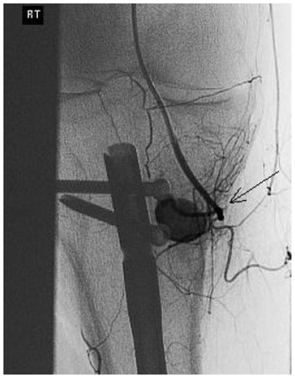
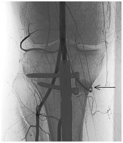
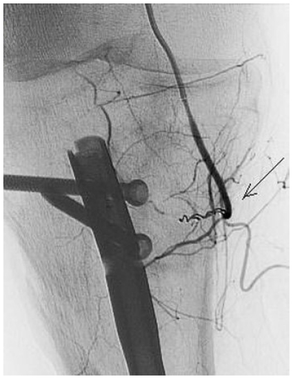
A few days after the procedure, a firm non-tender pulsatile swelling was noticed over the inferomedial aspect of the right knee. Digital subtraction angiography confirmed the palpable pseudoaneurysm to be arising from the inferomedial geniculate artery (Fig. 5,6). Successful embolisation of the pseudo-aneurysm was performed using a single 2/4 × 30 mm fibered helical coil after selective catheterisation of the feeding genicular artery. (Fig. 7).
Figure 5.
Case Report 2 - Selective digital subtraction Angiography demonstrating the pseudoaneurysm (black arrow) arising the inferomedial geniculate artery over the proximal locking screw.
Figure 6.
Case Report 2 - Digital Subtraction Angiography of the right popliteal artery demonstrating the tibial interlocking nail and static locking screws with the pseudoaneurysm (black arrow) arising from the inferomedial geniculate artery.
Figure 7.
Case Report 2 - Digital Subtraction Angiography after selective coil embolisation showing no contrast entering the pseudoaneurysm (black arrow).
The patient was subsequently discharged and at follow up there was no recurrence of the pseudoaneurysm.
DISCUSSION
Pseudoaneurysm formation is often secondary to trauma, leading to a focal disruption of the arterial vessel wall. As a consequence, blood flow continues to be maintained and both patient and surgeon are initially unaware of vessel injury. Extravasation of blood through the vessel wall leads to the formation of a pulsating haematoma, the cavity of which is still in direct continuity with the lumen of the artery. The clot liquefies and the pseudoaneurysm is subsequently contained by a fibrous capsule rather than the layers of the vessel wall itself. Unless action is taken to repair the pseudoaneurysm, it will progressively enlarge eroding surrounding structures or will rupture. As a consequence, presentation can vary from asymptomatic presentation to bruising, swelling, neurological symptoms (due to nerve compression) and even death.
Pseudoaneurysm formation is a rare complication following operations on the knee with the incidence of arterial complications following TKA found from 2 studies to be between 0.03 to 0.12% (9,10). There are few reports of pseudoaneurysm formation following both TKA (4,9,10) and following tibial nail placement (12, 18).
Pseudoaneurysm formation during knee replacement surgery may occur due to either direct intra-operative manipulation, especially during medial and lateral retinacular release, or indirectly by intimal plaque disruption of calcified atherosclerotic vessels by the pneumatic tourniquet or thermal injury by hot cement (4).
The descending genicular artery which lies within the medial aspect of the thigh is at risk during medial retinacular release. In case 1, the extensive debridement of the medial and lateral pseudomembranes combined with the difficulty with access and removal of the components predisposed to intra-operative injury to the surrounding vessels. However, as the pseudoaneurysm formed directly over the drain site it must be concluded that it was caused by direct vessel injury by the superomedial placement of the drain. The placement of suction drains superolaterally would avoid the potential injury to the vessel.
The inferomedial geniculate artery courses around the medial head of gastrocnemius, making it susceptible to injury when using a tibial interlocking nail. The pseudoaneurysm of the inferomedial geniculate artery in case 2 was probably caused by the initial drilling of the superior proximal locking screw.
If doubt remains in the clinicians mind regarding the diagnosis, duplex Doppler ultrasonography is a useful diagnostic aid in confirming the presence, size and location of the pseudoaneurysm.
Duplex Doppler ultrasonography is the imaging modality of choice if there is clinical doubt regarding the diagnosis between a haematoma or pseudoaneurysm. The definitive diagnosis on ultrasound is made by both the identification of the connection of the neck of the aneurysm and the injured artery, and the pathognomonic “to and fro” spectral waveform pattern within the neck due to the alternating flow direction in systole and diastole (19). Other diagnostic characteristics in triplex ultrasound include the “yin-yang” appearance (indicative of turbulent flow), expansile pulsatility, and the presence of haematoma of variable echogenicity, but these signs do not always distinguish between a pulsating haematoma and a pseudoaneurysm (20). In both our cases, a clinical diagnosis of pseudoaneurysm formation was made and the patients proceeded straight to angiography as this provides the ability to diagnose and treat simultaneously and subsequently negates the need for further diagnostic techniques.
Management of pseudoaneurysms around the knee is varied and adjusted according to the size and location of the pseudoaneurysm. Previously, open surgery would have been performed by gaining proximal and distal vascular control of the pseudoaneurysm, evacuating the thrombus and, if the artery was small and deemed non-essential can be ligated, or if essential can be repaired by primary anastomosis or patch repair by insertion of autologous vein graft.
With the continual advancement of interventional radiological techniques such as coil embolisation (14,21), thrombin injection (22,23) and stenting (11), patients may negate the need for further surgery, thus reducing the propensity for infection in the presence of metal implants and improving patient rehabilitation following orthopaedic surgery. With regards to both our cases, coil embolisation was used as the geniculate arteries form the genicular anastomosis surrounding the knee and can be sacrificed without subsequent circulatory compromise.
TEACHING POINT
Pseudoaneurysm formation is a rare but serious complication following elective and trauma surgery to the knee. To our knowledge, we have reported one of the few cases of a pseudoaneurysm of the descending genicular artery following drain placement and only the second case following placement of a tibial interlocking nail. To prevent the development of pseudoaneurysm formation we recommend the placement of drains superolaterally.
Pseudoaneurysms around the knee pose a significant challenge to the clinician, but if diagnosed early, can be successfully treated using radiological endovascular techniques negating the need for further surgery and without compromising patient rehabilitation.
ABBREVIATIONS
- TKA
Total knee arthroplasty
REFERENCES
- 1.Wilson JS, Miranda A, Johnson BL, Shames ML, Back MR, Bandyk DF. Vascular Injuries Associated with Elective Orthopedic Procedures. Annals of Vascular Surgery. 2003;17:641–644. doi: 10.1007/s10016-003-0074-2. [DOI] [PubMed] [Google Scholar]
- 2.Omary R, Stulberg D, Vogelzang RL. Therapeutic Embolization of False Aneurysms of the Superior Medial Genicular Artery After Operations on the Knee. J Bone Joint Surg Am. 1991;73:1257–1259. [PubMed] [Google Scholar]
- 3.Stanley D, Cumberland DC, Elson RA. Embolisation for Aneurysm After Knee Replacement: Brief Report. J. Bone and Joint Surg. 1989;71-B(1):138. doi: 10.1302/0301-620X.71B1.2914987. [DOI] [PubMed] [Google Scholar]
- 4.Law KY, Cheung KW, Chiu KH, Antonio GE. Pseudoaneurysm of the geniculate artery following total knee arthroplasty: a report of two cases. Journal of Orthopaedic Surgery. 2007;15(3):386–9. doi: 10.1177/230949900701500331. [DOI] [PubMed] [Google Scholar]
- 5.Rifaat MA, Massoud AF, Shafie MB. Post-Operative Aneurysm of the Descending Genicular Artery Presenting As a Pulsating Haemarthrosis of the Knee. J. Bone and Joint Surg. 1969;51- B(3):506–507. [PubMed] [Google Scholar]
- 6.Fairbank TJ, Jamieson ES. A complication of lateral meniscectomy. J Bone and Joint Surg. 1951;3-b(4):567–570. doi: 10.1302/0301-620X.33B4.567. [DOI] [PubMed] [Google Scholar]
- 7.Manning MP, Marshall JH. Aneurysm after arthroscopy. J Bone and Joint Surg. 1987;69-b(1):151. doi: 10.1302/0301-620X.69B1.3818725. [DOI] [PubMed] [Google Scholar]
- 8.Vincent GM, Stanish WD. False Aneurysm After Arthroscopic Meniscectomy. A Report of Two Cases. J. Bone and Joint Surg. 1990;72-A:770–772. [PubMed] [Google Scholar]
- 9.O'Connor JV, Stocks G, Crabtree JD, Jr, Galasso P, Wallsh E. Popliteal pseudoaneurysm following total knee arthroplasty. J Arthroplasty. 1998;13:830–2. doi: 10.1016/s0883-5403(98)90039-0. [DOI] [PubMed] [Google Scholar]
- 10.Karkos CD, Thomson GJ, D'Souza SP, Prasad V. False aneurysm of the popliteal artery: a rare complication of total knee replacement. Knee Surg Sports Traumatol Arthrosc. 2000;8:53–5. doi: 10.1007/s001670050011. [DOI] [PubMed] [Google Scholar]
- 11.Vaidhyanath R, Blanshard KS. Case report: insertion of a covered stent for treatment of a popliteal artery pseudoaneurysm following total knee arthroplasty. Br J Radiol. 2003;76:195–8. doi: 10.1259/bjr/32510074. [DOI] [PubMed] [Google Scholar]
- 12.Moran M, Hodgkinson J, Tait W. False aneurysm of the superior lateral geniculate artery following total knee replacement. Knee. 2002;9:349–51. doi: 10.1016/s0968-0160(02)00061-3. [DOI] [PubMed] [Google Scholar]
- 13.Kirschner S, Konrad T, Weil EJ, Buhler M. False aneurysm of the lateral superior genicular artery. A complication after the implantation of a knee prosthesis (in German) Orthopade. 2004;33:841–5. doi: 10.1007/s00132-004-0638-z. [DOI] [PubMed] [Google Scholar]
- 14.Pritsch T, Parnes N, Menachem A. A bleeding pseudoaneurysm of the lateral genicular artery after total knee arthroplasty: a case report. Acta Orthop. 2005;76:138–40. doi: 10.1080/00016470510030463. [DOI] [PubMed] [Google Scholar]
- 15.Pai VS. Traumatic aneurysm of the inferior lateral geniculate artery after total knee replacement. J Arthroplasty. 1999;14:633–4. doi: 10.1016/s0883-5403(99)90089-x. [DOI] [PubMed] [Google Scholar]
- 16.Sharma H, Singh GK, Cavanagh SP, Kay D. Pseudoaneurysm of the inferior medial geniculate artery following primary total knee arthroplasty: delayed presentation with recurrent haemorrhagic episodes. Knee Surg Sports Traumatol Arthrosc. 2006;14:153–5. doi: 10.1007/s00167-005-0639-4. [DOI] [PubMed] [Google Scholar]
- 17.Bennett FS, Born CT, Alexander J, Crincoli M. False aneurysm of the medial inferior genicular artery after intramedullary nailing of the tibia. J Orthop Trauma. 1994;8(1):73–5. doi: 10.1097/00005131-199402000-00016. [DOI] [PubMed] [Google Scholar]
- 18.Inamdar D, Alagappan M, Shyam L, Devadoss S, Devadoss A. Pseudoaneurysm of anterior tibial artery following tibial nailing: A case report. Journal of Orthopaedic Surgery. 2005;13(2):186–189. doi: 10.1177/230949900501300216. [DOI] [PubMed] [Google Scholar]
- 19.Abu-Yousef MM, Wiese JA, Shamma AR. The “to-and-fro” sign: duplex Doppler evidence of femoral artery pseudoaneurysm. AJR. 1988;150:632. doi: 10.2214/ajr.150.3.632. [DOI] [PubMed] [Google Scholar]
- 20.Carroll BA, Graif M, Orron DE. Vascular ultrasound. In: Kim DS, Orron DE, editors. Peripheral Vascular Imaging and Intervention. St. Louis: Mosby Yearbook; 1992. pp. 211–25. [Google Scholar]
- 21.Sugimoto T, Kitade T, Morimoto N, Terashima K. Pseudo aneurysms of peroneal artery: treatment with transcatheter platinum coil embolization. Ann Thorac Cardiovasc Surg. 2004;10(4):263–5. [PubMed] [Google Scholar]
- 22.Ibrahim M, Booth RE, Jr, Clark TW. Embolization of traumatic pseudoaneurysms after total knee arthroplasty. J Arthroplasty. 2004;19:123–8. doi: 10.1016/j.arth.2003.08.007. [DOI] [PubMed] [Google Scholar]
- 23.Grewe PH, Mugge A, Germing A, Harrer E. Occlusion of pseudoaneurysms using human or bovine thrombin using contrast-enhanced ultrasound guidance. Am J Cardiol. 2004;93(12):1540–2. doi: 10.1016/j.amjcard.2004.02.068. [DOI] [PubMed] [Google Scholar]


