Abstract
Median palatine cysts are rare, non-odontogenic fissural cysts of the hard palate. These cysts occur in the midline of the hard palate, behind the incisive canal. Only two case reports have documented these cysts on multi-detector computed tomography (MDCT), neither giving detailed descriptions of the cysts. Knowledge of their existence is important and should not be confused with malignant tumors. We present the first case describing the MDCT characteristics of the median palatine cyst.
Keywords: Median palatine cyst, Fissural cyst, Computed Tomography, Nasopalatine cyst
CASE REPORT
A 40-year old African American female presented to the emergency department after she experienced a tonic-clonic seizure. The patient had a history of seizures and was currently taking anti-seizure medication. Upon arrival to the emergency department, the patient complained only of headache. There was right peri-orbital soft tissue swelling on physical exam and her neurological exam was normal.
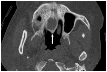
Contrast enhanced MDCT axial and coronal images (Figure 1 and 2) of the facial bones revealed a 16×13×10 mm expansile, ovoid cyst located in the midline of the hard palate. There was elevation of the floor of the nasal cavity and extension into the nares. Sagittal reformatted image (Figure 3) demonstrated no communication of the cyst with the incisive canal. All images failed to demonstrate an intimate relationship with a non-vital tooth.
Figure 1.
Contrast enhanced axial MDCT image in bone windows at the level of the maxilla. The median palatine cyst appears as a well circumscribed, expansile, ovoid cyst located in the midline of the hard palate (arrow).
Figure 2.
Contrast enhanced MDCT coronal reformatted image in bone windows through the paranasal sinuses. There is a well circumscribed, expansile, ovoid cyst located in the midline of the hard palate (arrow). Note the elevation of the floor of the nasal cavity and extension into the left nares.
Figure 3.
Contrast enhanced MDCT sagittal reformatted image in bone windows through the paranasal sinuses. Again noted is a well circumscribed, expansile, ovoid cyst located in the hard palate. There is no communication of the median palatine cyst (arrowhead) with the incisive canal (arrow).
Follow up imaging 10 months later for an unrelated complaint demonstrated stable size and contour of the median palatine cyst (Figure 4).
Figure 4.
Noncontrast axial MDCT image in bone windows at the level of the maxilla. Follow up imaging 10 months later demonstrated stable size and appearance of the median palatine cyst (arrow).
DISCUSSION
Median palatine cysts are rare, non-odontogenic fissural cysts of the hard palate (1–3). These lesions occur in the midline of the hard palate, behind the incisive canal. To date, only 19 cases have been reported in the surgical literature, with no cases reported in the radiology literature (3,4). Other fissural cysts include the median alveolar cyst, globulomaxillary cyst, nasoalveolar cyst, incisive canal papilla cyst and the nasopalatine cyst (4). Previously, median palatine cysts were distinguished from other fissural cysts based on histology and surgical confirmation demonstrating lack of incisive canal or non-vital tooth involvement. Since 1986 there have been only two case reports of median palatine cysts, the only two documented on computed tomography (CT). Neither report described the cyst’s relationship to the incisive canal, the presence or absence of non-vital teeth or additional CT characteristics. We present a case describing the MDCT findings of this rare entity, the median palatine cyst.
Median palatine cysts are generally asymptomatic and often are found incidentally during evaluation for an unrelated complaint. The pathogenesis is controversial and even its existence has been questioned (4–6). The most widely accepted theory suggests that it is the result of abnormal palatal development during embryogenesis. Fusion of the palatal shelves of the maxilla occurs during the sixth fetal week (2,7). Remnants of epithelium surrounding the two lateral maxillary processes that fuse to form the hard palate become entrapped. For unknown reasons, the epithelial remnants proliferate later in adult life and become cystic (2). Others suggest they are probably primordial cysts of supernumery tooth buds or redundant dental lamina (4). Rapidis et al suggest it is more likely that median palatine cysts are really nasopalatine cysts which have been displaced posteriorly to an unusual extent (6).
The most common clinical presentation is painless, fluctuant swelling along the lingual surface of the palate in the midline (3). However, most of cases have been discovered as an incidental finding during routine dental or radiological examination (8–10). The cysts may become painful if the nasopalatine nerve is involved by local extension or if the cyst becomes infected (4,7). The age at diagnosis, in the cases reported so far ranged from 20–50 years, and men were affected more often than women (4:1). Histologically, the cysts have a thick wall composed of dense, vascular, collagenous fibrous connective tissue lined by epithelium. The contents of the cyst include cellular debris, fluid and keratin (4). The cysts are usually treated by marsupialization or enucleation, and recurrence is rare (2).
The surgical literature has proposed diagnostic criteria including clinical, radiographic and histologic findings (1–3,8,11). The two main criteria for diagnosis of a median palatine cyst are; location in the median fissure of the palate behind the incisive canal, and the presence of an epithelial lined sac. Additional criteria are as follows: (1) asymptomatic swelling of the midline hard palate, (2) no association with a nonvital tooth, and (3) ovoid, pear or circular shape.
Globulomaxillary cysts and nasoalveolar cysts are located lateral to the midline. Nasopalatine duct cysts and the incisive canal cysts are midline, derived from the incisive duct. The median alveolar cyst is also midline, but related to the median fissure. Unlike the median palatine cyst, the median alveolar cyst appears anterior to the incisive canal, posterior to the maxillary incisors (7).
Our case clearly demonstrates that the cyst is a midline cyst and thus the lateral cysts (i.e. globulomaxillary and nasoalveolar) can be excluded. The cyst demonstrates no relationship to the incisive duct canal, excluding the nasopalatine duct cyst and incisive canal papilla cyst. Finally, the cyst arises from the median fissure, posterior to the incisive canal, thus excluding the median alveolar cyst.
TEACHING POINT
The median palatine cyst is a rare entity, knowledge that these and other benign palatal cysts is important and should not be confused with malignant tumors. There is a diagnostic criteria including clinical, radiographic and histologic findings.
ABBREVIATIONS
- MDCT
Multidetector computed tomography
- Mm
Millimeter
REFERENCES
- 1.Karacal N, Ambarcoglu O, Kutlu N. Median palatine cyst: Report of an unusual entity. Plast Reconstr Surg. 2005 Apr;115(4):1213–4. doi: 10.1097/01.prs.0000157510.37851.2a. [DOI] [PubMed] [Google Scholar]
- 2.Hadi U, Younes A, Ghosseini S, Tawil A. Median palatine cyst: An unusual presentation of a rare entity. Br J Oral Maxillofac Surg. 2001 Aug;39(4):278–81. doi: 10.1054/bjom.2001.0620. [DOI] [PubMed] [Google Scholar]
- 3.Gingell JC, Levy BA, Depaola LG. Median palatine cysts. J Oral Maxillofac Surg. 1985 Jan;43(1):47–51. doi: 10.1016/s0278-2391(85)80013-6. [DOI] [PubMed] [Google Scholar]
- 4.Zachariades N, Papanikolaou S. The median palatal cyst: does it exist? Report of three cases with oro-medical implications. J Oral Med. 1984 Jul-Sep;39(3):173–6. [PubMed] [Google Scholar]
- 5.Gordon NC, Swann NP, Hansen LS. Median palatine cyst and maxillary antral osteoma. J Oral Surg. 1980 May;38(5):361–5. [PubMed] [Google Scholar]
- 6.Rapidis AD, Langdon JD. Median cysts of the jaws-not a true clinical entity. Int J Oral Surg. 1982 Dec;11(6):360–3. doi: 10.1016/s0300-9785(82)80059-8. [DOI] [PubMed] [Google Scholar]
- 7.Cinberg JZ, Solomon MP. Median palatal cyst. A reminder of palate fusion. Ann Otol Rhinol Laryngol. 1979 May-Jun;88(3Pt1):377–81. doi: 10.1177/000348947908800314. [DOI] [PubMed] [Google Scholar]
- 8.Hatziotis J. Median palatine cyst: Report of case. J Oral Surg. 1966 Jul;24(4):343–6. [PubMed] [Google Scholar]
- 9.Courage GR, North AF, Hansen LS. Median palatine cysts. Review of the literature an report of a case. Oral Surg Oral Med Oral Path. 1974 May;37(5):745–53. doi: 10.1016/0030-4220(74)90140-6. [DOI] [PubMed] [Google Scholar]
- 10.Donnelly JC, Koudelka BM, Hartwell GR. Median palatal cyst. J Endod. 1986 Nov;12(11):549–9. doi: 10.1016/S0099-2399(86)80322-3. [DOI] [PubMed] [Google Scholar]
- 11.Thornton WE, Allen JW, Byrd DL. Median palatal cyst. Report of case. J Oral Surg. 1972 Sep;30(9):661–3. [PubMed] [Google Scholar]


