Abstract
A coumadin ridge is an occasionally observed, but seldom described structure seen in the left atrium during cardiac magnetic resonance (CMR) imaging. In this case, the coumadin ridge was particularly prominent and could easily have been mistaken for a tumour or thrombus. Using the combined assessment from different CMR pulse sequences, we were able to correctly identify it as the coumadin ridge. We make the reader aware of the location and the CMR imaging features of this structure so that misdiagnosis may be avoided.
Keywords: Coumadin ridge, pseudotumours, cardiac magnetic resonance
CASE REPORT
A 50 year old lady with a background history of cutaneous scleroderma presented to our hospital with symptoms of gradually increasing shortness of breath and palpitations. A cardiac magnetic resonance (CMR) scan was undertaken to look for evidence of cardiac involvement secondary to scleroderma.
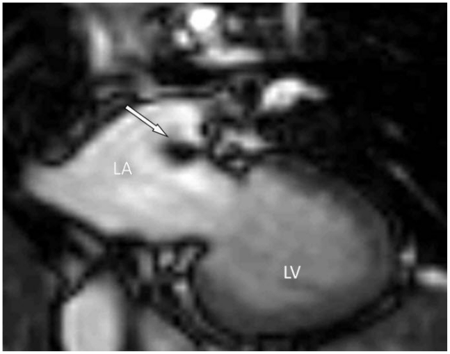
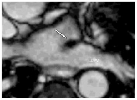
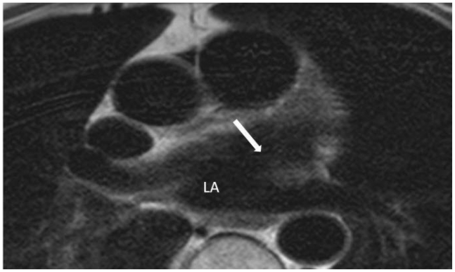
CMR demonstrated normal left ventricular and right ventricular cavity size and function. On cine imaging an incidental structure was identified in the roof of the left atrium (Fig. 1). Multi-plane, multi-phase cine imaging revealed the structure to be ridge-like in appearance and located adjacent to the left upper pulmonary vein (Fig. 2). On T1-weighted imaging pre- and post-contrast, the structure had similar signal intensity to adjacent myocardium. It did not enhance during first pass perfusion imaging with intra-venous gadolinium DTPA or on the late gadolinium enhancement images. It was concluded that the structure had all the characteristic appearances and location of a coumadin ridge.
Figure 1.
50 year old female with a prominent coumadin ridge. The study was performed using a 1.5 Tesla scanner (Inter CV, Philips, Best, The Netherlands). The CMR signals were received by a 5-element cardiac phased -array coil and ECG gating. This is a systolic frame of a multi-phase balanced SSFP cine (Echo time (TE) 1.7ms, Repetition time (TR) 3.5ms, flip angle 60 degrees, SENSE factor 2, matrix 192 × 192, field of view 320–460mm, slice thickness 6mm, 24 phases per cardiac cycle, 1 slice per breath-hold) acquired in a modified double oblique orientation demonstrating the incidental mass-like structure (arrow) in the left atrium (LA).
Figure 2.
In this 50 year old female with a prominent coumadin ridge, a customised axial view using a multi-phase balanced SSFP cine sequence (TE 1.7ms, TR 3.5ms, flip angle 60 degrees, SENSE factor 2, matrix 192 × 192, field of view 320–460mm, slice thickness 6mm, 24 phases per cardiac cycle, 1 slice per breath-hold) demonstrates the close proximity of the structure to the left upper pulmonary vein (LUPV).
DISCUSSION
CMR is rapidly developing as the imaging modality of choice for identification and assessment of cardiac and extra-cardiac tumours. It is an ideal imaging modality for this purpose because it doesn’t suffer from any imaging plane constraints and hence allows optimal visualisation of abnormal structures or ‘masses’ within the heart. The combined assessment from different CMR pulse sequences can also aid tissue characterisation of any mass seen, by using their inherent differences in vascularity and water content. It is important however for the reporter to be aware that there are normally occurring embryological remnants which may sometimes mimic tumours. Examples of these so called pseudotumours include the eustachian valve and the crista terminalis. The eustachian valve, also known as the valve of the inferior vena cava, is a thin flap- like structure located in the right atrium at the orifice of the inferior vena cava. The crista terminalis is a ridge of cardiac tissue which separates the right atrial appendage from the right atrium and when prominent, can be mistaken for a tumour. Lipomatous hypertrophy of the inter-atrial septum, characterised by fatty infiltration of the inter-atrial septum, is another benign condition which can mimic a malignant tumour (1,2).
The coumadin ridge has been described from echocardiographic studies as a ridge of atrial tissue separating the left atrial appendage from the left upper pulmonary vein (3,4). It can present as a linear structure or even sometimes as a nodular mass that protrudes into the left atrium. This ‘mass’ can undulate with cardiac motion and appear similar to a thrombus or atrial myxoma. In the past, this structure was often mistaken for thrombus and resulted in patient being prescribed anticoagulation therapy with warfarin (Coumadin) and it is from here that it derives its name.
Modern CMR methods, in particular cine imaging with steady state free precession (SSFP) techniques, provide improved morphological and functional assessment of such lesions and in combination with T1- and T2-weighted imaging and contrast-enhanced CMR, pseudotumours can be confidently identified by an experienced observer. On cine SSFP sequences, a prominent coumadin ridge can be identified by its typical location in the roof of the left atrium adjacent to the left upper pulmonary vein. As SSFP is a bright blood technique, a particularly prominent ridge can appear as a dark ‘mass’ protruding into the bright left atrial cavity. Important differential diagnoses, which need to be actively ruled out, include thrombus and myxoma. Fast spin echo techniques (FSE) with differential T1 and T2-weighting and use of contrast can help in making the correct diagnosis. T1-weighted images tend to highlight fat containing tissues whereas T2-weighted images are particularly useful to demonstrate areas with excessive water content (e.g cysts and edema). As the coumadin ridge is normal cardiac tissue it should have the same signal intensity as adjacent myocardial tissue on both T1 and T2-weighted imaging.
The technique of late gadolinium enhancement (LGE) after the intravenous injection of gadolinium chelates can further enhance tissue characterisation. The coumadin ridge typically does not show late enhancement. Thrombus on the other hand, is usually diagnosed on LGE images as a low signal-intensity mass surrounded by high intensity structures such as cavity blood or hyperenhanced myocardial scar. If the inversion time during the delay enhancement sequence is increased to 600 ms, avascular tissue such as thrombus is nulled and looks homogenously black in contrast to viable myocardium which will have intermediate signal intensity (5). Myxomas are usually but not always located in close proximity to the interatrial septum and demonstrate high signal on T2-weighted images. They also classically demonstrate heterogenous contrast enhancement following gadolinium injection (6,7).
In summary, a coumadin ridge is an occasionally observed structure on CMR images. If particularly prominent, as in this case, it can easily be mistaken for an atrial myxoma or thrombus. Awareness of the location and CMR imaging features of this structure will help prevent misdiagnosis.
TEACHING POINT
The coumadin ridge is a normally occurring embryological remnant in the left atrium which can easily be mistaken for a tumour or thrombus on CMR imaging. Awareness of the location and CMR imaging features of this structure will help prevent misdiagnosis.
Figure 3.
50 year old female with a prominent coumadin ridge. This ECG-gated breath-hold T1-weighted (TR msec/TE msec = 800/38) axial view demonstrates the signal characteristics of the ridge to be similar to adjacent myocardium on T1-weighted imaging.
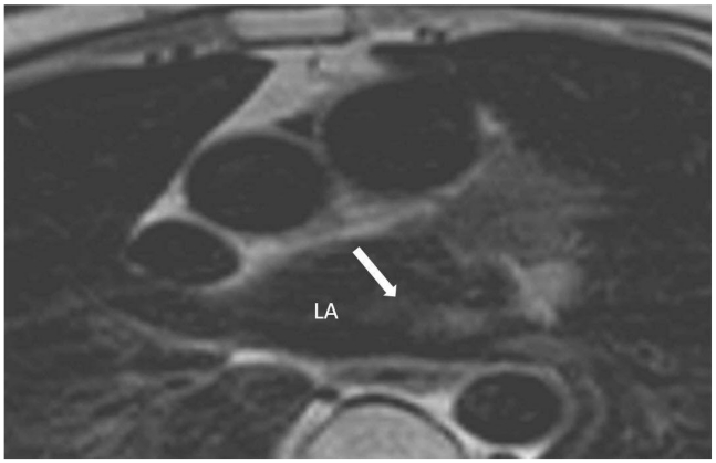
Figure 4.
50 year old female with a prominent coumadin ridge. The coumadin ridge is also of similar signal intensity to adjacent myocardium on T2-weighted imaging (TR msec/TE msec = 1,600/120).
ABBREVIATIONS
- CMR
Cardiac magnetic resonance
- SSFP
Steady state free precession
- LA
Left atrium
- LUPV
Left upper pulmonary vein
- TR
Repetition time
- TE
Echo time
REFERENCES
- 1.Mirowitz SA, Gutierrez FR. Fibromuscular elements of the right atrium: pseudomass at MR imaging. Radiology. 1992;182(1):231–3. doi: 10.1148/radiology.182.1.1727288. [DOI] [PubMed] [Google Scholar]
- 2.King MA, Vrachliotis TG, Bergin CJ. A left atrial pseudomass: potential pitfall at thoracic MR imaging. J Magn Reson Imaging. 1998;8(4):991–3. doi: 10.1002/jmri.1880080431. [DOI] [PubMed] [Google Scholar]
- 3.McKay T, Thomas L. ‘Coumadin ridge’ in the left atrium demonstrated on three dimensional transthoracic echocardiography. Eur J Echocardiogr. 2008;9(2):298–300. doi: 10.1016/j.euje.2006.10.002. [DOI] [PubMed] [Google Scholar]
- 4.Konstadt SN, Shernan SK, Oka Y. Clinical Transesophageal Echocardiography. 2003:511. [Google Scholar]
- 5.Weinsaft JW, Kim HW, Shah DJ, et al. Detection of left ventricular thrombus by delayed-enhancement cardiovascular magnetic resonance prevalence and markers in patients with systolic dysfunction. J. Am. Coll Cardiol. 2008;52(2):148–157. doi: 10.1016/j.jacc.2008.03.041. [DOI] [PubMed] [Google Scholar]
- 6.Fieno DS, Saouaf R, Thomson LEJ, et al. Cardiovascular magnetic resonance of primary tumors of the heart: A review. J Cardiovasc Magn Reson. 2006;8(6):839–853. doi: 10.1080/10976640600777975. [DOI] [PubMed] [Google Scholar]
- 7.Hoffmann U, Globits S, Frank H. Cardiac and paracardiac masses. Current opinion on diagnostic evaluation by magnetic resonance imaging. Eur Heart J. 1998;19(4):553–563. doi: 10.1053/euhj.1997.0788. [DOI] [PubMed] [Google Scholar]


