Abstract
Vicarious renal excretion of iodinated contrast introduced into the bowel is a known phenomenon that has rarely been reported. In clinical settings like Crohn's disease in which evaluation for recto-vesical fistula is frequently requested, vicarious excretion can cause misapprehension and error in diagnosis. We present a case of Crohn's disease in which gastrografin enema was performed to evaluate for fistula and initial interpretation was mistakenly positive, however, simple methods of elucidation were utilized to prevent error in diagnosis.
Keywords: Gastrografin, Crohn's, renal excretion, vicarious, enema
CASE REPORT
A 50-year-old female with a 17 years history of Crohn's disease, had underwent total colectomy and ileoanal anastomosis with Jejunal pouch reconstruction approximately 2.5 years prior to this examination. Since that time the patient had developed frequent bowel movements with up to 20 bowel movements a day. Multiple endoscopic evaluations were suspicious for chronic pouchitis and cuffitis. She was referred for a gastrografin enema to rule out the presence of a chronic anastomotic leak or chronic leak from the tip of the J-pouch, particularly because she was known to be significantly inflamed on endoscopy.
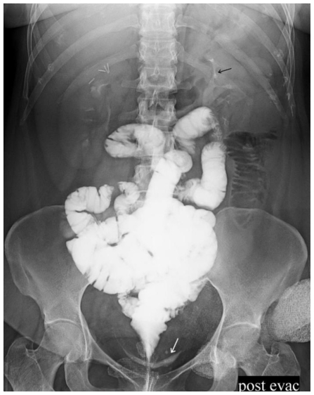
Initial scout image demonstrated surgical staple projecting over the pelvis. (Fig. 1) Initially digital rectal examination was done and afterwards, 480 ml (1320 mg diatrizoate meglumine and 200 ml diatrizoate sodium; 367 mg/ml) of non-diluted Gastrografin (Mallinckrodt. Inc., St. Louis, MO) was introduced into the J-pouch using a pediatric rectal catheter. Initial images demonstrated the site of anastomosis approximately 4 cm above the anal verge (Fig. 2). The J-pouch demonstrated normal expansion and there was no evidence of leaks. The entire small bowel was evaluated in a retrograde fashion in approximately 20 minutes and the residual contrast was evacuated. Post evacuation over-head images, taken approximately after 25 minutes from initial rectal introduction of gastrografin, demonstrated contrast in the patient's bladder (Fig. 3). Initial concern was the presence of a recto-vesical fistula, however, contrast leak due to anastomosis break down was among the differential diagnosis; therefore, additional dynamic views were obtained which also demonstrated contrast in the bladder. The images were once more reviewed and after meticulous attention to the images we realized that both kidneys were in nephrographic phase and bilateral ureters were opacified (Fig. 3). Therefore, we concluded that the contrast observed in the bladder is due to contrast excretion from the kidneys (Fig. 4). Gastrografin injected in the patient's bowel had been absorbed through her bowel and excreted through her renal collecting system.
Figure 1.
50-year-old female with Crohn's disease. Plain X-ray, AP film, scout image before rectal contrast introduction showing surgical staples from prior colectomy and ileoanal anastomosis (Black arrow).
Figure 2.
50-year-old female with Crohn's disease. Plain X-ray, lateral view after rectal injection of gastrografin. The contrast has passed through the ileoanal anastomosis with no evidence of leak or fistula. The contrast fills the small bowel. There is a bowel containing ventral hernia with no evidence of obstruction.
Figure 3.
50-year-old female with Crohn's disease. Plain X-ray, lateral view, post-evacuation after rectal injection of gastrografin. There is evidence of contrast adjacent to the ileoanal anastomosis, in the expected location of the bladder. There is evidence of contrast anterior to the ileoanal anastomosis, in the expected location of the bladder (Black arrow). Additionally, there is opacification of contrast in the kidneys.
Figure 4.
50-year-old female with Crohn's disease. Plain X-ray, AP view, post-evacuation after rectal injection of gastrografin. There is evidence of renal contrast excretion (black arrows) and contrast within the bladder (white arrow).
DISCUSSION
Gastrografin (sodium diatrizoate and meglumine diatrizoate) is an iodinated, water soluble contrast frequently used to investigate anastomosis leakage and/or breakdown in post-operative patients. Additionally, in patients suspected of having intestinal perforation, gastrografin is administrated orally or transrectally to evaluate suspected site of perforation.
Gastrografin is usually eliminated through the rectum and is absorbed from the bowel in very small quantities (1). It has been reported that approximate 2–3 % of the diluted ingested gastrografin is absorbed through an intact bowel (2, 3). This ingested contrast is then secreted through the kidney and will opacity the urinary tract. Urinary tract opacification secondary to absorption of orally or rectally administered iodinated contrast material has been recognized since 1955 (3). If gastrografin leaks into the peritoneum, unlike barium, it usually gets absorbed into the blood and is excreted by the kidney, while causing only a mild inflammatory response (4). Gastrografin will be absorbed more rapidly and to a larger extend in diseased bowel (3–7).
Absorption of gastrografin through the bowel and excretion through the kidneys is known as vicarious renal excretion. As mentioned above this could happen only in small amounts when the bowel is intact. However, it has been reported that this phenomenon occurs more extensively in patients with bowel perforation and in diseased bowel (3–7). Apter et al. reviewed the unenhanced CT scans of 82 patients with bowel disorders or perforation to assess the prevalence of urinary contrast material in various bowel diseases (4). They concluded that renal excretion of orally ingested gastrografin is significantly more likely to occur in diseases involving the bowel wall, such as inflammatory bowel disease, radiation enteritis, ischemia, and lymphoma of the bowel (4). Halme et al. also reported increased permeability of the bowel wall in patients with Crohn ileitis (5). In addition to the above mentioned situations, gastrografin absorption significantly increases in bowel obstruction (6).
Vicarious renal excretion of contrast may cause diagnostic error. Low et al. reported a case in which vicarious renal excretion had caused misinterpretation (7). They reported a 68-year-old man that had underwent an ultra-low anterior resection for rectal carcinoma, and was being evaluated for leak prior to reversal of his diverting ileostomy. Initial images had shown contrast posterior to his rectum with no definitive evidence of leak, however, they were primarily concerned for leak or fistula and therefore, CT scan was performed and contrast was detected in the patient's bladder with no definitive evidence of leak or fistula. Due to these findings the operation was delayed. One month later they repeated the gastrografin enema, which had demonstrated intact anastomosis with no evidence of fistula or leak. The patient had undergone reversion with no complication. This was a simple example in which such vicarious renal excretion was misinterpreted as a recto-vesical fistula and had resulted in unnecessary delay in the patient's management (7).
In our case, the patient was suspected of having chronic anastomosis leak and therefore, gastrografin was used. Our patient had Crohn's disease and presumably ileitis. Additionally, she had undergone total colectomy and therefore the contrast was introduced into her small bowel which resulted in a more rapid absorption of the contrast. Fortunately, in our case, meticulous attention to the renal collecting system prevented us from misinterpreting.
Multiple simple methods can be utilized in order to prevent misinterpreting vicarious renal excretion. Meticulous attention to the renal collecting system is a simple method that should be the first step in investigating these patients. The second method that can be utilized is consumption of diluted gastrografin instead of non-diluted contrast. This, however, will limit the evaluation. Previous studies indicated that renal excretion of gastrografin ranges from 3 to 21% in healthy normal bowels when undiluted gastrografin was used, whereas this rate significantly decreased while using diluted gastrografin (2, 4). In our case, the undiluted contrast was introduced into an already diseased small bowel, increasing the absorption significantly faster and more proficient.
Additionally, it has been reported that opacification of the urinary system is directly dependent on the concentration of iodine in urine rather than on the total amount of iodine excreted (7). Therefore, in situations in which prevention of vicarious renal excretion of contrast is strongly recommended hydration of the patient prior to the procedure would be a helpful method. Finally, we suggest performing the procedure as speedy and as prompt as possible. Prior studies have suggested that maximum concentration of contrast in the urine usually occurs between 2 and 4 h (8). This provides an acceptable window to perform the procedure. However, this time window extremely shortens in patients with abnormal bowels i.e. Crohn's ileitis.
TEACHING POINT
Absorption of gastrografin through the bowel and excretion through the kidneys is known as vicarious renal excretion. This could happen only in small amounts when the bowel is intact. Contrast in the urinary tract may cause confusion and misinterpreted as fistula. Meticulous attention to the renal collecting system, consumption of diluted gastrografin instead of non-diluted contrast, prehydration, and performing the procedure as speedy and as prompt as possible are simple methods that can help preventing misapprehensions.
ABBREVIATIONS
- CT scan
computed axial tomography
- J-pouch
Jejunal pouch
- AP
Antero-posterior
REFERENCES
- 1.Zissin R, Oscadchy A, Shapiro-Feinberg M. Renal excretion of oral gastrografin seen on an abdominal film in a patient with incomplete intestinal obstruction due to multiple bezoars. Isr Med Assoc J. 1999 Nov;1(3):200–1. [PubMed] [Google Scholar]
- 2.Sohn KM, Lee SY, Kwon OH. Renal excretion of ingested Gastrografin: clinical relevance in early postoperative treatment of patients who have undergone gastric surgery. AJR. 2002;178:1129–32. doi: 10.2214/ajr.178.5.1781129. [DOI] [PubMed] [Google Scholar]
- 3.Marinelli DL, Mintz MC. Absorption and excretion of dilute gastrografin during computed tomography in pseudomembranous colitis. J Comput Tomogr. 1987 Jul;11(3):236–8. doi: 10.1016/0149-936x(87)90088-9. [DOI] [PubMed] [Google Scholar]
- 4.Apter S, Gayer G, Amitai M, Hertz M. Urinary excretion of orally ingested gastrografin on CT. Abdom Imaging. 1998 May-Jun;23(3):297–300. doi: 10.1007/s002619900344. [DOI] [PubMed] [Google Scholar]
- 5.Halme L, Edgren K, von Smitten K, Linden H. Increased urinary excretion of iohexol after enteral administration in patients with ileal Crohn's disease: a new test for disease activity. Acta Radiol. 1993;34:237–241. [PubMed] [Google Scholar]
- 6.Laerum F, Stordahl A, Solheim KE, Haugstvedt JR, Roald HE, Skinningsrud K. Iodinated contrast agents and the gastrointestinal tract. Intestinal follow-through examination with iohexol and iopentol permeability. Alterations and efficacy in patients with small bowel obstruction. Invest Radiol. 1991;26:177–181. doi: 10.1097/00004424-199111001-00060. [DOI] [PubMed] [Google Scholar]
- 7.Low VH, Chu BK. Diagnostic error due to vicarious excretion of rectal iodinated contrast. Australas Radiol. 2006 Aug;50(4):369–72. doi: 10.1111/j.1440-1673.2006.01603.x. [DOI] [PubMed] [Google Scholar]
- 8.Mori P, Barrett H. A sign of intestinal perforation. Radiology. 1962;79:401–7. doi: 10.1148/79.3.401. [DOI] [PubMed] [Google Scholar]


