Abstract
A new generation of realistic, image-based anthropomorphic phantoms has been developed based on the reference masses and organ definitions given in the International Commission on Radiological Protection Publication 89. Specific absorbed fractions for internal radiation sources have been calculated for photon and electron sources for many body organs. Values are similar to those from the previous generation of ‘stylized’ (mathematical equation-based) models, but some differences are seen, particularly at low particle or photon energies, due to the more realistic organ geometries, with organs generally being closer together, and with some touching and overlapping. Extension of this work, to use these phantoms in Monte Carlo radiation transport simulation codes with external radiation sources, is an important area of investigation that should be undertaken.
INTRODUCTION
The current generation of anthropomorphic models used for dose calculations are based on the stylised anatomical models developed for the MIRD Committee of The Society of Nuclear Medicine in 1960s. Figure 1 shows exterior and cut-away views of the original MIRD model representing a reference adult(1). The body was defined in three sections: an elliptical cylinder representing the arm, torso and hips; a truncated elliptical cone representing the legs and feet and an elliptical cylinder representing the head and neck. Then, major organs were mathematically defined as occupying finite spaces within the body, and comprised three types of tissue: ‘soft tissue’, bone and lung. The mathematical descriptions of the organs were formulated based on descriptive and schematic materials from general anatomy references, and were modelled after the prescriptions of the ‘Reference Man’ defined by the International Commission on Radiological Protection (ICRP) in its Publication 23(2). Cristy and Eckerman(3) defined a ‘family’ of models representing reference individuals of several ages, and Stabin et al.(4) created a set of models representing the pregnant female at four stages of gestation. These two series of reference models were implemented in the MIRDOSE(5) and OLINDA/EXM 1.0(6) personal computer codes to facilitate calculation of internal dose calculations. To date, only a few limited calculations have been done with external sources using these new phantoms (to evaluate doses from some computed tomography (CT) examinations), and no results have been published.
Figure 1.
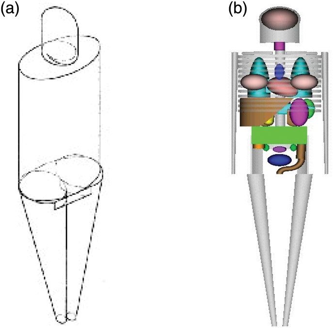
Stylised adult male model of Snyder et al.(1) showing (a) exterior view, (b) skeleton and internal organs.
The advent of medical image-based models now allows replacement of these ‘stylized’ body models with more anatomically realistic models. Paul Segars of Duke University developed human body models based on non-uniform rational b-spline (NURBS) models(7). These realistic renderings of the adult male and female are more anatomically accurate than the stylised models and furthermore can be rapidly and easily scaled to represent other ages and body sizes. Figure 2 shows the adult male and female NURBS models developed by Segars.
Figure 2.
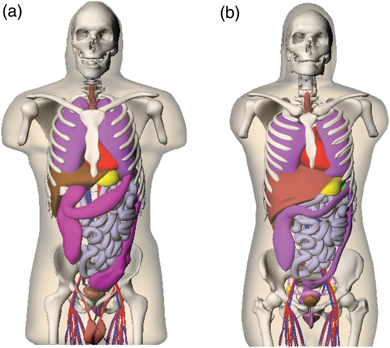
Anterior views of the NURBS models of the adult male (left) and adult female (right).
MATERIALS AND METHODS
The ICRP has issued a new report outlining reference masses for adults and children of different ages in its Publication 89(8). A software tool developed by Segars allows for the rapid deformation and scaling of human or animal NURBS models to match the ICRP reference mass values. One or more selected organs may be translated or rotated in any direction, scaled linearly in any direction, uniformly in three dimensions, from the center by a fixed factor and otherwise modified by the user. The viewing pane on the right then allows slice-by-slice inspection of results. Using the ICRP 89 recommended organ mass values for adults and children of the reference ages evaluated (newborn, 1-y, 5-y, 10-y, 15-y and adult), organ and whole body models for both genders at these reference ages were developed. The final models may be rendered in voxel format and used in Monte Carlo radiation transport simulation codes, employing internal or external simulated radiation sources.
The GEANT4 (GEometry ANd Tracking) C++ particle transport toolkit(9) was used to perform radiation transport calculations in the voxel-based models that were derived from the NURBS models. The NURBS models are easily voxelised at most resolutions and matrix sizes. For most organs, the difference between the NURBS reported and voxelised volumes was ∼3–5 %, but for some small organs, differences were greater.
RESULTS
Figure 3 shows images of selected models from the series. The complete ICRP 89 tables of recommended organ masses are large, and will not be reproduced here. Figures 4–8 show sample plots of absorbed fractions for selected internal sources from the models, some with comparison to values from the Cristy/Eckerman phantoms(3). These plots typify the major trends seen in the results. Figure 4 shows photon specific absorbed fractions (SAFs) for liver irradiating liver in the adult male phantom—results are similar to those of Cristy/Eckerman, as the organs are similar in mass, and minor differences in shape do not affect these numbers significantly. Figure 5 gives results for photons in the adult male phantom for liver irradiating lungs—here the values for the NURBS models are higher at all energies, owing to the close geometrical proximity of the two organs in the more realistic NURBS models, compared with the necessary separation which occurs in stylised phantoms. Figure 6 shows photon SAFs for liver irradiating kidneys for different reference age models (males), in which the usual trend of SAFs increasing with decreasing age is seen. Figure 7 shows electron AFs (not SAFs) for electron sources in the kidneys of the adult female irradiating either the kidneys or liver. Figure 8 shows electron AFs for the testes of the male newborn, showing the significant departure from unity for this small organ at moderate-to-high energies.
Figure 3.
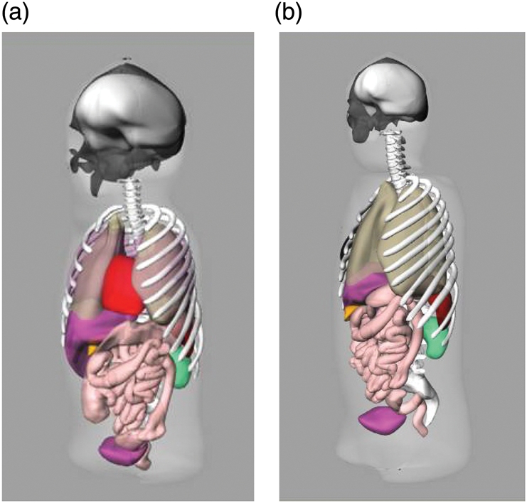
Sample images of NURBS standardised models, scaled to the ICRP 89 recommended reference organ masses, developed for internal and external dose assessment: (a) newborn female model, (b) 15-y-old male model.
Figure 4.
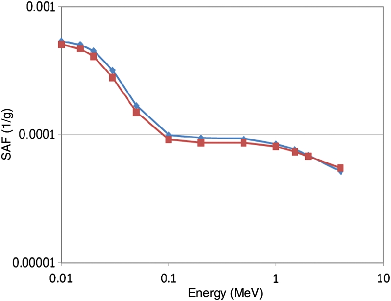
Sample photon SAF plot, showing comparison of NURBS model values (diamonds) with values from the Cristy/Eckerman stylised series (squares): Adult male (liver←liver).
Figure 8.
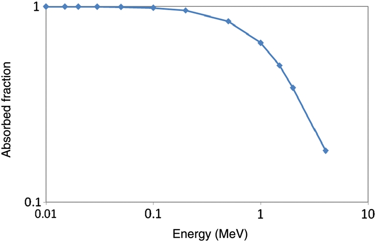
Electron absorbed fractions in the testes of the newborn male.
Figure 5.
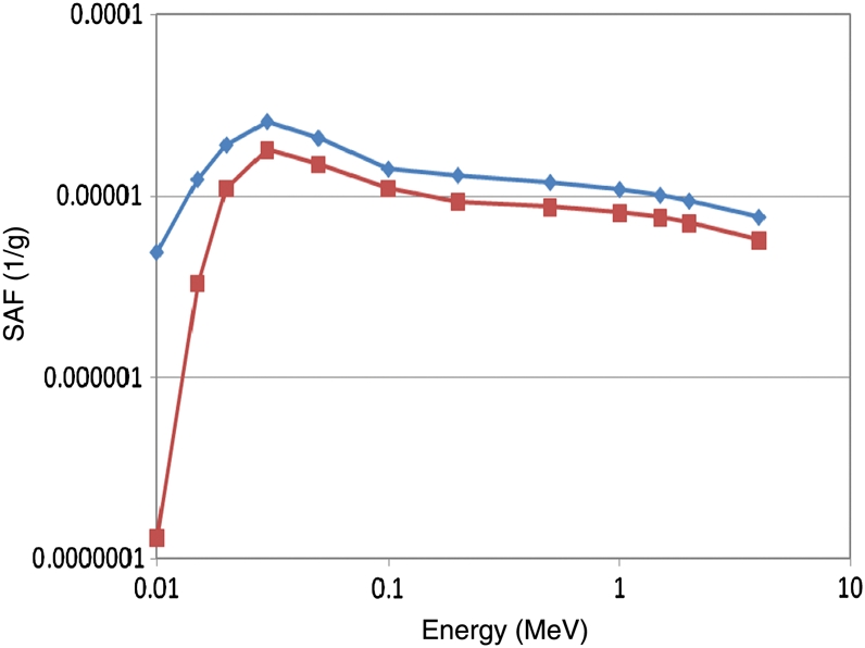
Sample photon SAF plot, showing comparison of NURBS model values (diamonds) with values from the Cristy/Eckerman stylised series (squares): Adult male (lungs←liver).
Figure 6.
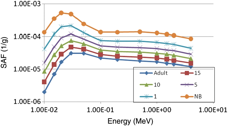
Sample photon SAF plot, showing comparison of NURBS model values for different reference age models: males, (kidneys ← liver).
Figure 7.
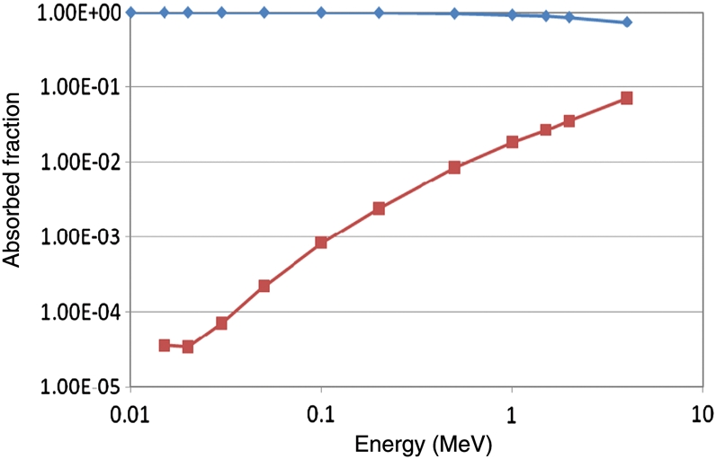
Electron absorbed fractions in the kidneys of the adult female, kidneys as a target (diamonds) and liver as a target (squares).
DISCUSSION
The previous generation of anthropomorphic models has served the dosimetry community well for over 30 y. Original body models used in dosimetry were spherical in nature(10); this was mathematically convenient and provided conservative estimates of SAF and dose, but was not realistic. The stylised phantom series by Cristy and Eckerman(3) represented a considerable improvement over simple spherical models for dose calculations. Monte Carlo methods were needed to estimate SAF values for the organs, and the ability to calculate cross-irradiation SAFs was introduced, and thus the science of dose calculation, for either internal or external sources of radiation, was greatly improved. The NURBS models developed here are intended as updates to the Cristy/Eckerman series, with the organ masses based on those recommended by the ICRP for standardised individuals of various ages(8). Changes in actual SAF values from the Cristy/Eckerman model series are notable, but mostly small in magnitude. These phantoms, in conjunction with those of the Rensselaer Polytechnic Institute (RPI) group for pregnant females(11) which provide an update on the Stabin et al.(4) stylised series of phantoms, will be used in new versions of the OLINDA/EXM computer code and made available to the user community through the Consortium of Computational Human Phantoms hosted at RPI. These models may be used in any standard Monte Carlo codes for calculation of dose estimates to standard individuals from internal or external sources of photons, neutrons or electrons. Furthermore, the NURBS models can be easily deformed to represent individuals of different body sizes, e.g. different normal weight individuals(12) or obese individuals(13), thus allowing investigation of how doses change in different circumstances with body size and shape.
Traditionally, all of the AFs for electron self-irradiation have been assumed to be 1.0 for an organ irradiating itself and 0.0 for other organs. Although this trend is generally true, when electron transport is performed, there is some departure from this assumption at higher energies for all organs, and the departure is more important for the smaller organs, particularly in the smaller phantoms, e.g. the testes of a newborn male, as shown in Figure 8. Explicit treatment of how electron energy may escape from one organ and irradiate another is an important advance in the treatment of internal radiation sources. It will cause some challenges in interpretation of the calculated doses, however. In most cases, the electron dose from one organ to another is only due to bremsstrahlung radiation, which is very low relative to normal photon and electron doses received by the organ. When the organs are touching, there is also dose from electrons leaving the source and striking the target, but this dose is not received uniformly by all parts of the organ, as traditional dose estimates assume. The dose is localised over the small portion of the organ where the source and target touch, and drops to zero at the range of the electron.
Although few calculations have been done with external sources using these new phantoms to date, any kind of source can be modelled with Monte Carlo radiation transport simulation codes, and further investigations using external sources with these phantoms have been planned. Interesting differences have been seen in the SAF values for internal sources with organs being physically closer and touching in many cases. For external radiation sources (e.g. CT scanners, photon or neutron fields), similar differences in absorbed fraction and dose values will be seen due to the more realistic modelling of the organ shapes and positions. As with internal sources, profound differences are not expected in final dose estimates, and in particular effective doses, (in the latter case because of the summing of contributions from many organs, some of which may report higher values than with previous ‘stylized’ models and others lower values). For internal sources, notable differences in SAFs were observed for different normal-weight adults (∼10–30 % differences in many SAFs from a 10th to a 90th percentile normal weight individual), but the differences were also within the uncertainties always associated with internal dose estimates(12). Differences were almost not perceptible for internal sources in moderately or severely obese adults(13). However, for external sources, obesity will clearly be an issue, as the radiation has to penetrate more layers of adipose tissue before reaching the internal organs. This is an important area for future investigations with these phantoms.
FUNDING
This work was supported partially by grant 1R42CA115122-01 from the National Institutes of Health.
REFERENCES
- 1.Snyder W. S., Ford M. R., Warner G. G. Society of Nuclear Medicine; 1978. Estimates of specific absorbed fractions for photon sources uniformly distributed in various organs of a heterogeneous phantom. MIRD Pamphlet No. 5, Revised. [PubMed] [Google Scholar]
- 2.Pergamon Press; 1975. International Commission On Radiological Protection: report of the task group on reference man. ICRP Publication 23. [Google Scholar]
- 3.Cristy M., Eckerman K. Oak Ridge National Laboratory; 1987. Specific absorbed fractions of energy at various ages from internal photons sources. ORNL/TM-8381 V1-V7. [Google Scholar]
- 4.Stabin M., Watson E., Cristy M., Ryman J., Eckerman K., Davis J., Marshall D., Gehlen K. Mathematical models and specific absorbed fractions of photon energy in the nonpregnant adult female and at the end of each trimester of pregnancy. 1995. ORNL Report ORNL/TM-12907. Oak Ridge National Laboratory.
- 5.Stabin M. MIRDOSE—the personal computer software for use in internal dose assessment in nuclear medicine. J. Nucl. Med. 1996;37:538. [PubMed] [Google Scholar]
- 6.Stabin M. G., Sparks R. B., Crowe E. OLINDA/EXM: the second-generation personal computer software for internal dose assessment in nuclear medicine. J. Nucl. Med. 2005;46:1023. [PubMed] [Google Scholar]
- 7.Segars J. P. The University of North Carolina; 2001. Development and application of the new dynamic NURBS-based cardiac-torso (NCAT) phantom. Ph.D. dissertation. [Google Scholar]
- 8.International Commission on Radiological Protection. Elsevier Health; 2003. Basic anatomical and physiological data for use in radiological protection: reference values. ICRP Publication 89. [Google Scholar]
- 9.Geant4—a simulation toolkit. Nucl. Instrum. Methods A. 2003;506:250. [Google Scholar]
- 10.International Commission on Radiological Protection. Pergamon Press; 1959. Report of committee II on permissible dose for internal radiation. ICRP Publication 2. [Google Scholar]
- 11.Xu X. G., Shi C., Stabin M. G., Taranenko V. Xu X. G., Eckerman K. F. Handbook of Anatomical Models for Radiation Dosimetry. CRC Press; 2009. Pregnant female/fetus computational phantoms and the latest RPI-P series representing 3, 6, and 9 months gestational periods; pp. 305–336. [Google Scholar]
- 12.Marine P. M., Stabin M. G., Fernald M. J., Brill A. B. Changes in radiation dose with variations in human anatomy: larger and smaller normal-stature adults. J. Nucl. Med. 2010;51:806–811. doi: 10.2967/jnumed.109.073007. [DOI] [PMC free article] [PubMed] [Google Scholar]
- 13.Clark L. D., Stabin M. G., Fernald M. J., Brill A. B. Changes in radiation dose with variations in human anatomy: moderately and severely obese adults. J. Nucl. Med. 2010;51:929–932. doi: 10.2967/jnumed.109.073015. [DOI] [PMC free article] [PubMed] [Google Scholar]


