Abstract
Oral erythema multiforme (EM) is considered as a third category of EM other than EM minor and major. Patients present with oral and lip ulcerations typical of EM but without any skin target lesions. It has been reported that primary attacks of oral EM is confined to the oral mucosa but the subsequent attacks can produce more severe forms of EM involving the skin. Hence, it is important to identify and distinguish them from other ulcerative disorders involving oral cavity for early management. This article reports two cases of oral EM that presented with oral and lip ulcerations typical of EM without any skin lesions and highlights the importance of early diagnosis and proper management.
Keywords: Drug reaction, lips, oral erythema multiforme, oral mucosa, ulcerations
INTRODUCTION
Adverse reactions to systemic drug administration can have different clinical patterns such as erythema multiforme minor, major, Steven Johnsons syndrome, anaphylactic stomatitis, intraoral fixed drug eruptions, lichenoid drug reactions, and pemphigoid-like drug reactions.[1] Erythema multiforme (EM) shows typical clinical patterns. Based on the severity and the number of mucosal sites involved, the disease has been subclassified into EM minor and major. EM minor shows ulcerations involving a single mucosal site with typical skin target lesions. EM major shows ulcerations involving more than one mucous membrane with skin target lesions. These lesions can be triggered by HSV infections or adverse drug reactions. Steven Johnson syndrome is a more severe condition characterized by wide spread small blisters on torso and mucosal ulcerations with atypical skin target lesions triggered by drug intake. Typical target skin lesions are necessary along with mucosal ulcerations to consider diagnosing them as EM minor and major. Many investigators have reported cases of oral mucosal ulcerations and lip lesions typical of EM without any skin lesions. They have classified them into a new category called oral EM.[2] It has been reported that even if the primary attacks of oral EM are confined to the oral mucosa the subsequent attacks can produce more severe forms of EM involving the skin and hence it is important to identify and distinguish them from other ulcerative disorders involving oral cavity for early management and proper follow up.[3,4] This article reports two cases of oral EM highlighting the importance of distinguishing this disorder.
CASE REPORTS
Case 1
A 21-year-old female patient visited the dental OPD with the complaint of extensive ulceration of oral cavity and pain and inability to eat for the past 4 days. She gave a history of leg sprain for which she took diclofenac sodium subsequent to which she developed multiple small ulcerations that later transformed into extensive, irregular ulcerations of the oral mucosa.
On extra oral examination, both upper and lower lips showed extensive irregular ulcerations, showing cracking and fissuring with blood encrustation. Bilateral submandibular lymph nodes were enlarged and tender. Intraoral examination showed extensive, irregular ulcerations with yellow base and erythematous borders on buccal mucosa, palate, dorsal and ventral surfaces of the tongue [Figures 1 and 2].
Figure 1.
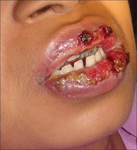
Case 1 irregular lip ulcerations with blood encrustations
Figure 2.
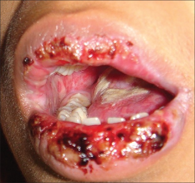
Case 1 irregular buccal mucosal and tongue ulcerations with lip lesions
The sudden onset, positive drug history, extensive ulcerations of the oral cavity, cracking, and fissuring of lips with bloody crusting lead to the diagnosis of oral Erythema multiforme.
The patient was advised to stop the diclofenac sodium medication and was treated with topical corticosteroids, mild analgesics, and local application of lignocaine gel to facilitate oral fluid intake. Healing was noticed on the third day and the lesions were completely cleared without scarring in 10 days time [Figure 3]. Patient was advised not to take any drug from the Diclofenac group.
Figure 3.
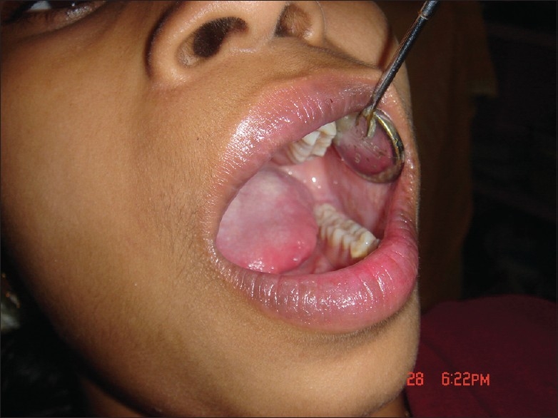
Case 1 after 10 days of treatment the oral mucosal and lip ulcerations are healed
Case 2
A 23-year-old female patient presented to the dental OP with the complaint of painful ulcerations of the oral cavity for the past 5 days. She gave a history of bronchial asthma for which she took homeopathy medicine few weeks back, within days she developed oral ulcerations.
She gave a history of multiple vesicles of the oral mucosa, buccal, and labial mucosa, which ruptured to form painful ulcerations. After 2 days she developed ulceration of lips and tongue. Patient was unable to eat any hot and spicy food and was on liquid diet for the last 2 days.
Extraoral examination showed extensive ulcerations with bloody crustations on the upper and lower lip. Intraorally multiple ulcerations of the buccal and labial mucosa and palate were seen [Figure 4]. Tongue showed white coating on the dorsal surface with irregular ulcerations of the right lateral border.
Figure 4.
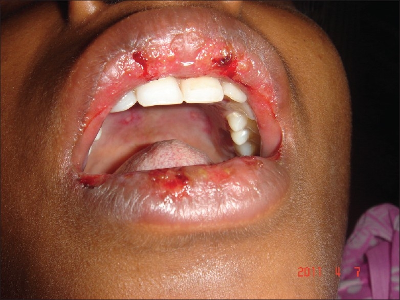
Case 2 palatal ulcerations with irregular blood encrusted lip lesions
Patient was advised to discontinue the homeopathic medicine and treated with cortico steroids (prednisolone 10 mg) twice a day for 3 days followed by tapering dose for 10 days and local application of topical anesthetic gel for pain relief. Patient responded well to the treatment and healing of the lesions occurred within a week [Figure 5]. The patient was strictly advised, not to take the same homeopathic medications again.
Figure 5.
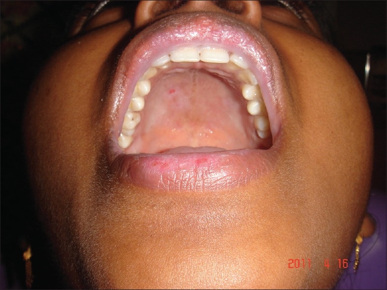
Case 2 after 10 days of treatment the healing palatal and healed lip ulcerations
Positive association between the drug intake and incidence of the lesions and the clinical appearance of the lesions lead to the diagnosis of oral erythema multiforme.
DISCUSSION
Erythema multiforme (EM) is an inflammatory disorder that affects the skin or mucous membrane or both.[5] According to von Hebra, who first described the disease in 1866, the patients with erythema multiforme should have acrally distributed typical target lesions or raised edematous skin papules with or without mucosal involvement.[6] In 1968, Kenneth described an inflammatory oral disorder with oral lesions typical of EM but without any skin involvement. He reported nine cases seen at the East Man Dental hospital. The common sites involved were lips, cheeks, and tongue. These patients had irregular large ulcers with necrotic tags attached to the borders. When lips are involved the typical blood encrusted lesions were seen. In this series of cases, the typical target skin lesions were seen during the recurrences not in their initial attacks.[3] Many investigators have suggested this as a third category of EM known as oral EM that are characterized by typical oral lesions of EM but no target skin lesions. Oral EM is a distinct but less well-recognized variant of EM. The diagnosis has to be established by excluding other oral inflammatory and vescicullobullous lesions.[2]
Our two cases showed extensive irregular erythematous ulcerations in the buccal mucosa, labial mucosa, tongue, and palate along with blood encrusted lip ulcerations. Biopsies are advised only in early vesicular lesions of erythema multiforme not in ulcerated ones since histopathologic appearances are nonspecific and nondiagnostic.[3] Our patients reported to us with advanced ulcerated lesions and hence the diagnosis had to be established based on the positive drug history, clinical appearance, and distribution of the lesion and exclusion of other ulcerative lesions.
We were able to establish a temporal relationship between the drug intake and occurrence of the oral mucosal lesions. The oral ulcerations in our cases started within a few days of the drug intake and were resolved upon cessation of the drug. Erythema multiforme is usually triggered by herpes simplex infections, but rarely by drug intake.
When the lesions are confined only to the oral cavity the different differential diagnosis that has to be considered are herpes, autoimmune vescicullobullous lesions such as pemphigus vulgaris or bullous pemphigoid and other patterns of drug reactions.
The most important of them is acute herpetic stomatitis. Herpetic lesions are more common in the keratinized mucosa especially the gingiva. Our cases did not have any gingival ulceration. Herpetic ulcers are smaller with regular borders than ulcers associated with EM. Extensive irregular ulcerations in the lining nonkeratinized mucosa as seen in our patients were typical of EM and are not a feature of herpes infection. The presence of a temporal relationship between the drug intake and onset of the disease excludes the possibility of any infectious aetiologies.[3]
The positive drug histories associated with onset of ulcerations in our cases ruled out the possibility of other autoimmune vescicullobullous lesions like pemphigus vulgaris. Unlike pemphigus vulgaris oral EM have an acute onset and does not show any desquamative gingivitis.[4] Bullous lichen planus lesions that may have similar ulcerations should have Wickham's striae, which were absent in our cases excluding it as the diagnosis.[3]
Other patterns of drug reactions like lichenoid drug reactions, pemphigoid-like drug reactions that resemble their namesake can be easily differentiated based on the clinical patterns as above mentioned. Anaphylactic stomatitis often shows urticarial skin reactions with other signs and symptoms of anaphylaxis which were absent in our cases. In mucosal fixed drug eruptions the lesions are confined to localized areas of oral mucosa but in our cases there were wide spread lesions affecting labial, buccal, palatal, and tongue mucosa along with lip involvement.[1]
Lesions of EM minor are characterized by single mucosal ulcerations and typical target lesions of skin. The oral mucosal ulcerations are usually irregular and large with necrotic tissue tags. Lip ulcerations are blood encrusted. Erythema multiforme major is considered to be a more aggressive form characterized by involvement of multiple mucosa accompanied by typical target skin lesions.[5–7] The third category of EM, also described by many investigators as oral EM has the lesions confined to the oral mucosa and lips with no skin involvement.[2] Since our cases were evidently triggered by drug intake and they had typical lesions of EM in the oral mucosa and lips with no skin involvement we came to a diagnosis of oral EM.
The most common drugs that trigger EM lesions are long acting sulfa drugs especially sulphonamides, co-trimoxazole, phenytoin, carbamazepine and nonsteroidal antiinflammatory drugs such as diclofenac, ibuprofen, and salicylates.
Management of oral EM involves identification of triggering agent. If it is found to be HSV infection patients have to be put on antiviral medications. If HSV is ruled out as a triggering agent and the culprit is an adverse drug reaction, the drug is immediately stopped. Usually lesions of oral EM can be treated palliatively with analgesics for oral pain, viscous lidocaine rinses, soothening mouth rinses, bland soft diet, avoidance of acidic and spicy food, systemic and topical antibiotics to prevent secondary infection.[2] Lesions of EM usually respond to topical steroids, for more severe cases systemic corticosteroids are recommended.[8]
CONCLUSION
Oral EM is a rare and less described variant of EM. Oral EM is often triggered by HSV infections and rarely by adverse drug reactions. Even though primary attacks of oral EM are confined to the oral mucosa the subsequent attacks can produce more severe forms of EM (EM minor and major) involving the skin.[3] Hence, it is important to distinguish oral EM for their early diagnosis, prompt management, and proper follow up.
Footnotes
Source of Support: Nil.
Conflict of Interest: None declared.
REFERENCES
- 1.Neville BW, Damm D, Allan CM, Bouquot JE, editors. Oral and Maxillofacial Pathology. 2nd ed. Philadelphia: Saunders; 2002. Allergic and immunologic diseases; pp. 285–314. [Google Scholar]
- 2.Ayangco L, Rogers RS., 3rd Oral manifestations of erythema multiforme. Dermatol Clin. 2003;21:195–205. doi: 10.1016/s0733-8635(02)00062-1. [DOI] [PubMed] [Google Scholar]
- 3.Kennett S. Erythema multiforme affecting the oral cavity. Oral Surg Oral Med Oral Pathol. 1968;25:366–73. doi: 10.1016/0030-4220(68)90010-8. [DOI] [PubMed] [Google Scholar]
- 4.Bean SF, Quezada RK. Recurrent oral erythema multiforme clinical experience with 11 Patients. JAMA. 1983;249:2810–2. [PubMed] [Google Scholar]
- 5.Scully C, Bagan J. Oral mucosal diseases: Erythema multiforme. Br J Oral Maxillofac Surg. 2008;46:90–5. doi: 10.1016/j.bjoms.2007.07.202. [DOI] [PubMed] [Google Scholar]
- 6.Assier H, Bastuji-Garin S, Revuz J, Roujeau J. Erythema multiforme with mucous membrane involvement and steven-johnson syndrome are clinically different disorders with distinct disorders. Arch Dermatol. 1995;131:539–43. [PubMed] [Google Scholar]
- 7.Bastuji-Garin S, Razny B, Stern RS, Shear NH, Naldi L, Roujeau JC. Clinical classification of cases of toxic epidermal necrolysis, Steven-Johnson syndrome and erythema multiforme. Arch Dermatol. 1993;129:92–6. [PubMed] [Google Scholar]
- 8.Williams RM, Cocklin RC. Erythema multiformea. Review and contrast from Stevens-Johnson syndrome/toxic epidermal necrolysis. Dent Clin North AM. 2005;49:67–76. doi: 10.1016/j.cden.2004.08.003. [DOI] [PubMed] [Google Scholar]


