Abstract
Objective
To quantify the cumulative effective dose (cED) of radiation due to repeated CT and PET/CT examinations after curative resection of gastric cancer and to assess the lifetime attributable risk (LAR) estimates based on Biological Effects of Ionizing Radiation VII models.
Subjects and Methods
Patients who underwent a curative resection for gastric cancer between January 2006 and December 2006 and were followed-up until May 2010 were included in this study. The cED was calculated by using the dose-length product values and conversion factors for quantitative risk assessment of radiation exposure. cED and LAR were compared between early and advanced gastric cancer patients and among American Joint Committee on Cancer TNM stage groups (stage I, II, and III). The nonparametric Mann-Whitney U and Kruskal-Wallis tests, followed by a post-hoc analysis with Bonferroni adjustment, were employed as part of the statistical analysis.
Results
The overall median cED was 57.8 mSv (interquartile range [IQR], 43.9-74.7). The cED was significantly higher in the advanced (median, 67.0; IQR, 49.1-102.3) than in the early gastric cancer group (median, 52.3; IQR, 41.5-67.9) (p < 0.001), and increased as the TNM stage increased. For radiation exposure, 62% of all patients received an estimated cED of over 50 mSv, while 11% of patients received over 100 mSv. The median LAR of cancer incidence was 0.28% (IQR, 0.20-0.40) and there were significant differences between the early gastric cancer and advanced gastric cancer group (p < 0.001) as well as among the three TNM stage groups (p = 0.015). The LAR of cancer incidence exceeded 1% in 2.4% of the patients.
Conclusion
The cED increases proportionally along with tumor stage and, even in early gastric cancer or stage I patients, cED is much higher than that found among the general population. Considering the very good prognosis of early gastric cancer after curative surgery, the cED should be considered when designing a postoperative follow-up CT protocol.
Keywords: Computed tomography, Effective dose, Lifetime attributable risk, Gastric cancer, Radiation exposure, Follow-up
INTRODUCTION
Although gastric cancer has decreased in incidence over the past few decades, it remains the fourth most common cancer in the world and the second most common cause of cancer-related deaths (1). The treatment of choice for gastric cancer is surgical resection, which is the curative treatment for gastric cancer. Recently, the technical development of diagnostic tools such as endoscopy or computed tomography (CT) and widespread application of surveillance programs, especially in Korea and Japan where gastric cancer is particularly common, more than 60-80% of patients are diagnosed at potentially curative stages and 5-year survival rates are approaching 70% for R0 resections (2, 3). As a result, the number of patients who require postoperative follow-up is rapidly increasing. However, guidelines for postoperative follow-up have not been established, and different follow-up protocols are used by each institution (2, 4). According to recent National Comprehensive Cancer Network (NCCN) guidelines, postoperative follow-up with medical history-taking and physical examinations are recommended specifically based on the follow-up period after surgery. However, recommendations for follow-up radiologic imaging only require that the imaging be clinically indicated, without any guidance regarding the follow-up period or imaging modalities. Presently, due to improvements in diagnostic performance and objectivity, most imaging follow-up is performed by CT with a variable follow-up interval, based on each institution's strategy (2, 4).
Public concern about radiation exposure has recently increased due to the rapid growth of CT use in medical applications, as well as nuclear catastrophes such as those at Chernobyl and in Japan. In 2007, the International Commission on Radiological Protection (ICRP) recommended dose limitations for occupational exposure (20 mSV per year) and for public exposure (1 mSv per year) (5). However, there are no established radiation dose limits for patients undergoing radiographic imaging, and risk-benefit evaluations should be performed to establish such guidelines. In a large-scale dose estimation study in the United States, more than 70% of patients received doses of less than 3 mSv per year, which is equivalent to the background levels of natural radiation in the United States (6). In contrast, a long-term retrospective cohort study demonstrated that patients with histories of malignancy or active malignancies experienced much higher radiation exposure than patients without malignancies (7). Considering that about 14.9-19.5% of gastric cancer patients are younger than 45 years of age, with a 5-year overall survival rate of about 70%, and rates that reach 90% in stage I and II cases (3, 8), there are growing concerns regarding cumulative radiation exposure due to lifelong radiologic surveillance. Therefore, it is necessary to estimate the current state of radiation exposure due to repeated follow-up CT or PET/CT imaging in gastric cancer patients for the risk-benefit analysis of postoperative follow-up imaging. Therefore, the purpose of this study was to quantify the cumulative effective dose (cED) from repeated CT and PET/CT examinations after curative resection of gastric cancer and to assess lifetime attributable risk based on the "Biological Effects of Ionizing Radiation" (BEIR) VII models.
SUBJECTS AND METHODS
Study Population
This retrospective study was approved by the institutional review board of the Yonsei University Health System and the requirement of informed consent was waived. Inclusion criteria were as follows: (a) patients who were diagnosed with gastric cancer and had undergone surgery with a curative intent between January 2006 and December 2006, (b) patients who were alive through May 2010, (c), patients who had no evidence of tumor recurrence in clinical and radiologic assessment, and (d) patients who had been regularly followed-up until May 2010. Patients were considered as a "follow-up loss" if their last visit did not occur within the last 18 months prior to May 2010. Patients were usually followed up at intervals of less than 1 year, but some patients visited the hospital a few months later than their prearranged follow-up dates due to personal reasons, and therefore we defined 18 months as the longest interval of regular follow-up.
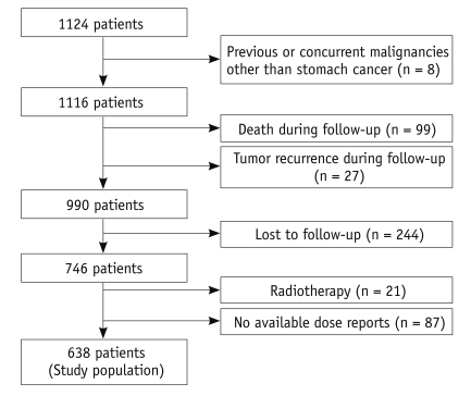
Between January 2006 and December 2006, 1124 patients underwent curative surgery for gastric cancer at our institution. Among these patients, eight who had previous or concurrent malignancies were excluded. In total, 99 patients were excluded due to death during the follow-up period. Another 27 patients, who experienced tumor recurrence during follow-up, were also excluded. Of of the remaining 990 patients, 244 were lost to follow-up. An additional 87 patients were excluded because CT dose profile reports were not available in the picture archive and communication system (PACS; Centricity, GE Medical Systems). Twenty-one patients who received neoadjuvant or adjuvant radiotherapy were also excluded, because radiotherapy deliberately uses a very high dose (between 20 to 50 Gy) of radiation to produce deterministic effects (i.e., killing tumor cells). After all, 638 patients (415 men, 223 women; average age, 60.1 years; range, 27-89 years) were included in our study (Fig. 1). Gastric cancer was pathologically confirmed by either a total gastrectomy (n = 194) or subtotal gastrectomy (n = 444). All patients were divided into two groups according to the definitions proposed by the Japanese Research Society for Gastric Cancer as follows: early gastric cancer (EGC), defined as an adenocarcinoma of the stomach confined to the mucosa or submucosa, irrespective of lymph node involvement, as opposed to advanced gastric cancer (AGC), which is beyond the submucosa of the stomach (9). Furthermore, we divided patients into three groups according to TNM stage (Stage I, II, and III) based on the American Joint Committee on Cancer (AJCC) 7th edition (1).
Fig. 1.
Flow chart of patient inclusion, with reason for exclusion and total study population.
Postoperative Follow-up Protocols
After curative surgery, most patients who had not received adjuvant chemotherapy were followed-up by surgeons. In our institution, the routine follow-up protocols after curative resection of gastric cancer include abdominal-pelvic CT every 6 months for the first 2 years and every 12 months for the next 3 years. A PET/CT scan was performed if the clinician thought that a whole body evaluation was needed. Routine imaging surveillance was not performed 5 years after surgery, and CT or PET/CT was only performed when clinicians needed to evaluate patient symptoms and laboratory results. Patients (n = 77) who had received adjuvant chemotherapy were followed up by medical oncologists with more intense monitoring. CT or PET/CT scans were performed every 3 months for the first 6 months and every 6 months to assess the response. In patients who had been enrolled in clinical trials, the follow-up frequency and duration of the imaging study varied according to the various protocols.
CT Effective Dose Estimates
Dose reports from PACS for all CT scans were reviewed by two radiologists (a faculty member with 10 years of experience in abdominal radiology and a senior radiology resident). The numbers of CT scans per patient and dose-length product (DLP) values per CT scan were recorded for all patients. CT scans that were performed for medical conditions unrelated to gastric cancer, such as trauma, were excluded.
Effective dose is used as a measure of quantitative risk assessment for radiation exposure. We calculated the effective dose of each CT examination using DLP values and the normalized values of conversion factors (EDLP) (abdomen-pelvis, 0.015 mSv/mGy·cm; chest, 0.017 mSv/mGy·cm; and head, 0.023 mSv/mGy·cm) proposed by the European Guidelines on Quality Criteria in Computed Tomography (5, 10). The formula for calculating the effective dose is as follows:
Effective dose (mSv) = DLP × EDLP
According to Gunner et al. (11), the estimated effective dose values for PET/CT protocols followed by four different hospitals were nearly identical, and the average effective dose was 7 mSv (5.7-7.0 mSv) for PET and 25 mSv (23.7-26.4 mSv) for PET/CT. Because dose reports for PET and PET/CT were not available and the PET and PET/CT protocols of our hospital were similar to those of the previous article, we assumed an average effective dose of 7 mSv for PET and 25 mSv for PET/CT (11). cED estimates for each patient were calculated by summing the effective doses for all CT and PET/CT scans.
Estimation of Lifetime Attributable Risk
To quantify the risks of radiation exposure, we estimated the lifetime attributable risk (LAR) of radiation-induced cancer incidence and mortality by using the Biological Effects of Ionizing Radiation (BEIR) VII methodology on the basis of the patient's sex and age (5). BEIR VII data points were interpolated to the nearest integer age of exposure by linear interpolation. No extrapolation was performed for patients older than 80 years, resulting in an assigned LAR of zero for all exposures after 80 years of age (7).
Statistical Analysis
Statistical analyses were performed using SPSS version 18.0.0 (SPSS Inc., Chicago, IL, USA). Each data set was tested for normality by means of the Shapiro-Wilk's test and by normality plots. For non-normal variables, medians are presented with interquartile ranges (IQR). Nonparametric Mann-Whitney U-tests and Kruskal-Wallis tests were used to compare the total CT and PET exam counts, cED, and LAR between groups. Chi-square and Fisher's exact tests were used to compare categorical variables. We used the Mann-Whitney U-test with a Bonferroni correction for post hoc analysis. Differences with p values less than 0.05 were considered statistically significant.
RESULTS
Patients
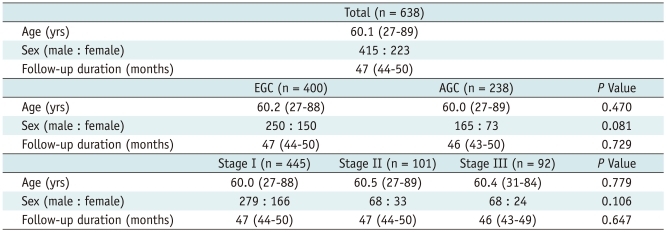
Of the 638 patients, 400 patients (250 men, 150 women; average age, 60.2 years; range, 27-88 years) were confirmed to have EGC, while the remaining 238 patients (165 men, 73 women; average age, 60.0 years; range, 27-89 years) were diagnosed with AGC after surgery. In terms of AJCC TNM staging, 445, 101, and 92 patients were assigned to stage I, stage II, and stage III, respectively (Table 1). There were no significant differences in age or sex between the EGC and AGC groups (p = 0.470 and p = 0.081) or among the three TNM stage groups (p = 0.779 and p = 0.106). The overall median follow-up period was 47 months (IQR, 44-50 months). The median follow up-periods of the EGC and AGC groups (p = 0.729) and among the three TNM stage groups (p = 0.647) were not significantly different (Table 1).
Table 1.
Patient Characteristics
Note.- EGC = early gastric cancer, AGC = advanced gastric cancer, IQR = interquartile range
Number of Imaging Studies
A total of 3035 abdomen-pelvis CT, 68 chest CT, 2 brain CT, and 315 PET/CT scans were performed during the follow-up period. Among the 638 patients, 253 (39.7%, 104 AGC patients and 149 EGC patients) underwent PET or PET/CT examination at least once during the follow-up period. The number of total imaging studies per patient was significantly different between the EGC and AGC groups (p < 0.001) and among the three TNM groups (p < 0.001) based on the AJCC staging system (Table 2).
Table 2.
Numbers of CT and PET/CT Studies and Median Cumulative Effective Doses (cED) during Follow-up Period
Note.- *Mann-Whitney U-test with Bonferroni correction: between stage I and II, p < 0.001; between stage II and III, p < 0.001; and between stage I and III: p <0.001, †Mann-Whitney U-test with Bonferroni correction: between stage I and II, p < 0.001; between stage II and III, p = 0.003; and between stage I and III, p < 0.001. cED = cumulative effective dose, IQR = interquartile range
cED Estimation
The median cED was 57.8 mSv (IQR, 43.9-74.7) per person in all patients. The median cED was 52.3 mSv (IQR, 41.5-67.9) for the EGC group and 67.0 mSv (IQR, 49.1-102.3) for the AGC group, and this difference was significant (p < 0.001). For TNM staging, the median cED was significantly different among the three groups (p < 0.001) (Table 2). For the post hoc analysis, the median cED was significantly different between stage I and stage II (p < 0.001), between stage II and stage III (p < 0.001), and between stage I and stage III (p = 0.003) (Table 2).
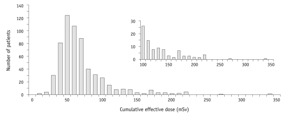
Among the 638 patients followed, 69 (10.8%) received an estimated cED of over 100 mSv and 328 (51.4%) received between 50 to 100 mSv during the follow-up period (Fig. 2) (Table 3). The remaining 241 patients (37.8%) received an estimated cED of < 50 mSv. In patients who received an estimated cED of over 100 mSv, there were significant differences in the median cED between the EGC and AGC groups (p = 0.033) and among three TNM stages (p < 0.001). For the post hoc analysis, stage I and stage II (p < 0.001) and stage I and stage III (p < 0.001) showed significant differences, but there was no difference between stage II and stage III (p = 0.096). In patients who received an estimated cED between 50 to 100 mSv, there were no significant differences among the three TNM stage groups (p = 0.228).
Fig. 2.
Histogram of cumulative CT effective dose per patient. Inset includes expanded y-axis to display right tail and contains number of patients who received more than 100 mSv.
Table 3.
Patients with Median Cumulative Effective Doses (cED) over 50 mSv/100 mSv by CT and PET/CT
Note.- *Chi-square and Fisher's exact test, followed by post hoc analysis with Bonferroni correction: between stage I and II, p < 0.001; between stage II and III, p = 0.096; and between stage I and III, p < 0.001. cED = cumulative effective dose
Estimates of LAR
The median estimated LAR of cancer incidence among all 638 patients was 0.28% (IQR: 0.20-0.40). Fifteen (2.4%) of the 638 patients (all AGC patients) had LARs in excess of 1%, and only one AGC patient exceeded 2% during the follow-up period after surgery. The estimates of LAR for both cancer incidence and mortality were higher in the AGC group than in the EGC group (p < 0.001 and p < 0.001, respectively) and increased with higher TNM stage (p < 0.001) (Table 4).
Table 4.
Lifetime Attributable Risk (LAR) of Cancer by CT and PET/CT Examination
Note.- *Mann-Whitney U-test with Bonferroni correction: between stage I and II, p < 0.001; between stage II and III, p = 0.003; and between stage I and III, p < 0.001, †Mann-Whitney U-test with Bonferroni correction: between stage I and II, p < 0.001; between stage II and III, p < 0.001 and between stage I and III, p < 0.001. IQR = interquartile range
DISCUSSION
Our findings show that the median cED was 57.8 mSv over the course of 47 months of the postoperative follow-up period in patients with gastric cancer and who underwent gastrectomy with curative intent. Furthermore, patients with AGC and patients with advanced TNM stage gastric cancer received greater radiation doses than patients with EGC or less advanced stage gastric cancer during the follow-up period. The median cumulative dose was higher than 50 mSv in 328 (51.4%) and 100 mSv in 69 (10.8%) of 638 patients.
The overall median cumulative dose might be dependent on the proportion of EGC and AGC as well as the follow-up protocol after a curative intent gastrectomy. Hence, the median cED estimated in our study could not be applied directly to other institutions or countries. For example, the proportion of EGC is relatively higher in Korea than in Western countries because of the widespread surveillance program for gastric cancer. Thus, the overall medial cumulative dose might be higher in Western countries than in Korea (12). Nevertheless, our results showed that cED increased with higher tumor stage and that cED was much higher, even in stage I or patients with EGC than in the normal population. This result could be the basis for determining the follow-up protocol in the future.
Berrington de Gonzalez stated that the cumulative cancer risk to the average 75-year-old patient attributable to diagnostic imaging was 3.2% in Japan and 1.5% in Germany and that, among diagnostic imaging modalities, CT scans were responsible for the most radiation exposure (13). Brenner et al. (14) reported that about 1.5 to 2.0% of all cancers might be attributable to the widespread use of CT in the United States. In our study, the overall median LAR was only 0.28%. However, considering that the study period was only 47 months and that these patients might undergo periodic surveillance imaging for the rest of their lives, the LAR may increase over their remaining lifespan. Furthermore, according to the BEIR VII methodology, the LAR of cancer incidence increases as patient age decreases and is higher in females than males. When the young female population is exposed, the probability of radiation-induced cancer increases because of their higher radiosensitivity and longer expected lifetime, making it possible for cancers with a long latency period to develop (5). Consequently, radiation exposure during postoperative follow-up should be monitored closely, especially in younger female patients (3, 8, 15).
When performing CT scans, the conventional wisdom has been that the exposure of a patient to radiation is justifiable when the individual benefit outweighs the risk posed by the radiation. According to previous reports, intensive follow-ups after surgery for colorectal cancer can be attributed to a 5-year survival benefit (16). However, there is still no evidence that postoperative follow-up can improve overall survival in gastric cancer (2, 14, 17-19). Based on these results, recent NCCN guidelines recommend imaging studies only if clinically indicated (20). However, most clinicians perform regular post-operative follow-up due to legal issues for themselves and patients (2), and because locally recurring tumors or secondary gastric cancer after a gastrectomy can be cured by surgical resection in up to 80% of cases with early detection. In such cases, even if the recurrent tumor is beyond the curable stage by surgery, patients may receive chemotherapy or participate in clinical trials for newly developed drugs as early as possible (4, 21). However, there is a lack of evidence that postoperative imaging follow-up extends patient survival, and therefore, the risks of cumulative radiation exposure must be considered in balance with the anticipated benefits of recurrent imaging at the level of the individual patient (7).
A previous nationwide multicenter survey investigating current follow-up practices after curative resection of gastric cancer showed that CT is the most popular follow-up imaging method (88.5% of responders), followed by PET/CT (60.4%) (4). This may be because CT is more objective and provides better diagnostic accuracy than ultrasound, including diagnosis of peritoneal recurrence (2, 4). Recently, PET/CT has become more widely used because it can diagnose small recurrent masses or peritoneal recurrence that may remain undetected by CT (2, 4). However, PET/CT studies also use ionizing radiation to generate images, and according to Brix et al. (11), the average effective dose of PET/CT is estimated to be 25 mSv (23.7-26.4 mSv), which is no less than that of CT studies. However, unlike CT or PET/CT, ultrasound or magnetic resonance (MR) imaging does not generate ionizing radiation. Since contrast-enhanced ultrasound and MR have limited diagnostic performance for detecting peritoneal recurrence or deep-seated lymph node recurrence, ultrasound and liver MR are not used as sole imaging modalities for postoperative follow-up. However, in a limited set of patients with TNM stage I or EGC, ultrasound and MR could be used as alternative follow-up imaging modalities combined with CT, especially 2-3 years after surgery because over two-thirds of tumor recurrence after surgery occurs in the first 2-3 years, and less than 10% recur after 5 years. If ultrasound and MR were to be used as alternative follow-up imaging modalities in conjunction with CT, the cED for patients with expected long-term survival could be reduced.
There are several limitations to our study. First, we assessed the estimated radiation induced lifetime cancer risk using the BEIR VII model, which is based on a linear non-threshold dose-response curve. Although recent reports from the National Research Council (BEIR VII) and International Commission on Radiological Protection have supported the appropriateness of the use of the linear non-threshold model, there is still possibility that other models might be more appropriate for evaluating LAR for radiation-induced cancer incidence and mortality (22).
Second, the BEIR VII model is based on general population data from the United States, whereas our study included patients with gastric cancer. As a result, the predicted LAR in our study may have been overestimated; in particular for patients with AGC, because they may have a shorter life expectancy compared to an age- and gender-matched population.
Third, there were statistical uncertainties when estimating cED and LAR in our study, because these values are calculated from DLP values, which are measured based on the absorbed dose to an acrylic phantom, and the different scanner models can result in some variability in DLP values (23).
Fourth, the conversion factors vary according to not only the types of CT scanners or phantoms used to calculate the conversion factor, but also age, sex, tube voltage, and ICRP-recommended tissue-weighting factors for the conversion factors, used to determine the effective dose from the DLP (5, 24). However, the most recent studies used published conversion factors and our results could show approximate values and the tendencies of radiation exposure to patients who underwent regular follow-ups after gastric surgery (10).
Fifth, the cED might be underestimated because we did not include data from before each operation, such as preoperative imaging studies, as well as other diagnostic or therapeutic imaging studies such as interventional radiology, fluoroscopy, and radiography.
Finally, the follow-up period in this study was relatively short. A future study with a longer duration of follow-up is warranted.
In conclusion, the median cumulative dose of radiation increases according to gastric cancer stage and is much higher, even in patients with EGC or stage I gastric cancer than among the general population. Considering the good prognosis of EGC after curative surgery, cED should be considered as an important factor when designing postoperative follow-up protocols.
References
- 1.Edge SB Cancer AJCo. Ajcc cancer staging manual. 7th ed. New York: Springer; 2010. [Google Scholar]
- 2.Whiting J, Sano T, Saka M, Fukagawa T, Katai H, Sasako M. Follow-up of gastric cancer: A review. Gastric Cancer. 2006;9:74–81. doi: 10.1007/s10120-006-0360-0. [DOI] [PubMed] [Google Scholar]
- 3.Park JC, Lee YC, Kim JH, Kim YJ, Lee SK, Hyung WJ, et al. Clinicopathological aspects and prognostic value with respect to age: An analysis of 3,362 consecutive gastric cancer patients. J Surg Oncol. 2009;99:395–401. doi: 10.1002/jso.21281. [DOI] [PubMed] [Google Scholar]
- 4.Hur H, Song KY, Park CH, Jeon HM. Follow-up strategy after curative resection of gastric cancer: A nationwide survey in Korea. Ann Surg Oncol. 2010;17:54–64. doi: 10.1245/s10434-009-0676-1. [DOI] [PubMed] [Google Scholar]
- 5.The 2007 recommendations of the international commission on radiological protection. Icrp publication 103. Ann ICRP. 2007;37:1–332. doi: 10.1016/j.icrp.2007.10.003. [DOI] [PubMed] [Google Scholar]
- 6.Fazel R, Krumholz HM, Wang Y, Ross JS, Chen J, Ting HH, et al. Exposure to low-dose ionizing radiation from medical imaging procedures. N Engl J Med. 2009;361:849–857. doi: 10.1056/NEJMoa0901249. [DOI] [PMC free article] [PubMed] [Google Scholar]
- 7.Sodickson A, Baeyens PF, Andriole KP, Prevedello LM, Nawfel RD, Hanson R, et al. Recurrent ct, cumulative radiation exposure, and associated radiation-induced cancer risks from ct of adults. Radiology. 2009;251:175–184. doi: 10.1148/radiol.2511081296. [DOI] [PubMed] [Google Scholar]
- 8.Chung HW, Noh SH, Lim JB. Analysis of demographic characteristics in 3242 young age gastric cancer patients in Korea. World J Gastroenterol. 2010;16:256–263. doi: 10.3748/wjg.v16.i2.256. [DOI] [PMC free article] [PubMed] [Google Scholar]
- 9.Murakami T, Yasui A, Tazaki H, Hirase Y, Maeda S. [gastric cancer] Saishin Igaku. 1971;27:115–119. [PubMed] [Google Scholar]
- 10.Office for Official Publications of the European Communities. European guidelines on quality criteria for computed tomography Luxembourg: Office for Official Publications of the European Communities. 1999. [Google Scholar]
- 11.Brix G, Lechel U, Glatting G, Ziegler SI, Munzing W, Muller SP, et al. Radiation exposure of patients undergoing whole-body dual-modality 18f-fdg pet/ct examinations. J Nucl Med. 2005;46:608–613. [PubMed] [Google Scholar]
- 12.Association KGC. 2004 nationwide gastric cancer report in Korea. J Korean Gastric Cancer Assoc. 2007;7:47–54. [Google Scholar]
- 13.Kirpalani H, Nahmias C. Radiation risk to children from computed tomography. Pediatrics. 2008;121:449–450. doi: 10.1542/peds.2007-3394. [DOI] [PubMed] [Google Scholar]
- 14.Kodera Y, Ito S, Yamamura Y, Mochizuki Y, Fujiwara M, Hibi K, et al. Follow-up surveillance for recurrence after curative gastric cancer surgery lacks survival benefit. Ann Surg Oncol. 2003;10:898–902. doi: 10.1245/aso.2003.12.009. [DOI] [PubMed] [Google Scholar]
- 15.Youn HG, An JY, Choi MG, Noh JH, Sohn TS, Kim S. Recurrence after curative resection of early gastric cancer. Ann Surg Oncol. 2010;17:448–454. doi: 10.1245/s10434-009-0772-2. [DOI] [PubMed] [Google Scholar]
- 16.Renehan AG, Egger M, Saunders MP, O'Dwyer ST. Impact on survival of intensive follow up after curative resection for colorectal cancer: Systematic review and meta-analysis of randomised trials. BMJ. 2002;324:813. doi: 10.1136/bmj.324.7341.813. [DOI] [PMC free article] [PubMed] [Google Scholar]
- 17.Okines A, Verheij M, Allum W, Cunningham D, Cervantes A. Gastric cancer: Esmo clinical practice guidelines for diagnosis, treatment and follow-up. Ann Oncol. 2010;21(Suppl 5):v50–v54. doi: 10.1093/annonc/mdq164. [DOI] [PubMed] [Google Scholar]
- 18.Kim JH, Jang YJ, Park SS, Park SH, Mok YJ. Benefit of post-operative surveillance for recurrence after curative resection for gastric cancer. J Gastrointest Surg. 2010;14:969–976. doi: 10.1007/s11605-010-1200-4. [DOI] [PubMed] [Google Scholar]
- 19.Park CH, Song KY, Kim SN. Treatment results for gastric cancer surgery: 12 years' experience at a single institute in Korea. Eur J Surg Oncol. 2008;34:36–41. doi: 10.1016/j.ejso.2007.03.004. [DOI] [PubMed] [Google Scholar]
- 20.Maehara Y, Kakeji Y, Oda S, Takahashi I, Akazawa K, Sugimachi K. Time trends of surgical treatment and the prognosis for japanese patients with gastric cancer. Br J Cancer. 2000;83:986–991. doi: 10.1054/bjoc.2000.1427. [DOI] [PMC free article] [PubMed] [Google Scholar]
- 21.Nunobe S, Hiki N, Ohyama S, Aikou S, Sano T, Yamaguchi T. Outcome of surgical treatment for patients with locoregional recurrence of gastric cancer. Langenbecks Arch Surg. 2011;396:161–166. doi: 10.1007/s00423-010-0730-2. [DOI] [PubMed] [Google Scholar]
- 22.National Research Council (U.S.) Health risks from exposure to low levels of ionizing radiation: Beir vii phase 2. Washington, D.C.: National Academies Press; 2006. Committee to Assess Health Risks from Exposure to Low Level of Ionizing Radiation. [PubMed] [Google Scholar]
- 23.Alessio AM, Phillips GS. A pediatric ct dose and risk estimator. Pediatr Radiol. 2010;40:1816–1821. doi: 10.1007/s00247-010-1761-0. [DOI] [PubMed] [Google Scholar]
- 24.Deak PD, Smal Y, Kalender WA. Multisection ct protocols: Sex- and age-specific conversion factors used to determine effective dose from dose-length product. Radiology. 2010;257:158–166. doi: 10.1148/radiol.10100047. [DOI] [PubMed] [Google Scholar]


