Abstract
Aim:
Demand from implant patients for quality and efficient treatment is increasing. Fortunately dental implant treatment is evolving with patients expectations.
Materials and Methods:
The study comprised of 45 patients for whom 89 implants were placed at different sites. Efficacy of the stents is evaluated in determining the position and diameter of the implants.
Conclusion:
this study shows the extreme accuracy of this surgical stents in implant installation in terms of position and diameter.
Keywords: Dental implants, diagnostic cast, stent, surgical procedure
INTRODUCTION
Osseointegrated implants are a practical alternative to traditional prosthodontics; however, designing an implant-supported prosthesis with function and esthetics is a challenge.[1] Malaligned implants often complicate the clinical laboratory procedures employed for fabrication of superstructures. Due to improper load distribution, an overall increase in stress concentration on supporting structures may occur. This may compromise the maintenance of the bone implant interface.[2] A stent is an appliance used for radiographic evaluation of height and width of the available bone during treatment planning for implant placement or during surgical procedures to provide site for optimum implant placement.[2] The stent can be of fixed type or variable positioning type. The variable type of stents includes vaccuform or acrylic resin templates adapted over duplicated casts of diagnostic wax-up. Fixed stents do not allow variation of implant location. These include plastic or metal tubes and channels in acrylic resin that dictate the position of implants.[3] Stents should be made of transparent material, stable and rigid when in position, should cover enough teeth to stabilize its position, and when no teeth are present, stents should extend onto unreflected soft tissue regions.[4] With the use of computed tomography (CT) and computer added designing (CAD)/ computer added machining (CAM) technology, it is now possible to construct a surgical implant stent that would allow the clinician to predetermine implant locations virtually and surgically place them without reflecting a tissue flap.[5–8] But the surgical stents prepared with CT and CAD/CAM technology are very much more cost effective than a conventional surgical stent and require enough time and special center and/or laboratories for their fabrication and later on their correction, if required. In this study, we used the conventional surgical stents to evaluate their efficacy in determining the position and diameter of implants.
Aim of the study
The aim of the study was to demonstrate the accuracy and clinical precision of conventional surgical stent protocol for optimum installation of dental implants in terms of position and diameter.
MATERIALS AND METHODS
The study was conducted in the out-patient Department of Prosthodontics and Dental Material Sciences in collaboration with Department of Oral and Maxillofacial Surgery, Faculty of Dental sciences, CSMMU, Lucknow. Patients were treated in this study with the conventional surgical stents; all the implants were placed to the desired depth as planned in virtual implant planning. All the patients (45 patients for whom 89 implants were placed at different sites) [Figures 1, 2] visiting the Department of Prosthodontics and Oral and Maxillofacial surgery for implant supported prosthesis placement and who followed the standard inclusion criteria for placement of implants were included in the study. First of all, diagnostic cast of the patients was made and acrylic teeth/tooth were waxed-up [Figure 3]. Then, the impression of that waxed-up cast was taken and poured to reproduce another cast with the missing teeth/tooth replaced by dental stone. Then, mesiodistal and buccolingual markings were drawn over these teeth/tooth to find a center point on the occlusal surface [Figure 4]. Later on, after applying the separating media over these regions and filling the undercuts with wax, transparent self-curing material was applied at the teeth to be replaced along with some adjacent area antero-posteriorly, which confers the stability to the stent during implant placement [Figure 5]. The holes were drilled in the stent so formed at the center point for the assessment of the diameter and position of implant and filled with gutta percha [Figure 6]. This stent was placed in the mouth and radiograph was taken to evaluate the position of gutta percha. The efficacy of stents was then evaluated at the time of surgical exposure for implant placement and after the surgery, with the help of radiographs [Figures 7, 8].
Figure 1.
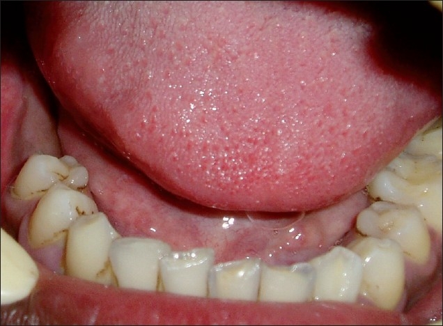
Intraoral view of the patient
Figure 2.
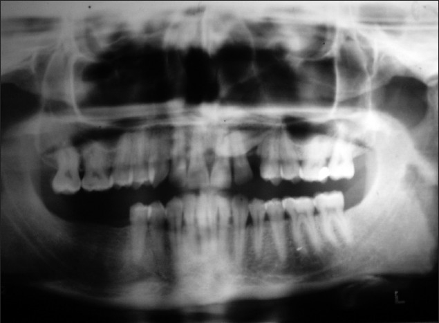
Radiograph of the patients
Figure 3.
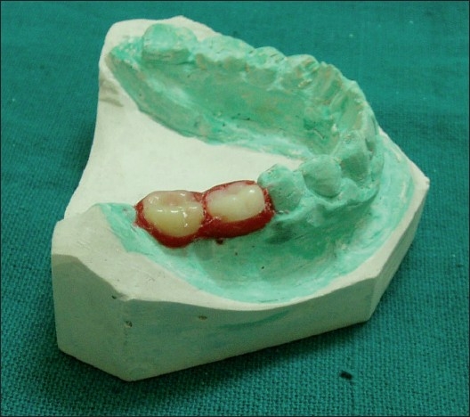
Waxed-up missing teeth
Figure 4.
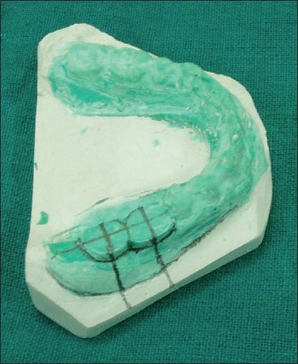
Missing teeth replaced by dental stone in the cast with markings
Figure 5.
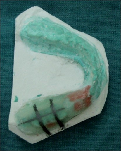
Stent made of transparent material
Figure 6.
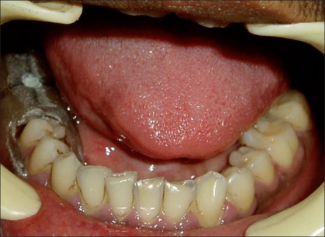
Stent in place with gutta percha filled in holes
Figure 7.
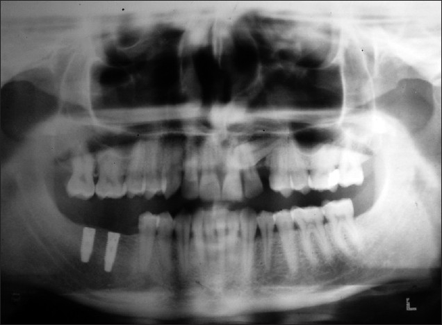
Post-operative radiograph
Figure 8.
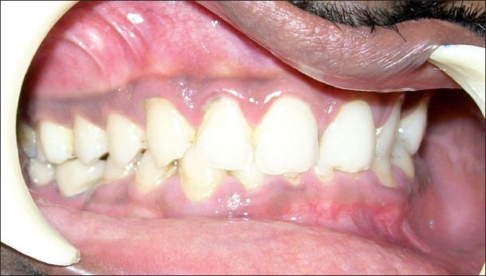
Teeth in occlusion
Review of literature
Philip J. described a technique to construct a surgical guide on mounted diagnostic casts. He also used these mounted casts to determine whether sufficient space exists for a fixed cantilevered implant prosthesis. Murat C cehreli suggested the fabrication of a bilaminar dual-purpose stent that facilitates implant placement with improved verification of implant positioning. The outer lamina is designed for use in the computed tomography (CT) evaluation using radiopaque markers.[9] Yen-Chen Ku presented a simple method of fabricating a vacuum-formed matrix filled with clear acrylic resin and a gutta-percha marker. The matrix can be used as not only a radiopaque marker for evaluation but also a surgical guide during the surgical stage for single implant therapy.[10] Roger A. Solow showed that a radiographic-surgical template can facilitate consultation with a surgeon and patient, when implant-supported restorations are planned. A template that provides radiographic evaluation of the implant site and precise or modified surgical placement is presented.[3] Kivanc Akea described a modified surgical stent that serves as a guide to proper mesiodistal paralleling of dental implants.[2]
DISCUSSION
The foremost advantage of the stent used in this study is its surgical ease, simplicity, precise accuracy and low cost. It can be fabricated with minimum laboratory procedures which are used in routine dental practice. On the other hand, the fixed type screw retained implant stents which are fabricated with the help of CT and CAD/CAM technology are very costly and require more time and laboratory procedures. The stents fabricated with the radiopaque markers provide radiographic as well as clinical ease for optimum implant installation. Along with all these advantages, they also provide liberty to flapless implant placement. The drill holes can directly be made through the stent, so the implant surgery may become less traumatic (with the preservation of soft tissue including the gingival margins of adjacent teeth and the interdental papilla) with decreased operative time which results in accelerated post-surgical healing, fewer post-operative complications and increased comfort and satisfaction for the patient. The possibility of displacement of stent during implant surgery was minimized by extension of the stent antero-posteriorly to cover enough area over the teeth/tooth present adjacent to edentulous area. In cases where no teeth are present, stabilization of stent was achieved by extending it over the unreflected areas like retromolar regions.
CONCLUSION
This study shows the extreme accuracy of this conventional surgical stent. If each step of this protocol is followed precisely, it is possible to deliver an optimum implant installation in terms of position and diameter and later on function at a very much reduced rate and time, as taken in CT and CAD/CAM technology which are the most accepted methods.
Footnotes
Source of Support: Nil.
Conflict of Interest: None declared.
REFERENCES
- 1.Verde MA, Morgano SM. A dual-purpose stent for the implant-supported prosthesis. J Prosthet Dent. 1993;69:276–80. doi: 10.1016/0022-3913(93)90106-x. [DOI] [PubMed] [Google Scholar]
- 2.Akça K, Iplikçioğlu H, Cehreli MC. A surgical guide for accurate mesiodistal paralleling of implants in posterior edentulous mandible. J Prosthet Dent. 2002;87:233–5. doi: 10.1067/mpr.2002.120900. [DOI] [PubMed] [Google Scholar]
- 3.Solow RA. Simplified radiographic-surgical template for placement of multiple, parallel implants. J Prosthet Dent. 2001;85:26–9. doi: 10.1067/mpr.2001.112793. [DOI] [PubMed] [Google Scholar]
- 4.Misch CE. St Louis: Mosby; 1993. contemporary implant dentistry; pp. 480–3. [Google Scholar]
- 5.Parel SM, Triplett RG. Interactive imaging for implants planning, placement and prosthesis construction. J Oral Maxillofac Surg. 2004;62:41–7. doi: 10.1016/j.joms.2004.05.207. [DOI] [PubMed] [Google Scholar]
- 6.van Steenberghe D, Naert I, Andersson M, Brajnovic I, Van Cleynenbreugel J, Suetens P. A costum template and definite prosthesis allowing immediate implant loading in the maxilla: A clinical report. Int J Oral Maxillofac Implants. 2002;17:663–70. [PubMed] [Google Scholar]
- 7.van Steenberghe D, Glauser R, Blombäck U, Andersson M, Schutyser F, Pettersson A, et al. A computed tomographic scan derived customized surgical template and implants in fully edentulous maxilla: A prospective multicentre study. Clin Implant Dent Relat Res. 2005;7:S111–20. doi: 10.1111/j.1708-8208.2005.tb00083.x. [DOI] [PubMed] [Google Scholar]
- 8.Balshi SF, Wolfinger GJ, Balshi TJ. Surgical planning and prosthesis construction using computer tomography, CAD/CAM technology and the internet for immediate loading of dental implants. J Esthat Restor Dent. 2006;18:235–47. doi: 10.1111/j.1708-8240.2006.00029.x. [DOI] [PubMed] [Google Scholar]
- 9.Cehreli MC. Bilaminar dual-purpose stent for placement of dental implants. J Prosthet Dent. 2000;84:55–8. doi: 10.1067/mpr.2000.107780. [DOI] [PubMed] [Google Scholar]
- 10.Ku YC, Shen YF. Fabrication of a radiographic and surgical stent for implants with a vacuum former. J Prosthet Dent. 2000;83:252–3. doi: 10.1016/s0022-3913(00)80019-0. [DOI] [PubMed] [Google Scholar]


