Abstract
Introduction:
The superficial veins, especially the external jugular vein (EJV), are increasingly being utilized for cannulation to conduct diagnostic procedures or intravenous therapies. EJV is also used in microsurgical procedures,used as a recipient for the free flaps.
Materials and Methods:
During routine dissection a variation was observed in the formation of EJV unilaterally on the left side.
Result:
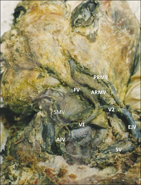
In the anterior triangle of the neck submandibular vein joined with the anterior jugular vein to form a large venous trunk (V1). Facial vein joined this venous trunk (V1) to form another common channel (V2). The retromandibular vein divided into unusually long anterior and posterior divisions. Anterior division did not join the facial vein but drained into the common channel V2.The posterior division of retromandibular vein also drained into V2 which further continued as EJV and drained into the subclavian vein.
Conclusion:
The knowledge of variations in the patterns of superficial veins is important for the surgeons to avoid any intraoperative error which might lead to unnecessary bleeding.
Keywords: Anomalous vein, development, external jugular vein
INTRODUCTION
The external jugular vein (EJV) is formed by the union of posterior division of retromandibular vein and posterior auricular vein. The anterior division of the retromandibular vein combines with facial vein to form common facial vein. The superficial venous system of head and neck displays great variability. These variations are very important for anatomists, anesthetists, surgeons and radiologists (Gupta et al,[1] Schummer et al,[2]). The EJV is used as a venous manometer and for catheterization. These veins are also significant for the surgeon doing head and neck surgery (Salmery et al, 1991). In the present case anomalous formation of EJV was observed. It was formed by union of facial vein, anterior and posterior divisions of retromandibular vein.
MATERIALS AND METHODs
During routine dissection in the Dissection Hall of Anatomy Department of C.S.M. Medical University, Lucknow, India, an anomalous EJV was found on the left side of a 60 years male cadaver. The vein was traced proximally as well as distally for its formation and termination respectively and its tributaries were also noted.
Observations
During the dissection of head and neck region a variation was observed in the formation of EJV unilaterally on the left side. In the anterior triangle of the neck submandibular vein joined with the anterior jugular vein to form a large 2.4-cm long venous trunk (V1). Distally anterior jugular vein was ending in the junction of subclavian vein and internal jugular vein. Facial vein draining the face region joined this venous trunk (V1) superficial to sternocleidomastoid to form a 1.5-cm long another common channel (V2). The retromandibular vein after emerging from the apex of parotid gland, behind the angle of mandible, divided into unusually long anterior and posterior divisions. The anterior division was 3.6-cm long whereas posterior division measured 4.2 cm. Anterior division did not join the facial vein but it drained into the common channel V2. The posterior division of retromandibular vein also drained into V2 a little farther away. Posterior auricular vein was not present. A very thin vein was present on the nape of neck draining the suboccipital plexus of veins. This vein was joining the EJV near its commencement. After travelling a short distance of approximately 2 cm the EJV pierced the investing layer of deep fascia of the posterior triangle and drained into the subclavian vein [Figure 1].
Figure 1.

Photograph showing anomalous formation of external jugular vein (FV- facial vein, PRMV- posterior division of retromandibular vein, ARMV-anterior division of retromandibular vein, SMV- submandibular vein, AJV-anterior Jugular vein, EJV- external jugular vein, SV- subclavian vein)
DISCUSSION
There is preponderance of venous variations on the right side of the face as observed in literature, but the present case reports an unusual venous pattern on the left side. The retromandibular vein has been reported to unite with the facial vein at a higher level in the right parotid gland (Kopuz et al[3]). Facial vein terminating into EJV has been reported into the literature (Choudhry et al[4]). The present case describes an unusual formation of EJV.
Anterior jugular vein and submandibular vein were joining to form a common venous trunk (V1). Distally anterior jugular vein was draining at the junction of subclavian vein and internal jugular vein [Figure 1].
This venous trunk (V1) joined the facial vein and continued as venous trunk V2, which subsequently received the anterior and posterior divisions of retromandibular vein to form EJV [Figure 1].
The present case hallmarks the absence of common facial vein (which usually drains into internal jugular vein, deep jugular system) and the facial vein is draining into EJV (superficial jugular venous system). An abnormal communicating channel is present between the proximal ends of anterior and external jugular venous systems (V1+V2) [Figure 1].
Posterior auricular vein was absent.
Superficial veins of head and neck develop from superficial plexus of the capillaries which will ultimately form primary head veins. Larger channels are formed by enlargement of individual capillaries, confluence of adjacent ones, regression of some form where the flow has been diverted. (Hamilton et al[5]). The anterior jugular vein normally develops from venous plexus around head mesenchyme. It communicates with primitive maxillary vein and separates from primitive head veins. Deep part of the cephalic venous ring develops along the medial end of clavicle and later on gives rise to subclavian vein which receives EJV as its tributary. Primary head vein also joins the cephalic venous ring and forms internal jugular vein.
In our case the communication of anterior jugular vein between primitive maxillary vein and primitive head veins may have persisted, so the anterior jugular vein is connected with EJV through venous trunk proximally and to the internal jugular vein distally.
Here possibly facial vein took over the territory of the primitive maxillary vein and retromandibular vein. Gupta et al,[1] studied facial vein draining into EJV. They found 9% cases in human cadavers. They explain the anamolus venous drainage of face on the bases of regression and retention of various parts of veins as found in ox, dog and horse where vein of face drain into EJV, the internal jugular vein being either absent or a small vessel accompanying the carotid artery.
The superficial veins, especially the EJV, are increasingly being utilized for cannulation to conduct diagnostic procedures or intravenous therapies. Ultrasound-guided venipuncture is a viable possibility in cases of variations in the patterns of superficial veins, and their knowledge is also important for surgeons doing reconstructive surgery (Gupta et al[1]). Awareness of these variations is important for the surgeons to avoid any intraoperative trial or error procedures which might lead to unnecessary bleeding (Nagase et al[6]). These veins are usually grafted into the carotid during endarterectomy and also used for surgery involving microvascular anastomosis especially in oral reconstruction procedures (Sabharwal and Mukherjee[7]).
Footnotes
Source of Support: Nil.
Conflict of Interest: None declared.
REFERENCES
- 1.Gupta V, Tuli A, Choudhry R, Agarwal S, Mangal A. Facial vein draining into external jugular vein in humans: Its variations, phylogenetic retention and clinical relevance. Surg Radiol Anat. 2003;25:36–41. doi: 10.1007/s00276-002-0080-z. [DOI] [PubMed] [Google Scholar]
- 2.Schummer W, Schummer C, Bredle D, Frober R. The anterior jugular venous system: Variability and clinical impact. Anesth Analg. 2004;99:1625–9. doi: 10.1213/01.ANE.0000138038.33738.32. [DOI] [PubMed] [Google Scholar]
- 3.Kopuz C, Yavuz S, Cumhur M, Tftik S, Ilgi S. An unusual coursing of the facial vein. Kaibogaku Zasshi. 1995;70:20–2. [PubMed] [Google Scholar]
- 4.Choudhry R, Tuli A, Choudhry S. Facial vein terminating in external jugular vein.An embryological interpretation. Surg Radiol Anat. 1997;19:73–7. [PubMed] [Google Scholar]
- 5.Hamilton, Boyd, Mossman . 4th ed. Heffer Cambbridge: 1972. Human Embryology; p. 261. [Google Scholar]
- 6.Nagase T, Kobayashi S, Sekiya S, Ohmori K. Anatomic evaluation of the facial artery and vein using color Doppler ultrasonography. Ann Plast Surg. 1997;39:64–7. doi: 10.1097/00000637-199707000-00011. [DOI] [PubMed] [Google Scholar]
- 7.Sabharwal P, Mukherjee D. Autogenous common facial vein or external jugular vein patch for carotid endarterectomy. Cardiovas Surg. 1998;6:594–7. doi: 10.1016/s0967-2109(98)00084-2. [DOI] [PubMed] [Google Scholar]


