Summary
Coil migration is a recognised but rare complication of endovascular coiling. Many techniques are available commercially for coil retrieval.
We report the case of an acute subarachnoid hemorrhage in a 54-year-old woman in which a migrated coil was successfully retrieved using an X6 MERCI device.
Key words: MERCI, coil migration
Introduction
Endovascular aneurysm occlusion, though certainly less invasive than open craniotomy does itself incur numerous potential risks. One of the less common, though of great significance is the occurrence of coil migration. The incidence of coil migration or displacement was previously estimated to be approximately 2.5% of all coilings1. Coil migration may result in vessel occlusion and stroke even with the use of anticoagulation.
Several methods of coil retrieval are available including open surgery and specific endovascular retrieval devices.Among these is the MERCI retrieval device (Concentric Medical, Inc, Mountain View, CA) originally designed as a mechanical thrombectomy device with FDA approval in 2005. Although there have been anecdotal reports of the use this original device there have been no documented reports of its use in coil retrieval. Recently Vora et Al. did publish on the use of the newer design, L5, in a case of stent and coil retrieval2. We report the successful use of the original X6 MERCI retriever in a case of coil migration.
Case Report
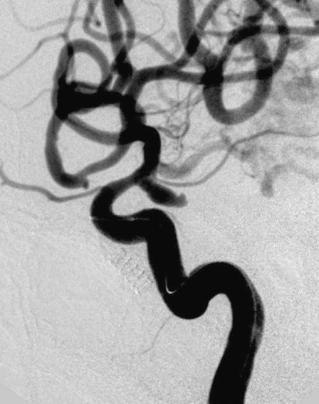
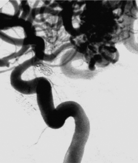
A 54-year-old woman presented with acute onset of headache to our emergency department on waking. After initial assessment in this department a non contrast CT demonstrated a small amount of right sided subarachnoid hemorrhage within the suprasellar cistern and basal cisterns. With this finding a CT angiogram (CTA) was also performed at the same time. CTA demonstrated two aneurysms, right posterior communicating and left paraclinoid. In addition the patient had a right temporal arteriovenous malformation measuring less than 3 cm and with superficial drainage. Based on the blood pattern the right posterior communicating artery aneurysm (Figure 1), was felt to be the source of hemorrhage. Clinically the patient was a Hunt and Hess grade 1 and was admitted to the clinical ward after appropriate initial management.
Figure 1.
Right internal carotid injection demonstrates an oval shaped posterior communicating artery aneurysm. The concomitant AVM can also be seen.
We proceeded to endovascular coiling of the offending right posterior communicating aneurysm. Under general anae-sthetic and systemic heparinisation Excelsior (Boston Scientific, Miami, FL) was placed over a Transcend guidewire (Boston Scientific, Miami, FL) into the aneurysm. On 3D angiography the aneurysm measured 3 mm x 7 mm with a 2.7 mm neck.
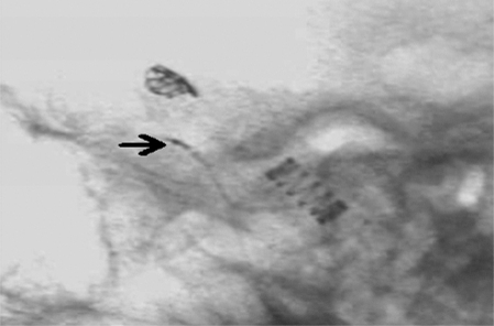
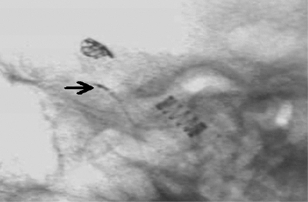
The first coil, a Microplex 10 3 mm x 7 mm complex coil, passed easily into the microcatheter and exited normally with a good shape within the aneurysm. Numerous attempts were made to detach the coil but on each occasion this was unsuccessful, despite the coil marker being in the correct position (Figure 2). Eventually we decided to retract the coil completely (Figure 3), but after re-entering the microcathether by 4cm it detached spontaneously from the pusher leaving the distal portion still within the aneurysm and the proximal free within the distal microcatheter.
Figure 2.
The original coil fully placed in the aneurysm and the coil marker (arrow) in correct position over the proximal microcatheter mark at the time of attempted release.
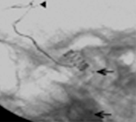
Figure 3.
The coil is now being pulled back into the microcatheter. Note the artefact from the endotracheal tube (arrow).
A left femoral puncture was performed and after suitable access was obtained the 14X 6F catheter was also placed in the right internal carotid artery. The 18L microcatheter was advanced beyond the coil and the X6 retriever deployed.
The initial two attempts were unsuccessful managing only to remove the coil completely from the microcatheter and pass distally into the middle cerebral artery. On the third attempt the migrated coil was ensnared (Figure 4), at this stage the coil was now out of the aneurysm and microcatheter and free in the middle cerebral artery (Figure 5).
Figure 4.
The MERCI device (arrow) is now deployed and open distal to the migrated coil ready to ensnare the coil.
Figure 5.
The microcatheter markers (arrows) show that the coil is now free from the catheter and has migrated towards the MCA. The MERCI (arrowhead) had wrapped around the coil whilst being pulled back.
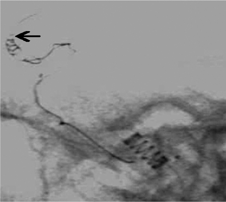
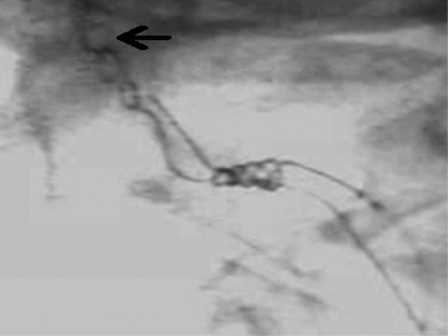
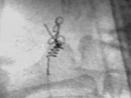
The coil and MERCI were pulled back to the level of the 14X catheter in the internal carotid artery (Figure 6). The entire MERCI system and coil was then removed from the patient under fluroscopic guidance (Figure 7). A standard anterior-posterior and lateral angiogram post procedure did not demonstrate any thrombus, stasis or extravasation of contrast.
Figure 6.
The free coils loops (arrow) being brought down the internal carotid artery by the MERCI.
Figure 7.
The MERCI and coil now distally and proximally over L5 on the way to exiting the left femoral sheath.
The microcatheter was then again placed in the aneurysm and we proceeded to place three coils to adequately occlude the aneurysm (Figure 8). The patient awoke from anaesthetic without any neurological deficit and the remainder of her inpatient stay was uneventful.
Figure 8.
The completed case shows complete occlusion of the aneurysm without vascular compromise.
The patient is due to be re-admitted for further endovascular treatment of the second aneurysm and AVM.
Discussion
Peri-procedural complications associated with endovascular coiling include perforation, thrombus formation and less frequently the herniation and ultimate migration of the detachable coil. Whilst the incidence is low, the potential adverse outcome is significant and primarily a result of the pro-thrombotic nature of the coils which make them effective at aneurysmal occlusion from the offset. This migration almost always occurs at the time of the procedure though the delayed migration of coils from an aneurysm has also been reported 3. This emphasises the fact that wide-necked aneurysms are the leading cause of coil herniation and migration, though in our case it was faulty detachment between the coil and pusher which resulted in the coil being partially within and partially outside the aneurysm.
The problem of coil migration has initiated the development of a number of devices specifically for the purpose of coil retrieval. The role of surgery is questionable and although there exist individual successful reports 4 on the whole it is quite invasive and does not always result in a favourable outcome 5. Likewise, dedicated retrieval devices are used in accordance with personal preference and knowledge of a particular device. The Alligator Retrieval Device (Chestnut Medical Technologies, Menlo Park, Calif) 6 and the Goose Snare 7 have had success in few small or individual patient groups.
The design of the original X6 MERCI device was that of a flexible nickel titanium wire (nitinol) which assumes a tapering helical shape as it is passed distally from the parent catheter.
The wire/catheter combination is passed distal to the foreign body/thrombus and the helical shape ensnares the foreign body which is then removed down the stabilising 8F guide catheter. This device received FDA approval on the basis of the MERCI trial in 2004 which examined its role in acute mechanical thrombectomy in 28 patients 8. While the primary approval was for the mechanical removal of thrombus it did also include the "use in the retrieval of foreign bodies in the peripheral, coronary or neuro vasculature"9. One of the difficulties we experienced personally with the X6 original design was the tendency to straighten out during clot retrieval thus requiring numerous attempts. This has been addressed by the introduction of the modified L5 device in 2006 which has increased stability by means of the addition of a suture filament.
Though there has already been a recent description of coil and stent retrieval with the modified MERCI there is still merit in our report. We have shown that this device even in its original format is in fact extremely effective in coil retrieval. This should certainly be considered a first line tool in the immediate management of coil migration. Furthermore in light of the final results of Multi MERCI trial10 recently published it is an interventional device that we should become more familiar with.
References
- 1.Henkes H, Fischer S, et al. Endovascular coil occlusion of 1811 intracranial aneurysms: early angiographic and clinical results. Neurosurgery. 2004;54:268–285. doi: 10.1227/01.neu.0000103221.16671.f0. [DOI] [PubMed] [Google Scholar]
- 2.Vora N, Thomas A, et al. Retrieval of a displaced detachable coil and intracranial stent with an L5 MERCI retriever during endovascular embolisation of an intracranial aneurysm. J Neuroimaging. 2008;18(1):81–84. doi: 10.1111/j.1552-6569.2007.00165.x. [DOI] [PubMed] [Google Scholar]
- 3.Gao BL, Li MH, et al. Delayed coil migration from a small wide necked aneurysm after stent assisted embolisation: case report and literature review. Neuroradiology. 2006;48(5):333–337. doi: 10.1007/s00234-005-0044-1. [DOI] [PubMed] [Google Scholar]
- 4.Shin YS, Lee KC, et al. Emergency surgical recanalisation of A1 segment occluded by a Guglielmi detachable coil. J Clin Neurosci. 2000;7(3):259–262. doi: 10.1054/jocn.1999.0207. [DOI] [PubMed] [Google Scholar]
- 5.Thornton J, Dovey Z, et al. Surgery following endovascular coiling of intracranial aneurysms. Surg Neurol. 2000;54:352–360. doi: 10.1016/s0090-3019(00)00337-2. [DOI] [PubMed] [Google Scholar]
- 6.Henkes H, Lowens S, et al. A new device for endovascular coil retrieval from intracranial vessels: Alligator Retrieval Device. Am J Neuroradiol. 2006;27:327–329. [PMC free article] [PubMed] [Google Scholar]
- 7.Koseoglu K, Parildar M, et al. Retrieval of intravascular foreign bodies with goose neck snare. Eur J Radiol. 2004;49(3):281–285. doi: 10.1016/S0720-048X(03)00078-0. [DOI] [PubMed] [Google Scholar]
- 8.Gobin YP, Starkman S, et al. MERCI 1: a phase 1 study of mechanical embolus removal in cerebral ischaemia. Stroke. 2004;35(12):2848–2854. doi: 10.1161/01.STR.0000147718.12954.60. [DOI] [PubMed] [Google Scholar]
- 9.Becker KJ, Brott TG. Approval of the MERCI clot retriever. Stroke. 2005;36(2):400–403. doi: 10.1161/01.STR.0000153056.25397.ff. [DOI] [PubMed] [Google Scholar]
- 10.Smith WS, Sung G, et al. Mechanical thrombectomy for acute ischaemic stroke: final results of the multi MERCI trial. Stroke. 2008;39(4):1205–1212. doi: 10.1161/STROKEAHA.107.497115. [DOI] [PubMed] [Google Scholar]


