Abstract
Sonic hedgehog (Shh) regulates neural progenitor cells in the adult brain but its role in postmitotic mature neurons is not well understood. Using immunoelectron microscopy, we have recently demonstrated the postsynaptic distribution of Patched (Ptch) and Smoothened (Smo), the receptors for Shh, in hippocampal neurons of the adult rat brain. In this study, we describe the distribution of Shh protein in these adult hippocampal neurons. We find that Shh is present in both presynaptic and postsynaptic terminals. In presynaptic terminals, Shh is located either at the center or on the side of the synaptic junction. In postsynaptic terminals, Shh is mostly located on the side of the synaptic junction. We also find Shh in dendrites. Synaptic and dendritic Shh often reside in or are associated with vesicular structures that include dense-cored vesicles, synaptic vesicles, and endosomes. Thus, our subcellular map of Shh and its receptors provides a foundation for elucidating the functional significance of Shh signaling in mature neurons.
Keywords: Sonic hedgehog, hippocampal neuron, synapse, synaptic vesicle, dense core vesicle, endosome, axon, dendrite
Sonic hedgehog (Shh) plays at least two important roles in the nervous system. One is to stimulate the production of stem/progenitor cells, which include granule cell precursors in the young cerebellum1-3 and neural stem cells in the specific brain regions of the adult brain.4-8 The other is to promote axon growth of young neurons, which include spinal cord commissural neurons,9,10 retinal ganglion cells,11 olfactory sensory neurons,12 and midbrain dopaminergic neurons.13 Evidence has indicated that Shh and its signaling components also exist in mature neurons, neurons that do not have progenitor properties.14-16 However, where Shh signaling takes place in these mature neurons and how Shh signaling affects them remain largely unknown.
We have recently described the subcellular distribution of Patched (Ptch) and Smoothened (Smo), the receptor and transducer for Shh respectively, in hippocampal neurons of adult rats.17 Multiple types of hippocampal neurons express Ptch and Smo, which are particularly concentrated in their dendrites, spines and postsynaptic terminals.17 Here, we studied the distribution of Shh protein within adult hippocampal neurons.
We used the same hippocampal tissue samples from adult rats that were used in our previous ultrastructural analysis of Ptch and Smo.17 We performed postembedding immunogold labeling (two animals) using monoclonal anti-Shh antibody (5E1; Developmental Studies Hybridoma Bank). The 5E1 antibody was generated against the N-terminus of Shh (aa1–198 of rat Shh)18 and its specificity has been characterized.10,15,18,19
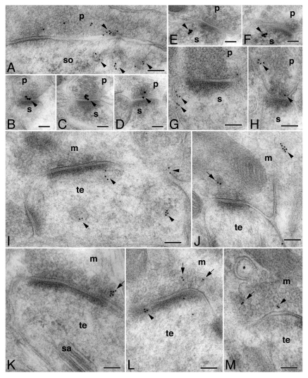
Because of the preferential distribution of Ptch and Smo in the postsynaptic terminals of hippocampal neurons,17 we wondered whether Shh also was located near or at the synapse. We examined several hippocampal regions that include the CA1 stratum pyramidal and stratum radiatum, the molecular layer of the dentate gyrus, and the CA3 stratum lucidum. Synapses from these regions exhibited different morphological characteristics. Nevertheless, we found Shh labeling present in all of these synapses (Fig. 1). Figure 1A is an example of an inhibitory synapse based on its symmetric appearance. Shh labeling was seen in the presynaptic compartment as well as in the postsynaptic compartment. Within both compartments, the Shh labeling was similarly distributed toward the side rather than the center of the synaptic junction (Fig. 1A). Upon closer examination, the postsynaptic Shh labeling appeared to be associated with pit-like structures, which were situated opposite or across from the presynaptic Shh labeling (Fig. 1A).
Figure 1.
Distribution of Shh at the synapses in CA1, CA3, and dentate gyrus of the adult rat hippocampus. Immunoelectron micrographs showing Shh immunolabeling at synaptic sites in: the CA1 stratum pyramidal (A,C), the CA1 stratum radiatum (B,D-G), the molecular layer of the dentate gyrus (H), and the CA3 stratum lucidum (I-M). Shh labeling (10 or 15 nm gold particles) is indicated by arrowheads. (A) shows an inhibitory synapse in which Shh labeling is in the presynaptic terminal (p) as well as the postsynaptic soma (so). In the case of both the presynaptic and postsynaptic compartment, Shh labeling is not in the active zone of the synaptic junction but is extrasynaptic. Also notice that the postsynaptic labeling is associated with pit-like structures, which are located opposite and across from the presynaptic labeling. (B)-(D) are examples of excitatory synapses in all of which Shh labeling is in the presynaptic terminal – near the center of the synaptic junction and directly contacting the presynaptic membrane. s, postsynaptic spine. (E)-(G) are also examples of excitatory synapses but in these cases, Shh labeling is found postsynaptically and it concentrates near the side of the postsynaptic membrane. (H) is an excitatory synapse in which Shh labeling is located both in the center and extrasynaptic side of the presynaptic membrane. (I)-(M) show Shh labeling in the mossy fiber terminal (m) and the postsynaptic thorny excrescence (te). Presynaptic Shh labeling is often associated with various vesicular structures (arrowheads in I,J,M) and dense-cored vesicles (arrows in J-M). Postsynaptic Shh labeling is seen in tubulovesicular organelles (I) or associated with the membrane (L). sa in K, spine apparatus; * in M marks a spinule. Scale bars are 100 nm.
Figure 1B-H show typical excitatory synapses based on their prominent postsynaptic density. In these synapses, presynaptic Shh labeling displayed a slightly different pattern from postsynaptic Shh labeling. Presynaptic Shh labeling could be found either directly at the membrane of the synaptic junction (Fig. 1B-D) or on the side of the terminal (Fig. 1H). Postsynaptic Shh labeling, on the other hand, was found mostly on the side, away from the center of the synaptic junction (Fig. 1E-G).
Figure 1I-M shows examples of mossy fiber synapses. As for other types of synapses, Shh was found in both the presynaptic mossy terminals and the postsynaptic thorny excrescences. Within the presynaptic mossy terminals, most Shh labeling was clearly seen associated with either dense-cored vesicles (arrows in Figure 1J-M), or other smaller vesicular structures (arrowheads in Figure 1I,J,M). Within the postsynaptic thorny excrescences, Shh was associated with tubulovesicular organelles in some cases (arrowheads in Figure 1I), or positioned quite close to the postsynaptic membrane in other cases (arrowhead in Figure 1L).
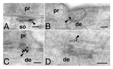
In addition to synaptic localizations, Shh labeling was found in various tubulovesicular organelles in the soma and dendrites of hippocampal neurons (Fig. 2). Interestingly, some of these Shh-labeled organelles made direct contact with the cell membrane surface, typically near contacts with adjacent processes (Fig. 2A-C).
Figure 2.
Shh (arrowheads; 10 or 15 nm gold particles) is found in tubulovesicular organelles that are located in soma (so) or dendrites (de). These Shh-containing organelles often make direct contact with the membrane surface of the neuron, near adjacent cell processes (pr in A-C). (A), the CA1 stratum pyramidal; (B), the CA1 stratum radiatum; (C), the molecular layer of the dentate gyrus; (D), the CA3 stratum lucidum. Scale bars are 100 nm.
Our previous immunoelectron microscopic work has described the postsynaptic localization of Ptch and Smo in adult hippocampal neurons.17 The present findings showing the presence of Shh in the presynaptic terminal, in particular localized in close vicinity to or even directly at the synaptic contact, raises the possibility that Shh signaling may occur across the synapse in these neurons. It is then tempting to ask what form of Shh protein is being released from the presynaptic terminal. In the photoreceptor neurons of the developing Drosophila retina, while the Hedgehog (Hh) C-terminus harbors the axonal targeting signal, the N-terminal domain, and a small amount of the full-length Hh, also travel along the axon.20 Our results obtained using the 5E1 antibody - specific to the Shh N-terminus, could reflect the full-length as well as the N-terminal domain of Shh. To definitively identify the Shh forms that are present and possibly released from the presynaptic terminal will require further studies, including the use of the Shh C-terminus specific antibody.
We also observed an interesting subcellular distribution of Shh in postsynaptic spines and dendrites of hippocampal neurons. Several studies have shown that the dendrite and the postsynaptic terminal of neurons can release transmitters21 or growth factors.22 Moreover, studies of other Hh-producing cells have shown that the release of Hh occurs on the apical and the basal side of the Drosophila photoreceptor neurons.20 Likewise, in Drosophila wing disk epithelium, Hh is found in the apical and basolateral plasma membranes.23 It will be interesting to investigate whether Shh in mammalian hippocampal neurons is also released from both pre- and post-synaptic sites. The mapping of Shh protein and its receptors at the subcellular level will advance the understanding of where and how Shh signaling occurs, and what roles Shh plays in the function and plasticity of mature neurons.
Acknowledgments
This work was supported by the Intramural Research Programs of the NIA/NIH and NIDCD/NIH.
Footnotes
Previously published online: www.landesbioscience.com/journals/cib/article/17832
References
- 1.Dahmane N, Ruiz I, Altaba A. Sonic hedgehog regulates the growth and patterning of the cerebellum. Development. 1999;126:3089–100. doi: 10.1242/dev.126.14.3089. [DOI] [PubMed] [Google Scholar]
- 2.Wallace VA. Purkinje-cell-derived Sonic hedgehog regulates granule neuron precursor cell proliferation in the developing mouse cerebellum. Curr Biol. 1999;9:445–8. doi: 10.1016/S0960-9822(99)80195-X. [DOI] [PubMed] [Google Scholar]
- 3.Wechsler-Reya RJ, Scott MP. Control of neuronal precursor proliferation in the cerebellum by Sonic Hedgehog. Neuron. 1999;22:103–14. doi: 10.1016/S0896-6273(00)80682-0. [DOI] [PubMed] [Google Scholar]
- 4.Palma V, Lim DA, Dahmane N, Sánchez P, Brionne TC, Herzberg CD, et al. Sonic hedgehog controls stem cell behavior in the postnatal and adult brain. Development. 2005;132:335–44. doi: 10.1242/dev.01567. [DOI] [PMC free article] [PubMed] [Google Scholar]
- 5.Breunig JJ, Sarkisian MR, Arellano JI, Morozov YM, Ayoub AE, Sojitra S, et al. Primary cilia regulate hippocampal neurogenesis by mediating sonic hedgehog signaling. Proc Natl Acad Sci USA. 2008;105:13127–32. doi: 10.1073/pnas.0804558105. [DOI] [PMC free article] [PubMed] [Google Scholar]
- 6.Ihrie RA, Shah JK, Harwell CC, Levine JH, Guinto CD, Lezameta M, et al. Persistent sonic hedgehog signaling in adult brain determines neural stem cell positional identity. Neuron. 2011;71:250–62. doi: 10.1016/j.neuron.2011.05.018. [DOI] [PMC free article] [PubMed] [Google Scholar]
- 7.Lai K, Kaspar BK, Gage FH, Schaffer DV. Sonic hedgehog regulates adult neural progenitor proliferation in vitro and in vivo. Nat Neurosci. 2003;6:21–7. doi: 10.1038/nn983. [DOI] [PubMed] [Google Scholar]
- 8.Han YG, Spassky N, Romaguera-Ros M, Garcia-Verdugo JM, Aguilar A, Schneider-Maunoury S, et al. Hedgehog signaling and primary cilia are required for the formation of adult neural stem cells. Nat Neurosci. 2008;11:277–84. doi: 10.1038/nn2059. [DOI] [PubMed] [Google Scholar]
- 9.Charron F, Stein E, Jeong J, McMahon AP, Tessier-Lavigne M. The morphogen sonic hedgehog is an axonal chemoattractant that collaborates with netrin-1 in midline axon guidance. Cell. 2003;113:11–23. doi: 10.1016/S0092-8674(03)00199-5. [DOI] [PubMed] [Google Scholar]
- 10.Parra LM, Zou Y. Sonic hedgehog induces response of commissural axons to Semaphorin repulsion during midline crossing. Nat Neurosci. 2010;13:29–35. doi: 10.1038/nn.2457. [DOI] [PubMed] [Google Scholar]
- 11.Trousse F, Martí E, Gruss P, Torres M, Bovolenta P. Control of retinal ganglion cell axon growth: a new role for Sonic hedgehog. Development. 2001;128:3927–36. doi: 10.1242/dev.128.20.3927. [DOI] [PubMed] [Google Scholar]
- 12.Gong Q, Chen H, Farbman AI. Olfactory sensory axon growth and branching is influenced by sonic hedgehog. Dev Dyn. 2009;238:1768–76. doi: 10.1002/dvdy.22005. [DOI] [PMC free article] [PubMed] [Google Scholar]
- 13.Hammond R, Blaess S, Abeliovich A. Sonic hedgehog is a chemoattractant for midbrain dopaminergic axons. PLoS ONE. 2009;4:e7007. doi: 10.1371/journal.pone.0007007. [DOI] [PMC free article] [PubMed] [Google Scholar]
- 14.Traiffort E, Charytoniuk DA, Faure H, Ruat M. Regional distribution of Sonic Hedgehog, patched, and smoothened mRNA in the adult rat brain. J Neurochem. 1998;70:1327–30. doi: 10.1046/j.1471-4159.1998.70031327.x. [DOI] [PubMed] [Google Scholar]
- 15.Beug ST, Parks RJ, McBride HM, Wallace VA. Processing-dependent trafficking of Sonic hedgehog to the regulated secretory pathway in neurons. Mol Cell Neurosci. 2011;46:583–96. doi: 10.1016/j.mcn.2010.12.009. [DOI] [PubMed] [Google Scholar]
- 16.Allen Brain Atlas. http://www.brain-map.org/
- 17.Petralia RS, Schwartz CM, Wang YX, Mattson MP, Yao PJ. Subcellular localization of patched and smoothened, the receptors for sonic hedgehog signaling, in the hippocampal neuron. J Comp Neurol. 2011 doi: 10.1002/cne.22681. In press. [DOI] [PMC free article] [PubMed] [Google Scholar]
- 18.Ericson J, Morton S, Kawakami A, Roelink H, Jessell TM. Two critical periods of Sonic Hedgehog signaling required for the specification of motor neuron identity. Cell. 1996;87:661–73. doi: 10.1016/S0092-8674(00)81386-0. [DOI] [PubMed] [Google Scholar]
- 19.Cooper MK, Porter JA, Young KE, Beachy PA. Teratogen-mediated inhibition of target tissue response to Shh signaling. Science. 1998;280:1603–7. doi: 10.1126/science.280.5369.1603. [DOI] [PubMed] [Google Scholar]
- 20.Chu T, Chiu M, Zhang E, Kunes S. A C-terminal motif targets Hedgehog to axons, coordinating assembly of the Drosophila eye and brain. Dev Cell. 2006;10:635–46. doi: 10.1016/j.devcel.2006.03.003. [DOI] [PubMed] [Google Scholar]
- 21.Bergquist F, Ludwig M. Dendritic transmitter release: a comparison of two model systems. J Neuroendocrinol. 2008;20:677–86. doi: 10.1111/j.1365-2826.2008.01714.x. [DOI] [PubMed] [Google Scholar]
- 22.Matsuda N, Lu H, Fukata Y, Noritake J, Gao H, Mukherjee S, et al. Differential activity-dependent secretion of brain-derived neurotrophic factor from axon and dendrite. J Neurosci. 2009;29:14185–98. doi: 10.1523/JNEUROSCI.1863-09.2009. [DOI] [PMC free article] [PubMed] [Google Scholar]
- 23.Callejo A, Bilioni A, Mollica E, Gorfinkiel N, Andrés G, Ibáñez C, et al. Dispatched mediates Hedgehog basolateral release to form the long-range morphogenetic gradient in the Drosophila wing disk epithelium. Proc Natl Acad Sci USA. 2011;108:12591–8. doi: 10.1073/pnas.1106881108. [DOI] [PMC free article] [PubMed] [Google Scholar]


