Abstract
RhoGTPases are crucial molecules in neuronal plasticity and cognition, as confirmed by their role in non-syndromic mental retardation. Activation of brain RhoGTPases by the bacterial cytotoxic necrotizing factor 1 (CNF1) reshapes the actin cytoskeleton and enhances neurotransmission and synaptic plasticity in mouse brains. We evaluated the effects of a single CNF1 intracerebroventricular inoculation in a mouse model of Rett syndrome (RTT), a rare neurodevelopmental disorder and a genetic cause of mental retardation, for which no effective therapy is available. Fully symptomatic MeCP2-308 male mice were evaluated in a battery of tests specifically tailored to detect RTT-related impairments. At the end of behavioral testing, brain sections were immunohistochemically characterized. Magnetic resonance imaging and spectroscopy (MRS) were also applied to assess morphological and metabolic brain changes. The CNF1 administration markedly improved the behavioral phenotype of MeCP2-308 mice. CNF1 also dramatically reversed the evident signs of atrophy in astrocytes of mutant mice and restored wt-like levels of this cell population. A partial rescue of the overexpression of IL-6 cytokine was also observed in RTT brains. CNF1-induced brain metabolic changes detected by MRS analysis involved markers of glial integrity and bioenergetics, and point to improved mitochondria functionality in CNF1-treated mice. These results clearly indicate that modulation of brain RhoGTPases by CNF1 may constitute a totally innovative therapeutic approach for RTT and, possibly, for other disorders associated with mental retardation.
Keywords: neurodevelopmental disorders, mental retardation, transgenic mice, neural plasticity, mitochondria, brain imaging
INTRODUCTION
Proteins belonging to the RhoGTPases' family, including Rho, Rac, and Cdc42 subfamilies, have a crucial role in neural plasticity and act as molecular switches (Etienne-Manneville and Hall, 2002) that respond to extracellular stimuli and induce dynamic changes in neuronal and glial morphology and functionality (Feltri et al, 2008; Hall, 2005; Luo, 2000; Nakayama et al, 2000; Tashiro et al, 2000). In line with their central role in controlling structural plasticity and actin cytoskeleton dynamics (Etienne-Manneville and Hall, 2002; Hall, 2005), aberrant Rho signaling has been reported to be associated with abnormalities in dendrites and spines in non-syndromic mental retardation, and to be responsible for cognitive impairments (Ramakers, 2002; van Galen and Ramakers, 2005).
In line with these observations, activation of brain RhoGTPases by intracerebroventricular (icv) inoculation of the cytotoxic necrotizing factor 1 (CNF1), an Escherichia coli toxin, which catalyzes the deamidation of a single glutamine residue of the RhoGTPases (Lemichez et al, 1997; Schmidt et al, 1997), enhances learning and memory performances, induces a re-arrangement of cerebral actin cytoskeleton and enhances neurotransmission and synaptic plasticity in mice. All these effects persist for weeks and are strictly dependent on RhoGTPases activation (De Viti et al, 2010; Diana et al, 2007; Pavone et al, 2009).
Recently, Rho GTPases signaling pathways have been suggested to be involved in the pathophysiology of a clinical variant of Rett syndrome (RTT) (Chen et al, 2011b), a rare and severe neurodevelopmental disorder and a genetic cause of mental retardation. About 90% of classic RTT cases are caused by mutations in the methyl-CpG-binding protein 2 (MeCP2) gene (Amir et al, 1999; Chahrour and Zoghbi, 2007). MeCP2 has multifunctional roles at the cellular level, mainly acting as a regulator of transcription (Chahrour et al, 2008; Hite et al, 2009; Ogier and Katz, 2008). Anatomical features in brains of RTT patients and MeCP2-null mice include reduced dendritic arborization and dendritic spine formation as well as reduced synaptic plasticity (Belichenko et al, 2009; Gonzales and LaSalle, 2010; Moretti et al, 2006). Recently, functional abnormalities in cultured astrocytes have been ascribed to MeCP2 deficiency, which may impact neuronal dendritic maturation (Ballas et al, 2009; Cahoy et al, 2008; Maezawa et al, 2009; Nagai et al, 2005). No cure currently exists for this disabling disorder.
Notably, among the extracellular stimuli affecting RhoGTPases activity, well-established MeCP2 gene targets have been reported, such as brain-derived neurotrophic factor and corticotropin-releasing hormone (Chang et al, 2006; McGill et al, 2006; Miyamoto et al, 2006; Swinny and Valentino, 2006). The MeCP2-deficient state in RTT brains could therefore prevent RhoGTPases from properly carrying out their functions.
To assess whether pharmacological interventions targeting RhoGTPases may be an effective therapeutic strategy for RTT, we evaluated the effects of a single icv injection of CNF1 at an advanced stage of the disease on RTT-like symptomatology and pathophysiological features in MeCP2-308 hemizygous (hz) male mice, a model of RTT that expresses a truncated form of the MeCP2 gene (De Filippis et al, 2010; Moretti et al, 2005, 2006; Shahbazian et al, 2002). Magnetic resonance imaging (MRI) and spectroscopy (MRS), powerful non-invasive techniques, also allowed detailed in vivo detection of morphological, functional and metabolic features of the disease in selected brain areas as well as the effects of CNF1 treatment thereon.
MATERIALS AND METHODS
CNF1 Preparation
CNF1 was obtained from the 392 ISS strain kindly provided by V Falbo (Rome, Italy) and purified as previously described (Falzano et al, 1993). In all experiments, the recombinant protein CNF1 C866S, in which the enzymatic activity on RhoGTPases is abrogated by change of cystein with serine at position 866 (Schmidt et al, 1998), was used as a control. The plasmid coding for CNF1 C866S, purified as previously described (Falzano et al, 1993), was kindly provided by E Lemichez (U627 INSERM, Nice, France). For further details, see Supplementary Methods.
Animals
To evaluate the efficacy of CNF1 on advanced RTT symptomatology, the experimental subjects were 8 months old hz male MeCP2-308 mice (B6.129S-MeCP2tm1Hzo/J, stock number: 005439; backcrossed to C57BL/6J mice for at least 12 generations from the Jackson Laboratories (USA)) and wt littermates. All procedures were carried out in accordance with the European Communities Council Directive (86/609/EEC) and formally approved by Italian Ministry of Health. For further details, see Supplementary Methods.
Icv Injections of CNF1
Mice were icv injected with 2 μl of either the CNF1 solution (10−10 M) or the recombinant protein CNF1 C886S (10−10 M) (as in De Viti et al, 2010; Diana et al, 2007; Pavone et al, 2009) over 1 min at the following coordinates: AP=0.0;. ML=+0.72 DV=−2.0 relative to bregma to target lateral ventricles. For further details, see Supplementary Methods.
Behavioral Testing
A battery of behavioral tests was carried out to determine CNF1 effects on cognition and motor function, the most affected behavioral domains in RTT.
Contextual and cued fear-conditioning tests
To evaluate CNF1 effects on cognitive abilities, 2 weeks after icv injection, mice underwent a cued and contextual fear-conditioning test as described in (Moretti et al, 2006). For further details, see Supplementary Methods.
Nest-building behavior
To evaluate forelimb motor capacities in a nest-material manipulation task, one piece of filter paper (10 × 12 cm; 1 g) was provided to each singly housed mouse three weeks after icv injection (see also, Moretti et al, 2005). After 1, 48 and 72 h, quality of the nests was scored by a trained observer, blind to mouse genotype and treatment, using a four-point scale (Deacon, 2006).
Home-cage spontaneous activity and its circadian variation
Spontaneous locomotor activity in the home-cages was monitored continuously for 24 h for 7 days, 4 weeks after the icv injection (De Filippis et al, 2010). The assessment of daily spontaneous activity was carried out by means of an automatic device using small passive infrared sensors positioned on the top of each cage (ACTIVISCOPE system, NEWBEHAVIOR, website: www.newbehavior.com). For further details, see Supplementary Methods.
Immunofluorescence and Morphometric Analysis of Brain Sections
After behavioral testing, an immunohistochemical analysis was conducted to characterize neuronal and astrocytic components in brain sections from MeCP2-308 mice and their wt littermates and CNF1 effects thereon. The neuronal cell populations were characterized in the hippocampus (dentate gyrus and CA1) and motor cortex. Immunoreactivity for GFAP, a specific marker of astrocytic cytoskeleton, was analyzed and morphometrically measured in brain areas that contain the two principal types of astrocytes, that is, hippocampus, which contains protoplasmic (grey matter type) astrocytes, and corpus callosum, which contains fibrous (white matter types) astrocytes. Morphometric analysis was performed as already described (Popoli et al, 2002). GFAP-positive area of each field, expressed as μm2 gave the astrocytic mean cell area for that field. Values obtained for each field were pooled to obtain a single mean value for each animal. In dentate gyrus, where single astrocytic cells could be more easily identified, the number of astrocytes was also counted in each field. In frozen sections from control mice, receiving the placement of the needle or the injection of 2 μl of vehicle, differences in GFAP immunolabeling were also evaluated (see Supplementary Results and Supplementary Figure S3). For further details, see Supplementary Methods.
GFAP Western Blot Analysis
To confirm our immunohistochemical results, hippocampal GFAP content was determined by western blot. Twenty-five micrograms of total protein extracts were resolved on 10% SDS-PAGE and electrically transferred onto PVDF membranes. Following membranes blocking in TBS-T containing 5% skimmed milk, primary antibodies were added for 18 h at 4 °C. After washing, immunocomplexes were detected with HRP-conjugated species-specific secondary antibodies followed by enhanced chemiluminescence reaction. For further details, see Supplementary Methods.
Enzyme-Linked Immunosorbent Assay
Cytokines have an important physiological role in synaptic plasticity, neurogenesis, and neuromodulation (McAfoose and Baune, 2009). As Rho GTPases are known to negatively control inflammatory cytokine expression in astrocytes, we evaluated whether the expression of IL-6, IL-1β, and TNF-α is altered in MeCP2-308 mice and CNF1-induced activation of Rho GTPases can influence their expression in mouse brain tissue.
Brain levels of IL-6, IL-1β, and TNF-α were analyzed by using microtiter wells plates coated with anti-mouse IL antibodies (BioVendor-Laboratorni, Czeck Republic) according to the manufacturer's instructions. The absorbance was read on a spectro-photometer (Bio-Rad) using 450 nm as the primary wave length, with a sub-wave length of 650 nm.
MRS and MRI Experiments
Brain morphometric MRI analyses were carried out to determine whether the atrophic features of astrocytes were accompanied by a decreased brain volume in Mecp2-308 mice and CNF1 effects thereon. MRI evaluations were focused on brain areas that had been previously shown to be reduced in MeCP2-null mice: whole brain, motor cortex, and corpus callosum (Nag et al, 2009; Saywell et al, 2006). Given the well-recognized role of the striatum and the hippocampus in the control of motor function and cognition, respectively, metabolic features of the disease and the effects of CNF1 treatment were evaluated by quantitative MRS in these brain areas.
One month after CNF1 administration, animals were anaesthetized with 3.5% sevoflurane in oxygen 2 l/min (Sevoflo, Abbott SpA, Latina, Italy) within an induction chamber. All MRI and MRS experiments were conducted on a 4.7 T Varian Inova animal system (Varian, Palo Alto, CA), equipped with actively shielded gradient system (max 200 mT/m, 12 cm bore size). A 6-cm diameter volume coil was used for transmission in combination with an electronically decoupled receive-only surface coil (Rapid Biomedical, Rimpar, Germany). A quantitative MRS protocol, including water T2 measurements, was applied (Canese et al, 2011). Spectra were analyzed using LCModel (Provencher, 1993). Multislice fast spin echo axial images (TR/TEeff=3200/60 ms, ns=4, slice thickness 0.6 mm, 24 slices, matrix 256 × 256, FOV=25 × 25 mm2, which correspond to voxel resolution of 98 × 98 × 600 μm3) were acquired for corpus callosum analyses. For further details, see Supplementary Methods online.
Data Analysis
Data are mean±SEM. Parametric analyses of variance (ANOVA) were performed on all data. Post hoc comparisons were performed using Tukey's test, which can be also used in the absence of significant ANOVA results (Wilcox, 1987). For further details, see Supplementary Methods.
RESULTS
CNF1 Treatment Improves the Behavioral Phenotype of MeCP2-308 Mice
Cnf1 treatment significantly improved the cognitive performance of mutant mice in a fear-conditioning task
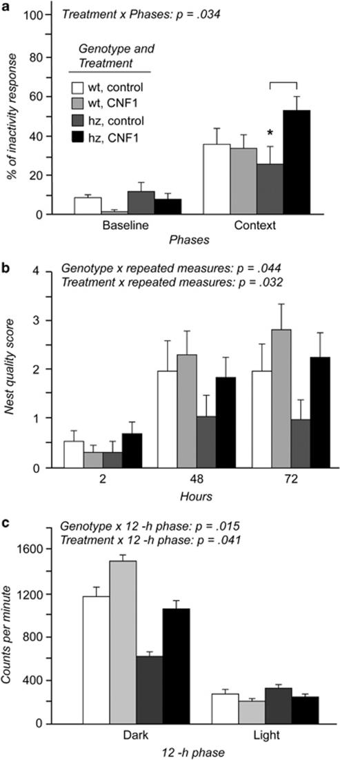
No significant changes were found as for inactivity levels during baseline (Figure 1a). In addition, the performance in the cued test was not affected by either genotype or treatment (% inactivity levels: wt control=34.3±6.7; wt CNF1=33.0±5.8; hz control=39.9±9.7; hz CNF1=36.1±6.7).
Figure 1.
CNF1 treatment improved the behavioral phenotype in a mouse model of RTT. (a) The cognitive performance of MeCP2-308 mice in a contextual fear-conditioning task was significantly improved by CNF1 treatment (12 days after CNF1 administration). The effects of icv inoculation of CNF1 C866S (control) or CNF1 on learning and memory abilities were evaluated as the increase in conditioned inactivity from the baseline. *p<0.05; N=7–9. The recombinant protein CNF1 C866S, in which the enzymatic activity on RhoGTPases is abrogated by change of cystein with serine at position 866, was used as a control. (b) CNF1 icv inoculation improved nest material manipulation and nest construction abilities in fully symptomatic MeCP2-308 mice, thus rescuing the deficit in the integrated use of mouth and forelimbs (20 days after icv administration). The quality of the nest (score=0–4) was evaluated at 2, 48 and 72 h after the introduction of nesting material into the cage. N=7–9. (c) CNF1 restored wt-like levels of home-cage spontaneous circadian locomotor activity in fully symptomatic MeCP2-308 hemizygous (hz) mice, thus rescuing circadian cyclicity (1 month after icv inoculation). The infrared sensors (20 Hz) detected any movement of mice with a frequency of 20 events per second. Scores were obtained as counts per minute (c.p.m.) expressed during 1-h periods, and the 24-h profile of daily activity was obtained by averaging 7 days of continuous registration. N=6–8.
Although inactivity levels were generally increased during the contextual test (in comparison with baseline values) (effect of phase: F(1,26)=54.57; p<0.001), mutant mice showed a poorer performance in this cognitive task: unlike all the other experimental groups (p<0.05), mutant control mice did not significantly increase their conditioned behavioral response during the test phase compared with baseline values (post hoc comparisons on the genotype*treatment*phase interaction: F(1,26)=2.82; p=0.105) (Figure 1a). These results are in line with a previous report on the same RTT mouse model (Moretti et al, 2006).
CNF1 significantly increased the conditioned response during the contextual fear-conditioning task (p<0.05 after post hoc comparisons on the treatment*phase interaction: p=0.034). However, post hoc comparisons on the genotype*treatment*phase interaction (F(1,26)=2.82; p=0.105) revealed that only the performance of MeCP2-308 mice was significantly improved by CNF1 treatment (p<0.05) (Figure 1a).
CNF1 improves mouth- and forelimb-dependent motor skills in mutant mice
As expected, when mice were followed for their ability to build a nest, the quality of the nests improved with time in all experimental groups (repeated measures: F(2,52)=40.64; p<0.001). However, in line with a previous report (Moretti et al, 2005), nest-material manipulation scores were generally lower in MeCP2-308 mice, suggesting a deficit in the integrated use of mouth and forelimbs (p<0.05 after post hoc comparison (wt control vs hz control at 72 h) on the genotype*repeated-measure interaction: F(2,52)=3.32; p=0.044) (Figure 1b).
Importantly, the poor performance shown by MeCP2-308 mice was efficaciously improved by CNF1 treatment, which significantly increased the quality of the nests in both genotypes (p<0.01 after post hoc comparison (wt control vs hz control at 72 h) on the treatment*repeated-measure interaction: F(2,52)=3.67; p=0.032) (Figure 1b).
Cnf1 restores wt-like levels of home-cage nocturnal activity in mutant mice
In line with previous reports in RTT mouse models (Moretti et al, 2005; Stearns et al, 2007), the evaluation of spontaneous locomotor activity during 24 h revealed that MeCP2-308 mice are generally less active than wt controls (genotype: F(1,24)=62.41; p=0.001) and fail to exhibit the hyperactivity profile usually emerging during the dark phase of the light/dark (L/D) cycle in rodents (p<0.01 after post hoc comparisons on the genotype*phase interaction: F(1,24)=6.84; p=0.015) (Figure 1c).
Levels of activity exhibited during the dark phase of the L/D cycle appeared significantly increased in CNF1-treated mice (p<0.01 after post hoc comparisons on the treatment*phase interaction: F(1,24)=4.63; p=0.041). The nocturnal peak of locomotor activity in mutant mice was thus restored (Figure 1c).
Astrocytes in MeCP2-308 Mice Show Atrophic Features, Which are Reversed by CNF1
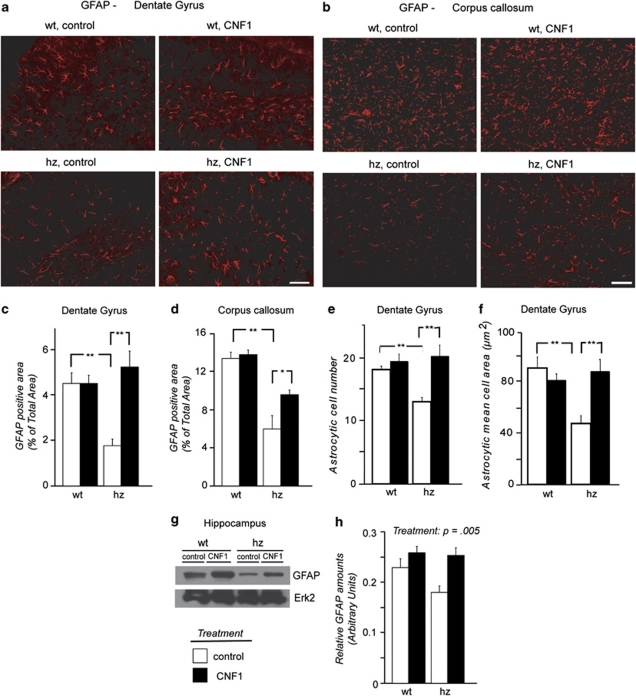
In line with a previous report (Moretti et al, 2006), no significant differences were observed between the genotypes in dendritic tree and synapses in hippocampus and motor cortex, and CNF1 did not have any effect on those markers (Supplementary Results and Supplementary Figure S2). Immunoreactivity of GFAP, a specific marker of astrocytic cytoskeleton, was analyzed in both dentate gyrus and corpus callosum, where astrocytes of grey and white matter types, respectively, are present. Both types of astrocytes in MeCP2-308 mice showed atrophic features, with fewer and poorly branched ramifications, when compared with wt animals. Morphometric analyses confirmed that GFAP-positive area was decreased in mutant mice. Although this difference was more marked in the dentate gyrus (p<0.01 after post hoc comparison on the genotype*treatment interaction: F(1,16)=11.65; p=0.004, Figure 2a and c; Supplementary Figure S4a), a similar effect of genotype was also evident in the corpus callosum (p<0.05 after post hoc comparison on the genotype*treatment interaction: F(1,18)=3.66; p=0.072, Figures 2b and d; Supplementary Figure S4b), thus suggesting that genotype effects are not limited to a single type of astrocytes. To better characterize the decrease in GFAP-positive area in mutant mice, astrocytic cell number (Figure 2e) and astrocytic mean cell area (Figure 2f) were also calculated in dentate gyrus. The results showed a decrease in both parameters in mutant brains (p<0.05 and p<0.01, respectively, after post hoc comparisons on the genotype*treatment interactions: cell number: F(1,16)=6.08; p=0.025 and mean cell area: F(1,15)=10.79; p=0.005) and suggest that they equally contributed to determine a smaller GFAP-positive area in mutant mice.
Figure 2.
CNF1 inoculation dramatically reversed astrocytic atrophy in a mouse model of RTT. The decrease in GFAP-immunoreactivity of astrocytes in MeCP2-308 hemizygous (hz) mice is counteracted by CNF1 treatment in dentate gyrus (a) and corpus callosum (b). CNF1 C866S was used as a control. Bar=50 μm. (c, d) Morphometric analysis of GFAP-positive area in dentate gyrus and corpus callosum, respectively: data are expressed as mean percentage of total area ±SEM. *p<0.05, **p<0.01. N=5–4. (e, f) Astrocytic cell number and astrocytic mean cell area in dentate gyrus. Data are expressed as mean±SEM. *p<0.05, **p<0.01. N=5–4. (g, h) Western blot analysis confirmed an increase in GFAP content in the hippocampus after CNF1 treatment.
Morphometric analysis of GFAP immunolabeling showed that CNF1 treatment increased GFAP-positive area in sections from both dentate gyrus and corpus callosum (treatment: F(1,16)=11.96; p=0.003 and F(1,18)=5.30; p=0.033, respectively, Figures 2a–d) and reversed the atrophic features of the two principal types of astrocytes in MeCP2-308 mice. In dentate gyrus, both astrocytic cell number (Figure 2e) and astrocytic mean cell area (Figure 2f) regained values close to wt after treatment with CNF1 (p<0.01 after post hoc comparisons on the genotype*treatment interactions: cell number: F(1,16)=6.08; p=0.025 and mean cell area: F(1,15)=10.79; p=0.005, respectively).
In line with immunohistochemical results, western blot analysis on hippocampal tissue from MeCP2-308 mice and their wt littermates confirmed an increase in GFAP content after CNF1 treatment (treatment: F(1,8)=14.22; p=0.005) (Figures 2g and h).
CNF1 Significantly Contrasts IL-6 Overexpression in MeCP2-308 Brains
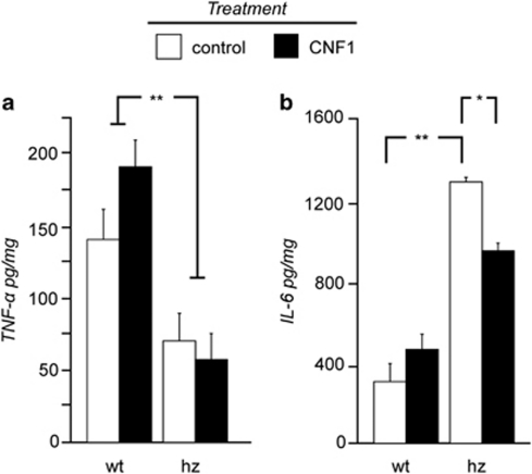
IL-1β levels were unaltered in mutant mice and CNF1 did not have any effect on this cytokine (data not shown). TNF-α was significantly decreased in MeCP2-308 mouse brains (genotype: F(1,13)=19.51; p<0.001, Figure 3a) and CNF1 did not modify it. By contrast, IL-6 expression was significantly increased in mutant brains when compared with wt mice (genotype: F(1,10)=107.88; p<0.001, Figure 3b). Icv inoculation of CNF1 selectively decreased IL-6 levels in MeCP2-308 mice, thus contrasting its overexpression (p<0.05 after post hoc comparisons on the genotype*treatment interaction: F(1,10)=10.34; p=0.009).
Figure 3.
Cytokines expression abnormalities in brain tissues of mutant mice are partially rescued by CNF1 treatment. CNF1 C866S was used as a control. (a) In fully symptomatic MeCP2-308 hemizygous (hz) mouse brains, TNF-α expression was significantly decreased in comparison with wt mice. No treatment effects were evidenced. Data are mean±SEM. **p<0.01. N=3–6. (b) CNF1 icv inoculation significantly contrasted the overexpression of IL-6 in mutant mice. *p<0.05, **p<0.01. N=3–4.
CNF1-Induced Brain Morphological and Metabolic Changes in Mutant Mice
Volumetric analyses
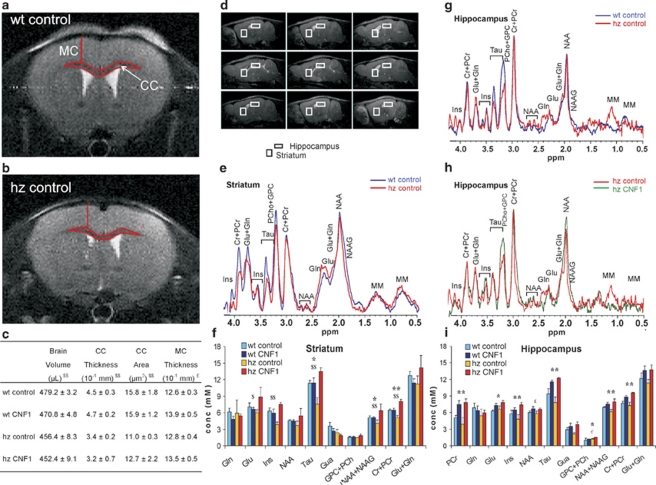
Microcephaly is a peculiar feature of RTT (Hagberg, 2002). Volumetric MRI analyses confirmed a reduction in the whole brain of MeCP2-308 mice (genotype: F(1,15)=10.81; p=0.005) (Figure 4c). In agreement with recent imaging studies, which demonstrated a reduction in corpus callosum thickness, associated with white matter pathologic features in RTT patients (Gotoh et al, 2001; Mahmood et al, 2010), a significant reduction in both area (genotype: F(1,8)=9.11; p=0.017) and thickness (genotype: F(1,8)=14.86; p=0.005) of the genu of the corpus callosum compared with wt values (Figures 4a and b) was also found in MeCP2-308 mice. No changes were found in motor cortex of mutant mice and only a trend for increased motor cortex thickness was observed in CNF1-treated mice (F(1,8)=4.69; p=0.062) (Figure 4c).
Figure 4.
MRI and MRS analyses revealed CNF1-induced brain morphological and metabolic changes. CNF1 C866S was used as a control. (a, b) Examples of axial T2-weighted MR images from wt mice and MeCP2-308 hemizygous (hz) littermates, respectively (TR/TEeff =3200/60 ms, ns=4, slice thickness 0.6 mm, 24 slices, matrix 256 × 256, FOV =25 × 25 mm2, which correspond to voxel resolution of 98 × 98 × 600 μm3). (c) Morphometric analyses included total brain volume, motor cortex (MC) thickness and corpus callosum (CC) thickness and area at +1.32 from bregma. Data are means±SEM. Effect of genotype: $p<0.05, $$p<0.01; Effect of treatment: £p=0.06. (d) Examples of in vivo sagittal T2-weighted spin-echo MR images (TR/TEeff=3000/60 ms, slice thickness 0.6 mm, ns=4, 17 slices, FOV=25 × 25 mm2, matrix 256 × 256). (e, g, h) Examples of in vivo 1H MR spectra (PRESS, TR/TE=4000/23 ms, NS=256) acquired from two voxels (indicated by the white rectangles in d) positioned on hippocampus and striatum. (f, i) Metabolite levels obtained from in vivo 1H spectra by using a quantitative protocol (which uses water as internal standard) and LCModel fitting program. Metabolite assignments: Cr, creatine; Gln, glutamine; Glu, glutamate; GPC, glicerophosphocholine; Gua, guanidoacetate; Ins, myo-inositol; NAA, nacetylaspartate; NAAG, nacetylaspartylglutamate; PCho, phosphocholine; PCr, phosphocreatine; Tau, taurine. Effect of genotype: €p<0.05; Effect of treatment: £p=0.06, *p<0.05, **p<0.01; genotype*treatment interaction: $p<0.05, $$p<0.01. Data are mean±SEM.
Brain metabolic profiles
In vivo MRI-guided 1H MRS analyses at 4.7 T detected differences in the spectra acquired in the striatum and the hippocampus of MeCP2-308, wt and treated mice. Water T2 analyses confirmed that no changes occurred in the T2s within the groups confirming that the alterations observed in the NMR signals correspond to changes in the levels of metabolites (Supplementary Table S1).
In the hippocampus, in line with immunohistochemical results, CNF1 significantly increased levels of the astrocytic marker myo-inositol (Ins) (F(1,13)=8.30; p=0.013), thus supporting our immunohistochemical results (Figures 4g–i). Notably, in this brain area CNF1 treatment concomitantly increased metabolites involved in: bioenergetics (phosphocreatine (PCr): F(1,9)=15.56; p=0.003 and total creatine (Cr) (ie, Cr+PCr): F(1,13)=17.08; p=0.001); synaptic efficiency (taurine (Tau): F(1,13)=24.23; p<0.001); neuronal health (nacetylaspartate (NAA): F(1,13)=3.91; p=0.069 and total NAA (ie, NAA+nacetylaspartylglutamate: F(1,13)=10.56; p=0.006)); phospholipid metabolism (phosphocoline (PCho) +glicerophosphocoline (GPC): F(1,13)=5.23; p<0.035) and the neurotrasmitter glutamate (Glu) (F(1,13)=5.22; p=0.026) (Figures 4g-i). In the hippocampus, the NAA/total NAA ratio was increased in mutant mice (p<0.05 after post hoc comparison on the genotype*treatment interaction: F(1,13)=4.96; p=0.044). CNF1 restored wt-levels in mutants, thus normalizing NAA metabolism (p<0.05 after post hoc comparison on the genotype*treatment interaction: p=0.044) (Supplementary Table S2). In line with a previous report (Saywell et al, 2006), mutant mice also showed increased choline-containing compounds (ie, PCho+GPC) (F(1,13)=5.56; p=0.040), suggesting abnormal phospholipid metabolism.
In line with our previous findings (Ricceri et al, 2011) suggesting a role for the striatum in the pathophysiology of RTT, CNF1-induced metabolic changes followed a different profile in this brain area: post hoc comparisons on the genotype*treatment interactions revealed that wt-like levels of Ins (F(1,11)=6.21; p=0.030), Tau (F(1,12)=7.69; p=0.017), Glu (F(1,12)=5.02; p=0.047), total NAA (F(1,11)=7.01; p=0.023), and total Cr (F(1,12)=12.60; p=0.004) were restored by CNF1 in mutant mice (p<0.05), in the absence of changes in wt controls (Figures 4e–f).
DISCUSSION
At present, no cure exists for RTT patients. This study provides the first evidence of the potential efficacy of a new treatment based on modulation of RhoGTPases for RTT and indicates that the beneficial effects on several RTT-related behavioral domains of CNF1 are mediated by a restoration of astrocytic function and involve mitochondria functionality. Our results also provide the first in vivo evidence of astrocytic atrophy in a neurodevelopmental disorder.
Intellectual disability is a core symptom of RTT (Hagberg, 2002). All the RTT mouse models so far available have been reported to show cognitive deficits (for a review, see Ricceri et al, 2008), including MeCP2-308 mice (Moretti et al, 2006). To evaluate CNF1 effects on cognitive performance, two tests were applied (fear-conditioning and novel object recognition), which assess different aspects of cognition and involve different brain areas. No changes were found in the cued version of the fear-conditioning test and in the novel object recognition task. Conversely, CNF1 treatment significantly increased the conditioned response of mutant mice in the contextual (hippocampal-dependent) version of the fear-conditioning test, thus improving their performance. As in mutant mice both anxiety-related profiles and activity levels in a novel environment are not affected by CNF1 administration (Supplementary Results and Supplementary Figure S1), treatment effects on the contextual fear-conditioning test are unlikely due to increased fear response per se or habituation to the environment. These results thus support a selective beneficial effect of CNF1 on hippocampal-dependent cognitive processes.
MeCP-308 mice were significantly impaired in nest-building abilities, thus confirming a previous report (Moretti et al, 2005). Such a deficit has been suggested to correspond to the apraxia of hand use described in RTT patients (Moretti et al, 2005), a disorder of motor planning, characterized by the loss of the ability to execute or carry out purposeful movements (Hagberg, 2002; Leiguarda and Marsden, 2000). Treatment with CNF1 significantly improved the poor performance of mutant mice in nest building, possibly improving the integrated and purpose-directed use of mouth and forelimbs.
In line with previous reports (Moretti et al, 2005; Stearns et al, 2007), the evaluation of spontaneous locomotor activity during 24 h revealed a hypoactive profile during the nocturnal/active phase in mutant mice. Interestingly, no changes were found in locomotion during 1-h open field in mutant mice (Supplementary Results), thus suggesting that motor deficits per se are unlikely to be responsible for the hypoactivity of RTT mice. Administration of CNF1 increased nocturnal levels of spontaneous locomotor activity, thus restoring a wt-like profile in mutant mice. In the open field test, CNF1 also restored wt-like levels of rearings, an exploratory behavior (Supplementary Results and Supplementary Figure S1). As CNF1 did not affect motor coordination in the Dowel test and general locomotor activity in the open field (Supplementary Results), as a whole these results suggest that motivational impairments, rather than motor deficits per se, may be contrasted by CNF1 treatment.
As already reported (Moretti et al, 2006), markers of neuronal dendritic tree and synapses were not affected in this mouse model characterized by a mild RTT phenotype. In line with recent in vitro studies that point to a critical role of astrocytes in the pathophysiology of RTT (Ballas et al, 2009; Maezawa et al, 2009), our immunohistochemical data revealed—for the first time in a RTT mouse model in vivo—a reduction in the number of the two principal types of astrocytes (protoplasmic and fibrous), analyzed in the hippocampus and the corpus callosum, respectively. Clear signs of atrophy were also detected in this cell population in mutants. Indeed, in cultured astrocytes, reducing MeCP2 levels using RNA interference leads to suppression of glial cell proliferation (Nagai et al, 2005), highlighting the possibility that a similar mechanism may account for the lower number of astrocytes here reported in mutant brains. Although atrophic or degenerative changes in astrocytes have been previously described in chronic neurodegenerative diseases (Rodriguez et al, 2009; Rossi and Volterra, 2009) and major depression (Gosselin et al, 2009; Rajkowska and Miguel-Hidalgo, 2007; Si et al, 2004), this is, to our knowledge, the first report of astrocytic deficits in a neurodevelopmental disorder.
Notably, astrocytic deficits were dramatically reversed five months after CNF1 administration, indicating persistent effects of a single CNF1 icv inoculation. Moreover, treatment effects were evident in both types of astrocytes (protoplasmic astrocytes in hippocampus and fibrous astrocytes in corpus callosum), thus suggesting a widespread effect of CNF1 on this cell population.
Our results are perfectly in line with a report showing that re-expressing Mecp2 in astrocytes of globally Mecp2-null mice improves their behavioral phenotype (Lioy et al, 2011). The results of this recent publication thus suggest that a link between the rescue of astrocytic deficits and the CNF1-induced improvement in RTT symptomatology can be attempted. First, CNF1 treatment significantly improved memory of fear in mutant mice, increasing their conditioned response in the contextual test, a paradigm primarily dependent on hippocampal integrity (Phillips and LeDoux, 1992). Considering the clear role played by astrocytes in hippocampal synaptic plasticity (Barker and Ullian, 2010; Henneberger et al, 2010), the rescue of astrocytic atrophy and the increase in astrocyte number observed in the hippocampus of mutant mice suggests that cognitive improvement induced by CNF1 in symptomatic MeCP2-308 mice may be mediated by its effects on astrocytes. Indeed, the performance of mutant mice was not affected by CNF1 in the cued version of the fear-conditioning task and the novel object recognition test (Supplementary Results), which are not hippocampal-dependent (Langston et al, 2010; Langston and Wood, 2009). In addition, hippocampal lesioned mice have been reported to build poorer nests (Deacon et al, 2002), thus further supporting a role for improved hippocampal functionality in CNF1-induced behavioral effects.
In MeCP2-308 mouse brains, an abnormal profile of cytokines was also observed (IL-6 was dramatically overexpressed, while TNF-α was downregulated), suggesting that the morphological deficits of astrocytes are accompanied by aberrant functionality. As high levels of IL-6 have also been reported to inhibit pro-inflammatory TNF-α (Petersen and Pedersen, 2005), overexpression of IL-6 is not necessarily in contrast with the observed downregulation of TNF-α and could have a role in RTT etiopathology. Indeed, exposure to high levels of IL-6, as observed during normal aging, in certain neurodegenerative diseases and in mice overexpressing IL-6 in glial cells, results in neuropathological and functional alterations, including decreased synaptic plasticity and impaired neurogenesis (Bellinger et al, 1995; Campbell, 1998; Campbell et al, 1997, 1998; Godbout and Johnson, 2004; Heyser et al, 1997; Vallieres et al, 2002; Vereyken et al, 2007).
In mutant mouse brains, CNF1 significantly counteracted IL-6 increase, but not TNF-α downregulation, suggesting a selective effect of CNF1 on the cytokine network. These results are in line with previous reports showing that the Rho-inhibiting toxin CdB upregulates inflammatory cytokine expression in astrocytes only in the presence of TLR-ligand challenges such as LPS (Borysiewicz et al, 2009; Konat et al, 2008), thus suggesting that regulation of Rho GTPase on cytokine expression is dependent on the pathophysiological status of astrocytes. Consistently, we found that Rho GTPases activation did decrease, thus normalizing, IL-6 expression in mutants, without affecting wt levels.
How CNF1 can improve the signs of morphological and functional deficit in astrocytes, however, remains to be defined. CNF1 controls, via RhoGTPases' modulation, the organization of the actin network (Fiorentini et al, 1997), whereas misregulation of actin cytoskeleton is associated with the disruption of GFAP meshwork and drugs preventing actin polymerization alter several physiological astrocytic functions (Lascola et al, 1998; Sergeeva et al, 2000). Moreover, expression of a dominant-negative mutant of the Rac1 GTPase reduces astrocytes process motility and thus functionality (Nishida and Okabe, 2007). Actin remodeling induced by CNF1 might thus have a role in CNF1-induced recovery of astrocytic function.
However, apart from cytoskeleton remodeling, RhoGTPases are known to have several other key roles (Heasman and Ridley, 2008). A complex cross-talk among RhoGTPases family members has been found to regulate generation of reactive oxygen species (ROS) (Bokoch and Diebold, 2002; Diebold et al, 2004; Heyworth et al, 1993; Selvakumar et al, 2008), a mechanism that has a pivotal role in host defense and bacterial killing (Koh et al, 2005; Miller and Britigan, 1997). Indeed, MRS analysis revealed that CNF1 increases brain content of Tau, an amino acid that has a role in preventing oxidative damage of mitochondria (Rodriguez-Martinez et al, 2004; Parvez et al, 2008; Schaffer et al, 2009), and increasing overall mitochondrial function (El Idrissi, 2008; El Idrissi and Trenkner, 1999). In RTT patients, oxidative stress markers are increased (De Felice et al, 2009). Consistently with a harmful role of ROS in lipid metabolism and mitochondria functionality, brain profile of choline-containing phospholipids is abnormal in MeCP2 mutant mice (Saywell et al, 2006 and present study) and in patients (Naidu et al, 2001) and structural and functional anomalies of mitochondria and impairments in cerebral energy metabolism have been reported in both RTT patients (Belichenko et al, 2009; Cornford et al, 1994; Dotti et al, 1993; Gibson et al, 2010) and mouse models (Kriaucionis et al, 2006; Saywell et al, 2006).
In full agreement with previous in vitro evidence of CNF1 effects on structure of the mitochondrial network (Miraglia et al, 2007) and in mitochondrial homeostasis (Fiorentini et al, 1998), a CNF1-induced buffering of intracellular energy levels is also confirmed in present study by the increase, in both hippocampal and striatal areas, in PCr content. MRS also detected an increase in Glu in CNF1-treated mice. Although it is not known whether such an increase is of extracellular or intracellular source, it is worth noting that a concomitant elevation in Tau occurred. Notably, the latter is known to counteract Glu-induced excitotoxicity through both the enhancement of mitochondrial function and the regulation of intra-mitochondrial calcium homeostasis (El Idrissi, 2008; El Idrissi and Trenkner, 1999).
As a whole MRS data therefore suggest a role for CNF1 in reducing ROS production and improving mitochondria functionality in RTT brains. Indeed, to resist host defense, several microbes have evolved defense mechanisms against oxidant production, which utilize Rho GTPases (Finlay, 2005; Siemsen et al, 2009). As in vivo studies suggest that CNF1 production by E. coli strains mediates bacterial invasion of the host (Rippere-Lampe et al, 2001), it is conceivable that the RhoGTPases-dependent mechanisms that are here proposed to be beneficial in RTT brains (eg, decreasing IL-6 expression and ROS production), may have evolved to modify central host cellular function and allow E. coli to resist host defense (Finlay, 2005; Siemsen et al, 2009).
In conclusion, our study provides the first evidence of the potential efficacy of CNF1 treatment in improving RTT symptomatology and suggests that different Rho-dependent pathways are responsible for the beneficial effects of the proposed treatment. Importantly, the use as control treatment of the mutant protein CNF1 C866S devoid of Rho enzymatic activity unequivocally confirms the pivotal role of RhoGTPases in the rescue of RTT symptoms, although further studies are needed to deeper understand the mechanisms of action of this bacterial toxin. Indeed, although RhoGTPases are critical for several cellular key processes and are involved in many pathological conditions, their pharmacological manipulation is so far still troublesome because of the lack of specific drugs. We believe that CNF1 could therefore represent a totally innovative therapeutic approach for RTT and, possibly, for other neurodevelopmental disorders (Chen et al, 2011a; Hayashi et al, 2007; Ide and Lewis, 2011; Mocholi et al, 2011).
Acknowledgments
We are grateful to Michela Di Nottia for her technical assistance and to Luigia Cancemi for animal care. This research was supported by Jerome Lejeune Foundation (France) to GL, ERARE Project (FP6 European Program) to GL, ISS-NIH 530F/52 to LR and Cure_FXS project under the E-Rare programme to CF and FM.
The authors declare no conflict of interest.
Footnotes
Supplementary Information accompanies the paper on the Neuropsychopharmacology website (http://www.nature.com/npp)
Supplementary Material
References
- Amir RE, Van den Veyver IB, Wan M, Tran CQ, Francke U, Zoghbi HY. Rett syndrome is caused by mutations in X-linked MECP2, encoding methyl-CpG-binding protein 2. Nat Genet. 1999;23:185–188. doi: 10.1038/13810. [DOI] [PubMed] [Google Scholar]
- Ballas N, Lioy DT, Grunseich C, Mandel G. Non-cell autonomous influence of MeCP2-deficient glia on neuronal dendritic morphology. Nat Neurosci. 2009;12:311–317. doi: 10.1038/nn.2275. [DOI] [PMC free article] [PubMed] [Google Scholar]
- Barker AJ, Ullian EM. Astrocytes and synaptic plasticity. Neuroscientist. 2010;16:40–50. doi: 10.1177/1073858409339215. [DOI] [PubMed] [Google Scholar]
- Belichenko PV, Wright EE, Belichenko NP, Masliah E, Li HH, Mobley WC, et al. Widespread changes in dendritic and axonal morphology in Mecp2-mutant mouse models of Rett syndrome: evidence for disruption of neuronal networks. J Comp Neurol. 2009;514:240–258. doi: 10.1002/cne.22009. [DOI] [PubMed] [Google Scholar]
- Bellinger FP, Madamba SG, Campbell IL, Siggins GR. Reduced long-term potentiation in the dentate gyrus of transgenic mice with cerebral overexpression of interleukin-6. Neurosci Lett. 1995;198:95–98. doi: 10.1016/0304-3940(95)11976-4. [DOI] [PubMed] [Google Scholar]
- Bokoch GM, Diebold BA. Current molecular models for NADPH oxidase regulation by Rac GTPase. Blood. 2002;100:2692–2696. doi: 10.1182/blood-2002-04-1149. [DOI] [PubMed] [Google Scholar]
- Borysiewicz E, Fil D, Konat GW. Rho proteins are negative regulators of TLR2, TLR3, and TLR4 signaling in astrocytes. J Neurosci Res. 2009;87:1565–1572. doi: 10.1002/jnr.21968. [DOI] [PubMed] [Google Scholar]
- Cahoy JD, Emery B, Kaushal A, Foo LC, Zamanian JL, Christopherson KS, et al. A transcriptome database for astrocytes, neurons, and oligodendrocytes: a new resource for understanding brain development and function. J Neurosci. 2008;28:264–278. doi: 10.1523/JNEUROSCI.4178-07.2008. [DOI] [PMC free article] [PubMed] [Google Scholar]
- Campbell IL. Transgenic mice and cytokine actions in the brain: bridging the gap between structural and functional neuropathology. Brain Res Brain Res Rev. 1998;26:327–336. doi: 10.1016/s0165-0173(97)00038-6. [DOI] [PubMed] [Google Scholar]
- Campbell IL, Stalder AK, Akwa Y, Pagenstecher A, Asensio VC. Transgenic models to study the actions of cytokines in the central nervous system. Neuroimmunomodulation. 1998;5:126–135. doi: 10.1159/000026329. [DOI] [PubMed] [Google Scholar]
- Campbell IL, Stalder AK, Chiang CS, Bellinger R, Heyser CJ, Steffensen S, et al. Transgenic models to assess the pathogenic actions of cytokines in the central nervous system. Mol Psychiatry. 1997;2:125–129. doi: 10.1038/sj.mp.4000225. [DOI] [PubMed] [Google Scholar]
- Canese R, Pisanu ME, Mezzanzanica D, Ricci A, Paris L, Bagnoli M, et al. 2011Characterization of in vivo ovarian cancer models by quantitative magnetic resonance imaging and spectroscopy NMR BiomedE-pub. [DOI] [PubMed]
- Chahrour M, Jung SY, Shaw C, Zhou X, Wong ST, Qin J, et al. MeCP2, a key contributor to neurological disease, activates and represses transcription. Science. 2008;320:1224–1229. doi: 10.1126/science.1153252. [DOI] [PMC free article] [PubMed] [Google Scholar]
- Chahrour M, Zoghbi HY. The story of Rett syndrome: from clinic to neurobiology. Neuron. 2007;56:422–437. doi: 10.1016/j.neuron.2007.10.001. [DOI] [PubMed] [Google Scholar]
- Chang Q, Khare G, Dani V, Nelson S, Jaenisch R. The disease progression of Mecp2 mutant mice is affected by the level of BDNF expression. Neuron. 2006;49:341–348. doi: 10.1016/j.neuron.2005.12.027. [DOI] [PubMed] [Google Scholar]
- Chen LY, Rex CS, Babayan AH, Kramar EA, Lynch G, Gall CM, et al. Physiological activation of synaptic Rac>PAK (p-21 activated kinase) signaling is defective in a mouse model of fragile X syndrome. J Neurosci. 2011a;30:10977–10984. doi: 10.1523/JNEUROSCI.1077-10.2010. [DOI] [PMC free article] [PubMed] [Google Scholar]
- Chen Q, Zhu YC, Yu J, Miao S, Zheng J, Xu L, et al. CDKL5, a protein associated with Rett syndrome, regulates neuronal morphogenesis via Rac1 signaling. J Neurosci. 2011b;30:12777–12786. doi: 10.1523/JNEUROSCI.1102-10.2010. [DOI] [PMC free article] [PubMed] [Google Scholar]
- Cornford ME, Philippart M, Jacobs B, Scheibel AB, Vinters HV. Neuropathology of Rett syndrome: case report with neuronal and mitochondrial abnormalities in the brain. J Child Neurol. 1994;9:424–431. doi: 10.1177/088307389400900419. [DOI] [PubMed] [Google Scholar]
- De Felice C, Ciccoli L, Leoncini S, Signorini C, Rossi M, Vannuccini L, et al. Systemic oxidative stress in classic Rett syndrome. Free Radic Biol Med. 2009;47:440–448. doi: 10.1016/j.freeradbiomed.2009.05.016. [DOI] [PubMed] [Google Scholar]
- De Filippis B, Ricceri L, Laviola G. Early postnatal behavioral changes in the Mecp2-308 truncation mouse model of Rett syndrome. Genes Brain Behav. 2010;9:213–223. doi: 10.1111/j.1601-183X.2009.00551.x. [DOI] [PubMed] [Google Scholar]
- De Viti S, Martino A, Musilli M, Fiorentini C, Diana G. The Rho GTPase activating CNF1 improves associative working memory for object-in-place. Behav Brain Res. 2010;212:78–83. doi: 10.1016/j.bbr.2010.03.049. [DOI] [PubMed] [Google Scholar]
- Deacon RM. Assessing nest building in mice. Nat Protoc. 2006;1:1117–1119. doi: 10.1038/nprot.2006.170. [DOI] [PubMed] [Google Scholar]
- Deacon RM, Croucher A, Rawlins JN. Hippocampal cytotoxic lesion effects on species-typical behaviours in mice. Behav Brain Res. 2002;132:203–213. doi: 10.1016/s0166-4328(01)00401-6. [DOI] [PubMed] [Google Scholar]
- Diana G, Valentini G, Travaglione S, Falzano L, Pieri M, Zona C, et al. Enhancement of learning and memory after activation of cerebral Rho GTPases. Proc Natl Acad Sci USA. 2007;104:636–641. doi: 10.1073/pnas.0610059104. [DOI] [PMC free article] [PubMed] [Google Scholar]
- Diebold BA, Fowler B, Lu J, Dinauer MC, Bokoch GM. Antagonistic cross-talk between Rac and Cdc42 GTPases regulates generation of reactive oxygen species. J Biol Chem. 2004;279:28136–28142. doi: 10.1074/jbc.M313891200. [DOI] [PubMed] [Google Scholar]
- Dotti MT, Manneschi L, Malandrini A, De Stefano N, Caznerale F, Federico A. Mitochondrial dysfunction in Rett syndrome. An ultrastructural and biochemical study. Brain Dev. 1993;15:103–106. doi: 10.1016/0387-7604(93)90045-a. [DOI] [PubMed] [Google Scholar]
- El Idrissi A. Taurine increases mitochondrial buffering of calcium: role in neuroprotection. Amino Acids. 2008;34:321–328. doi: 10.1007/s00726-006-0396-9. [DOI] [PubMed] [Google Scholar]
- El Idrissi A, Trenkner E. Growth factors and taurine protect against excitotoxicity by stabilizing calcium homeostasis and energy metabolism. J Neurosci. 1999;19:9459–9468. doi: 10.1523/JNEUROSCI.19-21-09459.1999. [DOI] [PMC free article] [PubMed] [Google Scholar]
- Etienne-Manneville S, Hall A. Rho GTPases in cell biology. Nature. 2002;420:629–635. doi: 10.1038/nature01148. [DOI] [PubMed] [Google Scholar]
- Falzano L, Fiorentini C, Donelli G, Michel E, Kocks C, Cossart P, et al. Induction of phagocytic behaviour in human epithelial cells by Escherichia coli cytotoxic necrotizing factor type 1. Mol Microbiol. 1993;9:1247–1254. doi: 10.1111/j.1365-2958.1993.tb01254.x. [DOI] [PubMed] [Google Scholar]
- Feltri ML, Suter U, Relvas JB. The function of RhoGTPases in axon ensheathment and myelination. Glia. 2008;56:1508–1517. doi: 10.1002/glia.20752. [DOI] [PMC free article] [PubMed] [Google Scholar]
- Finlay BB. Bacterial virulence strategies that utilize Rho GTPases. Curr Top Microbiol Immunol. 2005;291:1–10. doi: 10.1007/3-540-27511-8_1. [DOI] [PubMed] [Google Scholar]
- Fiorentini C, Fabbri A, Flatau G, Donelli G, Matarrese P, Lemichez E, et al. Escherichia coli cytotoxic necrotizing factor 1 (CNF1), a toxin that activates the Rho GTPase. J Biol Chem. 1997;272:19532–19537. doi: 10.1074/jbc.272.31.19532. [DOI] [PubMed] [Google Scholar]
- Fiorentini C, Matarrese P, Straface E, Falzano L, Fabbri A, Donelli G, et al. Toxin-induced activation of Rho GTP-binding protein increases Bcl-2 expression and influences mitochondrial homeostasis. Exp Cell Res. 1998;242:341–350. doi: 10.1006/excr.1998.4057. [DOI] [PubMed] [Google Scholar]
- Gibson JH, Slobedman B, K NH, Williamson SL, Minchenko D, El-Osta A, et al. Downstream targets of methyl CpG binding protein 2 and their abnormal expression in the frontal cortex of the human Rett syndrome brain. BMC Neurosci. 2010;11:53. doi: 10.1186/1471-2202-11-53. [DOI] [PMC free article] [PubMed] [Google Scholar]
- Godbout JP, Johnson RW. Interleukin-6 in the aging brain. J Neuroimmunol. 2004;147:141–144. doi: 10.1016/j.jneuroim.2003.10.031. [DOI] [PubMed] [Google Scholar]
- Gonzales ML, LaSalle JM. The role of MeCP2 in brain development and neurodevelopmental disorders. Curr Psychiatry Rep. 2010;12:127–134. doi: 10.1007/s11920-010-0097-7. [DOI] [PMC free article] [PubMed] [Google Scholar]
- Gosselin RD, Gibney S, O'Malley D, Dinan TG, Cryan JF. Region specific decrease in glial fibrillary acidic protein immunoreactivity in the brain of a rat model of depression. Neuroscience. 2009;159:915–925. doi: 10.1016/j.neuroscience.2008.10.018. [DOI] [PubMed] [Google Scholar]
- Gotoh H, Suzuki I, Maruki K, Mitomo M, Hirasawa K, Sasaki N. Magnetic resonance imaging and clinical findings examined in adulthood-studies on three adults with Rett syndrome. Brain Dev. 2001;23 (Suppl 1:S118–S121. doi: 10.1016/s0387-7604(01)00355-2. [DOI] [PubMed] [Google Scholar]
- Hagberg B. Clinical manifestations and stages of Rett syndrome. Ment Retard Dev Disabil Res Rev. 2002;8:61–65. doi: 10.1002/mrdd.10020. [DOI] [PubMed] [Google Scholar]
- Hall A. Rho GTPases and the control of cell behaviour. Biochem Soc Trans. 2005;33:891–895. doi: 10.1042/BST20050891. [DOI] [PubMed] [Google Scholar]
- Hayashi ML, Rao BS, Seo JS, Choi HS, Dolan BM, Choi SY, et al. Inhibition of p21-activated kinase rescues symptoms of fragile X syndrome in mice. Proc Natl Acad Sci USA. 2007;104:11489–11494. doi: 10.1073/pnas.0705003104. [DOI] [PMC free article] [PubMed] [Google Scholar]
- Heasman SJ, Ridley AJ. Mammalian Rho GTPases: new insights into their functions from in vivo studies. Nat Rev Mol Cell Biol. 2008;9:690–701. doi: 10.1038/nrm2476. [DOI] [PubMed] [Google Scholar]
- Henneberger C, Papouin T, Oliet SH, Rusakov DA. Long-term potentiation depends on release of D-serine from astrocytes. Nature. 2010;463:232–236. doi: 10.1038/nature08673. [DOI] [PMC free article] [PubMed] [Google Scholar]
- Heyser CJ, Masliah E, Samimi A, Campbell IL, Gold LH. Progressive decline in avoidance learning paralleled by inflammatory neurodegeneration in transgenic mice expressing interleukin 6 in the brain. Proc Natl Acad Sci USA. 1997;94:1500–1505. doi: 10.1073/pnas.94.4.1500. [DOI] [PMC free article] [PubMed] [Google Scholar]
- Heyworth PG, Knaus UG, Xu X, Uhlinger DJ, Conroy L, Bokoch GM, et al. Requirement for posttranslational processing of Rac GTP-binding proteins for activation of human neutrophil NADPH oxidase. Mol Biol Cell. 1993;4:261–269. doi: 10.1091/mbc.4.3.261. [DOI] [PMC free article] [PubMed] [Google Scholar]
- Hite KC, Adams VH, Hansen JC. Recent advances in MeCP2 structure and function. Biochem Cell Biol. 2009;87:219–227. doi: 10.1139/o08-115. [DOI] [PMC free article] [PubMed] [Google Scholar]
- Ide M, Lewis DA. Altered cortical CDC42 signaling pathways in schizophrenia: implications for dendritic spine deficits. Biol Psychiatry. 2011;68:25–32. doi: 10.1016/j.biopsych.2010.02.016. [DOI] [PMC free article] [PubMed] [Google Scholar]
- Koh AL, Sun CX, Zhu F, Glogauer M. The role of Rac1 and Rac2 in bacterial killing. Cell Immunol. 2005;235:92–97. doi: 10.1016/j.cellimm.2005.07.005. [DOI] [PubMed] [Google Scholar]
- Konat GW, Krasowska-Zoladek A, Kraszpulski M. Statins enhance toll-like receptor 4-mediated cytokine gene expression in astrocytes: implication of Rho proteins in negative feedback regulation. J Neurosci Res. 2008;86:603–609. doi: 10.1002/jnr.21509. [DOI] [PubMed] [Google Scholar]
- Kriaucionis S, Paterson A, Curtis J, Guy J, Macleod N, Bird A. Gene expression analysis exposes mitochondrial abnormalities in a mouse model of Rett syndrome. Mol Cell Biol. 2006;26:5033–5042. doi: 10.1128/MCB.01665-05. [DOI] [PMC free article] [PubMed] [Google Scholar]
- Langston RF, Stevenson CH, Wilson CL, Saunders I, Wood ER. The role of hippocampal subregions in memory for stimulus associations. Behav Brain Res. 2010;215:275–291. doi: 10.1016/j.bbr.2010.07.006. [DOI] [PubMed] [Google Scholar]
- Langston RF, Wood ER. Associative recognition and the hippocampus: differential effects of hippocampal lesions on object-place, object-context and object-place-context memory. Hippocampus. 2009;20:1139–1153. doi: 10.1002/hipo.20714. [DOI] [PubMed] [Google Scholar]
- Lascola CD, Nelson DJ, Kraig RP. Cytoskeletal actin gates a Cl- channel in neocortical astrocytes. J Neurosci. 1998;18:1679–1692. doi: 10.1523/JNEUROSCI.18-05-01679.1998. [DOI] [PMC free article] [PubMed] [Google Scholar]
- Leiguarda RC, Marsden CD. Limb apraxias: higher-order disorders of sensorimotor integration. Brain. 2000;123 (Part 5:860–879. doi: 10.1093/brain/123.5.860. [DOI] [PubMed] [Google Scholar]
- Lemichez E, Flatau G, Bruzzone M, Boquet P, Gauthier M. Molecular localization of the Escherichia coli cytotoxic necrotizing factor CNF1 cell-binding and catalytic domains. Mol Microbiol. 1997;24:1061–1070. doi: 10.1046/j.1365-2958.1997.4151781.x. [DOI] [PubMed] [Google Scholar]
- Lioy DT, Garg SK, Monaghan CE, Raber J, Foust KD, Kaspar BK, et al. A role for glia in the progression of Rett's syndrome. Nature. 2011;475:497–500. doi: 10.1038/nature10214. [DOI] [PMC free article] [PubMed] [Google Scholar]
- Luo L. Rho GTPases in neuronal morphogenesis. Nat Rev Neurosci. 2000;1:173–180. doi: 10.1038/35044547. [DOI] [PubMed] [Google Scholar]
- Maezawa I, Swanberg S, Harvey D, LaSalle JM, Jin LW. Rett syndrome astrocytes are abnormal and spread MeCP2 deficiency through gap junctions. J Neurosci. 2009;29:5051–5061. doi: 10.1523/JNEUROSCI.0324-09.2009. [DOI] [PMC free article] [PubMed] [Google Scholar]
- Mahmood A, Bibat G, Zhan AL, Izbudak I, Farage L, Horska A, et al. White matter impairment in Rett syndrome: diffusion tensor imaging study with clinical correlations. AJNR Am J Neuroradiol. 2010;31:295–299. doi: 10.3174/ajnr.A1792. [DOI] [PMC free article] [PubMed] [Google Scholar]
- McAfoose J, Baune BT. Evidence for a cytokine model of cognitive function. Neurosci Biobehav Rev. 2009;33:355–366. doi: 10.1016/j.neubiorev.2008.10.005. [DOI] [PubMed] [Google Scholar]
- McGill BE, Bundle SF, Yaylaoglu MB, Carson JP, Thaller C, Zoghbi HY. Enhanced anxiety and stress-induced corticosterone release are associated with increased Crh expression in a mouse model of Rett syndrome. Proc Natl Acad Sci USA. 2006;103:18267–18272. doi: 10.1073/pnas.0608702103. [DOI] [PMC free article] [PubMed] [Google Scholar]
- Miller RA, Britigan BE. Role of oxidants in microbial pathophysiology. Clin Microbiol Rev. 1997;10:1–18. doi: 10.1128/cmr.10.1.1. [DOI] [PMC free article] [PubMed] [Google Scholar]
- Miraglia AG, Travaglione S, Meschini S, Falzano L, Matarrese P, Quaranta MG, et al. Cytotoxic necrotizing factor 1 prevents apoptosis via the Akt/IkappaB kinase pathway: role of nuclear factor-kappaB and Bcl-2. Mol Biol Cell. 2007;18:2735–2744. doi: 10.1091/mbc.E06-10-0910. [DOI] [PMC free article] [PubMed] [Google Scholar]
- Miyamoto Y, Yamauchi J, Tanoue A, Wu C, Mobley WC. TrkB binds and tyrosine-phosphorylates Tiam1, leading to activation of Rac1 and induction of changes in cellular morphology. Proc Natl Acad Sci USA. 2006;103:10444–10449. doi: 10.1073/pnas.0603914103. [DOI] [PMC free article] [PubMed] [Google Scholar]
- Mocholi E, Ballester-Lurbe B, Arque G, Poch E, Peris B, Guerri C, et al. RhoE deficiency produces postnatal lethality, profound motor deficits and neurodevelopmental delay in mice. PLoS One. 2011;6:e19236. doi: 10.1371/journal.pone.0019236. [DOI] [PMC free article] [PubMed] [Google Scholar]
- Moretti P, Bouwknecht JA, Teague R, Paylor R, Zoghbi HY. Abnormalities of social interactions and home-cage behavior in a mouse model of Rett syndrome. Hum Mol Genet. 2005;14:205–220. doi: 10.1093/hmg/ddi016. [DOI] [PubMed] [Google Scholar]
- Moretti P, Levenson JM, Battaglia F, Atkinson R, Antalffy B, Amstrong D, et al. Learning and memory and synaptic plasticity are impaired in a mouse model of Rett syndrome. J Neurosci. 2006;26:319–327. doi: 10.1523/JNEUROSCI.2623-05.2006. [DOI] [PMC free article] [PubMed] [Google Scholar]
- Nag N, Moriuchi JM, Peitzman CG, Ward BC, Kolodny NH, Berger-Sweeney JE. Environmental enrichment alters locomotor behaviour and ventricular volume in Mecp2 1lox mice. Behav Brain Res. 2009;196:44–48. doi: 10.1016/j.bbr.2008.07.008. [DOI] [PubMed] [Google Scholar]
- Nagai K, Miyake K, Kubota T. A transcriptional repressor MeCP2 causing Rett syndrome is expressed in embryonic non-neuronal cells and controls their growth. Brain Res Dev Brain Res. 2005;157:103–106. doi: 10.1016/j.devbrainres.2005.03.011. [DOI] [PubMed] [Google Scholar]
- Naidu S, Kaufmann WE, Abrams MT, Pearlson GD, Lanham DC, Fredericksen KA, et al. Neuroimaging studies in Rett syndrome. Brain Dev. 2001;23 (Suppl 1:S62–S71. doi: 10.1016/s0387-7604(01)00381-3. [DOI] [PubMed] [Google Scholar]
- Nakayama AY, Harms MB, Luo L. Small GTPases Rac and Rho in the maintenance of dendritic spines and branches in hippocampal pyramidal neurons. J Neurosci. 2000;20:5329–5338. doi: 10.1523/JNEUROSCI.20-14-05329.2000. [DOI] [PMC free article] [PubMed] [Google Scholar]
- Nishida H, Okabe S. Direct astrocytic contacts regulate local maturation of dendritic spines. J Neurosci. 2007;27:331–340. doi: 10.1523/JNEUROSCI.4466-06.2007. [DOI] [PMC free article] [PubMed] [Google Scholar]
- Ogier M, Katz DM. Breathing dysfunction in Rett syndrome: understanding epigenetic regulation of the respiratory network. Respir Physiol Neurobiol. 2008;164:55–63. doi: 10.1016/j.resp.2008.04.005. [DOI] [PMC free article] [PubMed] [Google Scholar]
- Parvez S, Tabassum H, Banerjee BD, Raisuddin S. Taurine prevents tamoxifen-induced mitochondrial oxidative damage in mice. Basic Clin Pharmacol Toxicol. 2008;102:382–387. doi: 10.1111/j.1742-7843.2008.00208.x. [DOI] [PubMed] [Google Scholar]
- Pavone F, Luvisetto S, Marinelli S, Straface E, Fabbri A, Falzano L, et al. The Rac GTPase-activating bacterial protein toxin CNF1 induces analgesia up-regulating mu-opioid receptors. Pain. 2009;145:219–229. doi: 10.1016/j.pain.2009.06.026. [DOI] [PubMed] [Google Scholar]
- Petersen AM, Pedersen BK. The anti-inflammatory effect of exercise. J Appl Physiol. 2005;98:1154–1162. doi: 10.1152/japplphysiol.00164.2004. [DOI] [PubMed] [Google Scholar]
- Phillips RG, LeDoux JE. Differential contribution of amygdala and hippocampus to cued and contextual fear conditioning. Behav Neurosci. 1992;106:274–285. doi: 10.1037//0735-7044.106.2.274. [DOI] [PubMed] [Google Scholar]
- Popoli P, Pintor A, Domenici MR, Frank C, Tebano MT, Pezzola A, et al. Blockade of striatal adenosine A2A receptor reduces, through a presynaptic mechanism, quinolinic acid-induced excitotoxicity: possible relevance to neuroprotective interventions in neurodegenerative diseases of the striatum. J Neurosci. 2002;22:1967–1975. doi: 10.1523/JNEUROSCI.22-05-01967.2002. [DOI] [PMC free article] [PubMed] [Google Scholar]
- Provencher SW. Estimation of metabolite concentrations from localized in vivo proton NMR spectra. Magn Reson Med. 1993;30:672–679. doi: 10.1002/mrm.1910300604. [DOI] [PubMed] [Google Scholar]
- Rajkowska G, Miguel-Hidalgo JJ. Gliogenesis and glial pathology in depression. CNS Neurol Disord Drug Targets. 2007;6:219–233. doi: 10.2174/187152707780619326. [DOI] [PMC free article] [PubMed] [Google Scholar]
- Ramakers GJ. Rho proteins, mental retardation and the cellular basis of cognition. Trends Neurosci. 2002;25:191–199. doi: 10.1016/s0166-2236(00)02118-4. [DOI] [PubMed] [Google Scholar]
- Ricceri L, De Filippis B, Fuso A, Laviola G. Cholinergic hypofunction in MeCP2-308 mice: beneficial neurobehavioural effects of neonatal choline supplementation. Behav Brain Res. 2011;221:623–629. doi: 10.1016/j.bbr.2011.03.051. [DOI] [PubMed] [Google Scholar]
- Ricceri L, De Filippis B, Laviola G. Mouse models of Rett syndrome: from behavioural phenotyping to preclinical evaluation of new therapeutic approaches. Behav Pharmacol. 2008;19:501–517. doi: 10.1097/FBP.0b013e32830c3645. [DOI] [PubMed] [Google Scholar]
- Rippere-Lampe KE, O'Brien AD, Conran R, Lockman HA. Mutation of the gene encoding cytotoxic necrotizing factor type 1 (cnf(1)) attenuates the virulence of uropathogenic Escherichia coli. Infect Immun. 2001;69:3954–3964. doi: 10.1128/IAI.69.6.3954-3964.2001. [DOI] [PMC free article] [PubMed] [Google Scholar]
- Rodriguez-Martinez E, Rugerio-Vargas C, Rodriguez AI, Borgonio-Perez G, Rivas-Arancibia S. Antioxidant effects of taurine, vitamin C, and vitamin E on oxidative damage in hippocampus caused by the administration of 3-nitropropionic acid in rats. Int J Neurosci. 2004;114:1133–1145. doi: 10.1080/00207450490475959. [DOI] [PubMed] [Google Scholar]
- Rodriguez JJ, Olabarria M, Chvatal A, Verkhratsky A. Astroglia in dementia and Alzheimer's disease. Cell Death Differ. 2009;16:378–385. doi: 10.1038/cdd.2008.172. [DOI] [PubMed] [Google Scholar]
- Rossi D, Volterra A. Astrocytic dysfunction: insights on the role in neurodegeneration. Brain Res Bull. 2009;80:224–232. doi: 10.1016/j.brainresbull.2009.07.012. [DOI] [PubMed] [Google Scholar]
- Saywell V, Viola A, Confort-Gouny S, Le Fur Y, Villard L, Cozzone PJ. Brain magnetic resonance study of Mecp2 deletion effects on anatomy and metabolism. Biochem Biophys Res Commun. 2006;340:776–783. doi: 10.1016/j.bbrc.2005.12.080. [DOI] [PubMed] [Google Scholar]
- Schaffer SW, Azuma J, Mozaffari M. Role of antioxidant activity of taurine in diabetes. Can J Physiol Pharmacol. 2009;87:91–99. doi: 10.1139/Y08-110. [DOI] [PubMed] [Google Scholar]
- Schmidt G, Sehr P, Wilm M, Selzer J, Mann M, Aktories K. Gln 63 of Rho is deamidated by Escherichia coli cytotoxic necrotizing factor-1. Nature. 1997;387:725–729. doi: 10.1038/42735. [DOI] [PubMed] [Google Scholar]
- Schmidt G, Selzer J, Lerm M, Aktories K. The Rho-deamidating cytotoxic necrotizing factor 1 from Escherichia coli possesses transglutaminase activity. Cysteine 866 and histidine 881 are essential for enzyme activity. J Biol Chem. 1998;273:13669–13674. doi: 10.1074/jbc.273.22.13669. [DOI] [PubMed] [Google Scholar]
- Selvakumar B, Hess DT, Goldschmidt-Clermont PJ, Stamler JS. Co-regulation of constitutive nitric oxide synthases and NADPH oxidase by the small GTPase Rac. FEBS Lett. 2008;582:2195–2202. doi: 10.1016/j.febslet.2008.04.062. [DOI] [PMC free article] [PubMed] [Google Scholar]
- Sergeeva M, Ubl JJ, Reiser G. Disruption of actin cytoskeleton in cultured rat astrocytes suppresses ATP- and bradykinin-induced [Ca(2+)](i) oscillations by reducing the coupling efficiency between Ca(2+) release, capacitative Ca(2+) entry, and store refilling. Neuroscience. 2000;97:765–769. doi: 10.1016/s0306-4522(00)00062-2. [DOI] [PubMed] [Google Scholar]
- Shahbazian M, Young J, Yuva-Paylor L, Spencer C, Antalffy B, Noebels J, et al. Mice with truncated MeCP2 recapitulate many Rett syndrome features and display hyperacetylation of histone H3. Neuron. 2002;35:243–254. doi: 10.1016/s0896-6273(02)00768-7. [DOI] [PubMed] [Google Scholar]
- Si X, Miguel-Hidalgo JJ, O'Dwyer G, Stockmeier CA, Rajkowska G. Age-dependent reductions in the level of glial fibrillary acidic protein in the prefrontal cortex in major depression. Neuropsychopharmacology. 2004;29:2088–2096. doi: 10.1038/sj.npp.1300525. [DOI] [PMC free article] [PubMed] [Google Scholar]
- Siemsen DW, Kirpotina LN, Jutila MA, Quinn MT. Inhibition of the human neutrophil NADPH oxidase by Coxiella burnetii. Microbes Infect. 2009;11:671–679. doi: 10.1016/j.micinf.2009.04.005. [DOI] [PMC free article] [PubMed] [Google Scholar]
- Stearns NA, Schaevitz LR, Bowling H, Nag N, Berger UV, Berger-Sweeney J. Behavioral and anatomical abnormalities in Mecp2 mutant mice: a model for Rett syndrome. Neuroscience. 2007;146:907–921. doi: 10.1016/j.neuroscience.2007.02.009. [DOI] [PubMed] [Google Scholar]
- Swinny JD, Valentino RJ. Corticotropin-releasing factor promotes growth of brain norepinephrine neuronal processes through Rho GTPase regulators of the actin cytoskeleton in rat. Eur J Neurosci. 2006;24:2481–2490. doi: 10.1111/j.1460-9568.2006.05129.x. [DOI] [PubMed] [Google Scholar]
- Tashiro A, Minden A, Yuste R. Regulation of dendritic spine morphology by the rho family of small GTPases: antagonistic roles of Rac and Rho. Cereb Cortex. 2000;10:927–938. doi: 10.1093/cercor/10.10.927. [DOI] [PubMed] [Google Scholar]
- Vallieres L, Campbell IL, Gage FH, Sawchenko PE. Reduced hippocampal neurogenesis in adult transgenic mice with chronic astrocytic production of interleukin-6. J Neurosci. 2002;22:486–492. doi: 10.1523/JNEUROSCI.22-02-00486.2002. [DOI] [PMC free article] [PubMed] [Google Scholar]
- van Galen EJ, Ramakers GJ. Rho proteins, mental retardation and the neurobiological basis of intelligence. Prog Brain Res. 2005;147:295–317. doi: 10.1016/S0079-6123(04)47022-8. [DOI] [PubMed] [Google Scholar]
- Vereyken EJ, Bajova H, Chow S, de Graan PN, Gruol DL. Chronic interleukin-6 alters the level of synaptic proteins in hippocampus in culture and in vivo. Eur J Neurosci. 2007;25:3605–3616. doi: 10.1111/j.1460-9568.2007.05615.x. [DOI] [PubMed] [Google Scholar]
- Wilcox RG.(ed) (1987New Statistical Procedures for the Social Sciences Lawrence Erlbaum Associates, NJ; 187–189. [Google Scholar]
Associated Data
This section collects any data citations, data availability statements, or supplementary materials included in this article.


