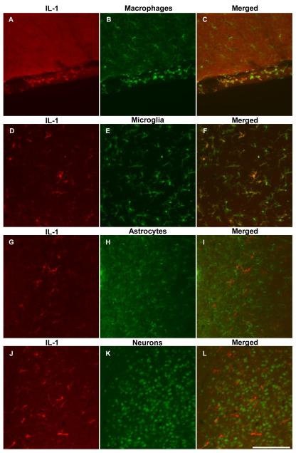
FIG. 2.
Representative photomicrographs showing dual fluorescence immunohistochemistry for IL-1β, with markers for microglia and macrophages, astrocytes and neurons in the rat brain after icv injection of 1.5 nmol galanin-like peptide (GALP). IL-1β-positive cells in the meninges (A-C) and the parenchyma of the peri-third ventricular region (D-F) and were found to be macrophages (Iba1-positive), and microglia (Iba1-positive) respectively. However, cells expressing IL-1β did not co-localize with markers for astrocytes (GFAP-positive; G-I) or neurons (NeuN-positive; J-L). Cells expressing IL-1α in the parenchyma were also found to be microglia (data not shown for clarity). Scale bar, 100μm

