Abstract
Rosai-Dorfman disease (RDD), also known as sinus histiocytosis with massive lymphadenopathy is a rare disease involving the lymph nodes. Extranodal RDD involving the thyroid is extremely rare. So far, six cases of RDD involving thyroid have been reported in the literature; all have occurred in females with a mean age of 56.3 years. Clinically, radiologically and cytologically, all the cases were initially diagnosed as thyroid malignancies with lymph nodal metastasis. The final diagnosis was made histologically only after total thyroidectomy. We herein, present a seventh case of RDD involving the thyroid in a 15-year-old female, diagnosed first on fine needle aspiration cytology (FNAC). We conclude that FNAC is a useful diagnostic procedure for RDD involving thyroid; it can avoid an unnecessary thyroidectomy.
Keywords: Fine needle aspiration cytology, Rosai–Dorfman disease, thyroid
Introduction
Rosai-Dorfman disease (RDD), also known as sinus histiocytosis with massive lymphadenopathy most often presents as a painless, massive bilateral enlargement of the cervical lymph nodes associated with fever, leucocytosis, elevated erythrocyte sedimentation rate (ESR), and polyclonal hypergammaglobulenemia.[1] Most cases occur during the first or second decade of life with male preponderance. It is characterized by marked lymphoplasmacytic and hystiocytic infiltration with emperipolesis (lymphophagocytosis) in the lymph nodes.
Although extranodal RDD (involving the orbit, upper respiratory tract, skin, subcutaneous tissue, skeletal system, central nervous system, etc.) is found in 40% of the patients with RDD, involvement of the thyroid has been reported rarely. So far, six cases of RDD involving the thyroid were reported in literature, all occurred in females with a mean age of 56.3 years.[2] All the cases were initially diagnosed as thyroid malignancies with lymph nodal metastasis, both clinicoradiologically and cytologically.[2–4] The cytological features of RDD involving the thyroid are virtually diagnostic and can obviate the need for a biopsy in most cases.
Case Report
A 15-year-old female presented with multiple, painless enlargement of the cervical and submandibular lymph nodes along with thyromegaly and fever since three months. She was initially diagnosed outside as having tuberculosis and was under antituberculous treatment since two months, to which there was no response. Routine laboratory investigations revealed hemoglobin of 10 gm%, erythrocyte sedimentation rate of 90 mm at the end of first hour, total leukocyte count of 14,600 cells/mm3 and a differential count of neutrophils-44%, lymphocytes-52%, monocytes-2%, eosinophils-2%.
Clinical diagnosis given was thyroid malignancy with metastasis. Ultrasound scan diagnosis was papillary carcinoma thyroid with cystic change and lymph nodal metastasis.
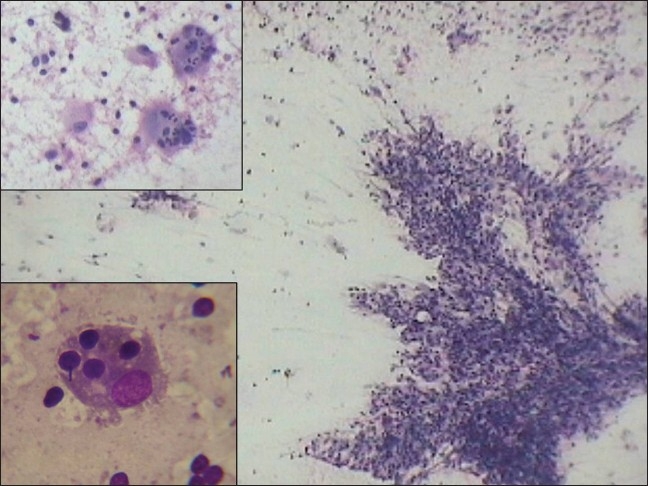
Fine needle aspiration cytology (FNAC) was performed from the thyroid as well as the lymph nodes. Smears were stained with Giemsa as well as hematoxylin and eosin and found to be richly cellular [Figure 1]. Smears from both the lesions, that is, thyroid and lymph nodes showed a similar cytomorphological picture. Microscopic examination revealed the presence of histiocytes with single to multiple nuclei, fine nuclear chromatin and inconspicuous-to-prominent nucleoli; no nuclear grooving or atypia was noted. The histiocytes had abundant pale cytoplasm containing numerous intact lymphocytes (emperipolesis) [Figure 1 lower inset], plasma cells and neutrophils [Figure 1 upper inset]. The background showed plenty of mature lymphocytes, plasma cells, and neutrophils. Based on this characteristic cytomorphology, a diagnosis of Rosai-Dorfman disease, nodal and extranodal, involving the thyroid, was made, ruling out malignancy. A subsequent histological examination of the lymph node biopsy confirmed the cytological diagnosis. The patient was treated with steroids with a good response and thus an unnecessary total thyroidectomy was prevented.
Figure 1.

Cellular smears with clusters of histiocytes over a lymphocytic background (H and E, ×100). Upper inset shows histiocytes with emperipolesis of neutrophils (H and E, ×400) and lower inset shows emperipolesis of lymphocytes (Giemsa, ×1000)
Discussion
Rosai-Dorfman disease is a rare histiocytic disorder of unknown etiology involving the lymph nodes. Extranodal RDD involving the thyroid is still rare. So far, in the literature six cases of RDD involving the thyroid have been reported.[2] We herein, report a seventh case.
Although most cases of nodal RDD occur during the first or second decade of life, with a male predominance, all the previously reported six cases of RDD involving the thyroid have occurred in elderly females with a mean age of 56.3 years.[2] Our case was a young female of 15 years.
The clinical presentation of RDD as described in the initial publications consists of painless, bilateral cervical lymphadenopathy associated with fever, leucocytosis, elevated ESR, and hypergammaglobulenemia.[1] A wide range of extranodal involvement described recently includes involvement of the eyes, respiratory tract, including nose, skin, subcutaneous tissue, bone, skeletal muscle, central nervous system, intestinal tract, salivary glands, genitourinary tract, thyroid, breast, liver, kidney, heart, uterine cervix and so on.[5]
Our case was a 15-year-old female, who presented with multiple cervical and submandibular lymphadenopathies, diffuse thyromegaly, fever, leucocytosis and elevated ESR. However, the possibility of RDD involving thyroid was not considered until FNAC was performed.
The cytological features of RDD usually reveal numerous large histiocytes with abundant pale cytoplasm and ingested lymphocytes (emperipolesis). Emperipolesis has always been described as the critical diagnostic feature of RDD. The background characteristically shows lymphocytes, plasma cells and neutrophils.[4,6,7] The histiocytes show positive immunostaining for S100 protein, CD14, CD11C, CD33 and CD68 antigens in the cytological smears.[5]
Differential diagnosis of RDD involving the thyroid include undifferentiated carcinoma, Langerhans cell histiocytosis, thyroiditis and lymphoma.[3,5] In undifferentiated thyroid carcinoma, the neoplastic cells show highly pleomorphic and hyperchromatic nuclei and neoplastic spindle cells. In the Langerhans cell histiocytosis, the Langerhans cells have grooved nuclei with eosinophils in the background. De Quervain thyroiditis should be differentiated from RDD on the basis of the presence of multinucleate giant cells, degenerating thyrocytes, inflammatory cells, macrophages, colloid and dirty background and absence of emperipolesis and lymphadenopathy. In Hodgkin lymphoma, the smears show lymphocytes, plasma cells, histiocytes, eosinophils and the characteristic Reed–Sternberg (R-S) cells. In RDD, the R-S cells and eosinophils are absent.[5,6] RDD involving the thyroid, although rare, can be diagnosed by the characteristic cytological features, obviating the need for a biopsy.
Ours is the first case of RDD involving the thyroid to be diagnosed by FNAC, to the best of our knowledge.
Footnotes
Source of Support: Nil
Conflict of Interest: None declared.
References
- 1.Das DK, Gulati A, Bhatt NC, Sethi GR. Sinus histiocytosis with massive lymphadenopathy: Report of two cases with fine needle aspiration cytology. Diagn Cytopathol. 2001;24:42–5. doi: 10.1002/1097-0339(200101)24:1<42::aid-dc1007>3.0.co;2-n. [DOI] [PubMed] [Google Scholar]
- 2.Lee FY, Jan YJ, Chou G, Wang J, Wang CC. Thyroid Involvement in Rosai-Dorfman disease. Thyroid. 2007;17:471–6. doi: 10.1089/thy.2006.0192. [DOI] [PubMed] [Google Scholar]
- 3.Mrad K, Charfi L, Dhouib R, Ghorbel I, Sassi S, Abbes I, et al. Extra-nodal Rosai-Dorfman disease: A case report with thyroid involvement. Ann Pathol. 2004;24:446–9. doi: 10.1016/s0242-6498(04)94002-3. [DOI] [PubMed] [Google Scholar]
- 4.Powell JG, Goellner JR, Nowak LE, mciver B. Rosai-Dorfman Disease of the thyroid masquerading as anaplastic carcinoma. Thyroid. 2003;13:217–21. doi: 10.1089/105072503321319549. [DOI] [PubMed] [Google Scholar]
- 5.Kumar B, Karki S, Paudyal P. Diagnosis of sinus histiocytosis with massive lymphadenopathy by fine needle aspiration cytology. Diagn Cytopathol. 2008;36:691–5. doi: 10.1002/dc.20904. [DOI] [PubMed] [Google Scholar]
- 6.Deshpande AH, Nayak S, Munshi MM. Cytology sinus histiocytosis with massive lymphadenopathy. Diagn Cytopathol. 2000;22:181–5. doi: 10.1002/(sici)1097-0339(20000301)22:3<181::aid-dc10>3.0.co;2-6. [DOI] [PubMed] [Google Scholar]
- 7.Iyer VK, Handa KK, Sharma MC. Variable extent of emperipolesis in the evolution of Rosai Dorfman disease: Diagnostic and pathogenetic implipications. J Cytol. 2009;26:111–6. doi: 10.4103/0970-9371.59398. [DOI] [PMC free article] [PubMed] [Google Scholar]


