

Abstract
Hemangioendothelioma is a rare vascular tumor of intermediate malignancy. Cytologically, it can simulate a non-vascular malignant tumor. We report two cases of this tumor, which were misdiagnosed at cytology. In the first case, a 27-year-old man presented with an anterior abdominal wall tumor. Fine needle aspiration cytology (FNAC) of the tumor showed polygonal cells with vacuolated cytoplasm in clusters having moderate nuclear atypia in a background of necrosis. A diagnosis of metastatic carcinoma was made. The histological examination showed features of epithelioid hemangioendothelioma. In the second case, a 13-year-old female child presented with unilateral enlargement of the right tonsil. At ultrasound-guided FNAC, a diagnosis of, ‘small round cell tumor, could be consistent with alveolar rhabdomyosarcoma,’ was made. The histological examination showed features of papillary intralymphatic angioendothelioma (Dabska's tumor). We conclude that epithelioid hemangioendothelioma should be considered in the differential diagnosis of metastatic carcinoma and small round cell tumor even at unusual sites.
Keywords: Carcinoma, epithelioid hemangioendothelioma, small round cell tumor
Introduction
Hemangioendotheliomas are slow growing vascular neoplasms, with intermediate malignant potential. However, the recent (2002) World Health Organisation (WHO) classification does not strictly define these lesions as having intermediate behavior, but instead describes them as lesions that fall into the category of locally aggressive tumors and those with metastatic potential.[1] Reports of aspiration cytology of these unusual vascular tumors are very few in literature.
The cytology of vascular tumors can simulate non-vascular malignant tumors. The diagnostic difficulty is compounded when the presentation occurs at unusual sites.
We describe the cytological features of epithelioid hemangioendothelioma of soft tissue, and papillary intralymphatic angioendothelioma in two young patients at unusual sites.
Case Reports
Case 1
A 27-year-old male presented with an eight-month history of painful anterior abdominal wall swelling. Physical examination showed a superficial tumor, measuring 4 × 3 × 2 cm in size with ulceration. FNAC was performed.
Cytological findings
The smears were moderately cellular. The cells were arranged singly and in clusters, were polygonal in shape, with moderate amount of vacuolated cytoplasm and enlarged hyperchromatic nuclei. Few cells were multinucleated. Intranuclear inclusions were noted in some cells [Figure 1a]. There was a myxoid stromal component in a necrotic background, with abundant neutrophils. There were no clear-cut intracytoplasmic lumina within the erythrocytes. A provisional diagnosis of poorly differentiated carcinoma, possibly metastatic, was made, and a biopsy was advised.
Figure 1.
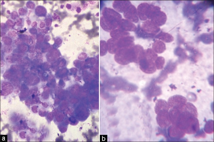
Epithelioid hemangioendothelioma. (a) Cytology shows a cluster of polygonal cells with moderate atypia (MGG ×400). Papillary intralymphatic angioendothelioma. (b) Cytology shows small round cells in an acini-like pattern (MGG ×400)
Histopathological findings
The biopsy showed a tumor composed of round to spindle-shaped cells in solid nests. The cells were embedded in a light blue to deep pink myxohyaline matrix. The cells were seen extending centrifugally from large vessels into the soft tissue. The tumor had a blistered, vacuolated appearance due to the intracytoplasmic lumina, many of which contained erythrocytes. The cells had moderate-to-abundant cytoplasm, almost uniform with a vesicular nucleus and prominent nucleoli in some. There was no increase in mitotic activity.
Immunohistochemistry
CD 34 was positive revealing the endothelial nature of the cells.
A diagnosis of epithelioid hemangioendothelioma was made.
Case 2
A 13-year-old girl presented to the Ear, Nose and Throat (ENT) outpatient department (OPD) with unilateral enlargement of the right tonsil. A computed tomography (CT) scan showed a vascular mass around the carotid artery extending up to the base of the skull. Some degree of bone destruction was also seen at the base of the skull. Ultrasonography (USG) guided FNAC was done.
Cytological findings
The smears were moderately cellular and diluted with blood. The cells were small and round, with scanty cytoplasm. They were arranged in small clusters as well as singly. Occasional acini-like structures were also seen [Figure 1b]. The chromatin was finely granular. A provisional diagnosis of a ‘small round cell tumor, could be consistent with alveolar rhabdomyosarcoma,’ was made.
Histopathological findings
An effort was made to remove the tumor. Sections showed a tumor mass composed of intra-vascular papillary structures lined by hobnail endothelial cells. Some glomeruloid bodies were also noted. The cells had scanty eosinophilic cytoplasm with a prominent nucleus and little or no cytological atypia. Mitotic figures were rare.
Immunohistochemistry
CD 34 was positive revealing the endothelial nature of the cells.
A diagnosis of papillary intralymphatic angioendothelioma was made.
Discussion
Epithelioid hemangioendothelioma of soft tissue is most often a solitary lesion, in either the superficial or deep tissue. It occurs mainly in mid-adult life, affecting patients of either sex equally.[1]
Papillary intralymphatic angioendothelioma (Dabska's tumor) was first described in infants and children, but the age of the patients ranges from birth to 83 years and there is no sex predilection. Besides skin and subcutaneous tissue the tumor also occurs in other deep locations.[2]
Histopathological examination remains the mainstay of diagnosis for this rare tumor. Currently there is an increasing use of FNAC in the diagnosis of visceral organs and soft tissue masses. Hence, cytopathologists should be aware of the cytological picture of these unusual tumors.
Case 1, on review, depicts most of the cytological features of an Epithelioid hemangioendothelioma. As described in earlier reports, we observed the tumor cells in clusters and singly. The cells had vacuolated cytoplasm with a background of myxoid material as observed by the other workers.[3,4] In angiosarcoma, the aspirates are varied in cellularity and the degree of nuclear atypia. However, the vasoformative features are more remarkable. Thus, there are microacinar formations, vascular channels, prominent intracytoplasmic eosinophilic inclusions and a bloody background. There can be numerous single cells and a presence of spindle cells. The myxoid stromal component has not been reported in angiosarcoma.[5,6]
Occasional cells showed intranuclear inclusion as described by Gupta et al.[7] However, intracytoplasmic lumina with erythrocytes seen on histology were rarely seen in cytology.[7] Cell blocks and immunocytochemistry could be helpful.
Although epithelioid hemangioendothelioma is capable of producing regional and distant metastasis, it does so far less frequently than the conventional angiosarcoma. In a recent series by Andrea et al. the rate of metastases in epithelioid hemangioendothelioma was found to be 22%.[8]
The majority of epithelioid hemangioendotheliomas have innocuous histological features and have a good clinical course. Atypical hemangioendothelioma has cellular atypia, mitotic activity (>1 mitosis/10 HPF), necrosis, extensive spindling or solid areas of overt angiosarcoma. These have a more aggressive course, with a higher rate of metastasis.[8]
Dabska's type hemangioendothelioma was first described in 1969, in a small series of six patients. All occurred in the skin or subcutis of infants and young children. Dabska's Tumor or Endovascular papillary angioendotheliomas are rare tumors, which lack extensive studies. These usually occur in distal extremities of children, but other deep locations like the spleen, soft tissues, bone, tongue, and neck region may also be affected.[9]
The tumors are composed of dilated vascular spaces with prominent intraluminal papillary tufts with hyaline cores lined by hobnail endothelial cells. There is no or minimal cytological atypia.[1] Generally, it presents as a slow growing, painless, usually intradermal nodule. In our case the tumor presented as a subtonsillar mass. This presentation has not been described in previous studies.
We have found no reports of cytological description of this tumor. In deep sites, FNAC of these lesions can cause confusion with malignant small round cell tumors. However, careful attention to the bland chromatin pattern can be helpful. The occasional acini-like pattern observed in the smears is possibly an attempt at vessel formation. Additionally there is a low cellular yield and a hemorrhagic background.
Papillary intralymphatic angioendothelioma, although believed to have a favourable prognosis can be locally invasive with a potential to metastasise.[10] Therefore, further longitudinal studies are required in this regard.
Our cases highlight the difficulty in diagnosing hemangioendothelioma on FNAC, the importance of considering this in the differential diagnosis and the need for more studies on the cytomorphological presentation of these tumors.
Footnotes
Source of Support: Nil
Conflict of Interest: None declared.
References
- 1.Fletcher DM, Unni KK, Mortens F. WHO-Pathology and genetics of tumours of soft tissue and bone. Lyon (France): IARC Press; 2002. [Google Scholar]
- 2.Long XD, Qu DY, Huang YZ, Lu YM. Endovascular papillary angioendothelioma in soft tissue of the gluteal region. [Last accessed on 2011 Jan 22];Internet J Pathol. 2008 7 [about 2p] Available from: http://www.ispub.com/journal/the_internet_journal_of_pathology/volume_7_number_2_15/article/endovascular_papillary_angioendothelioma_in_soft_tissue_of_the_gluteal_region.html . [Google Scholar]
- 3.Evans HL, Raymond AK, Ayala AG. Vascular tumours of bone: A study of 17 cases other than ordinary hemangioma with an evaluation of the relationship of haemangioendothelioma of bone to epithelioid hemangioma, epithelioid haemangioendothelioma and high grade angiosarcoma. Hum Pathol. 2003;34:680–9. doi: 10.1016/s0046-8177(03)00249-1. [DOI] [PubMed] [Google Scholar]
- 4.Hristova EN, Krisnamurthy S, Ro JY, Ayala AG. Pulmonary epithelioid haemangioendothelioma with prominent signet ring cell features mimicking metastatic adenocarcinoma. Ann Diagn Pathol. 2003;7:160–4. doi: 10.1016/s1092-9134(03)00014-5. [DOI] [PubMed] [Google Scholar]
- 5.Boucher LD, Swanson PE, Stanley MW, Silverman JF, Raab SS, Geisinger KR. Cytology of angiosarcoma.Findings in 14 fine needle aspiration biopsy specimens and one pleural fluid specimen. Am J Clin Pathol. 2000;114:210–9. doi: 10.1309/PXMU-LF05-3894-W29F. [DOI] [PubMed] [Google Scholar]
- 6.Pai MR, Upadhyaya K, Naik R, Malhotra S. Bilateral angiosarcoma breast diagnosed by fine needle aspiration cytology. Indian J Pathol Microbiol. 2008;51:421–3. doi: 10.4103/0377-4929.42549. [DOI] [PubMed] [Google Scholar]
- 7.Gupta R, Mathur SR, Gupta SD, Durgapal P, Iyer VK, Das CJ, et al. Hepatic epithelioid haemangioendothelioma: A diagnostic pitfall in aspiration cytology. Cytojournal. 2009;6:25. doi: 10.4103/1742-6413.58951. [DOI] [PMC free article] [PubMed] [Google Scholar]
- 8.Deyrup AT, Tighiouart M, Montag AG, Weiss SW. Epithelioid haemangioendothelioma of soft tissue: A proposal for risk stratification based on 49 cases. Am J Surg Pathol. 2008;32:924–7. doi: 10.1097/pas.0b013e31815bf8e6. [DOI] [PubMed] [Google Scholar]
- 9.Weiss SW, Goldblum JR. Enzinger and Weiss's soft tissue tumours. 5th ed. Philadelphia: Mosby Elsevier; 2008. [Google Scholar]
- 10.Bhatia A, Nada R, Kumar Y, Menon P. Dabska tumour (Endovascular papillary angioendothelioma) of testis: A case report with brief review of literature. Diagn Pathol. 2006;1:12. doi: 10.1186/1746-1596-1-12. [DOI] [PMC free article] [PubMed] [Google Scholar]