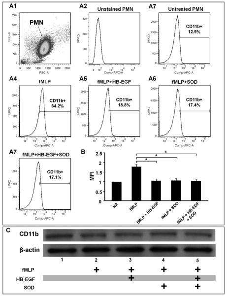
Figure 6.
HB-EGF inhibits CD11b mobilization in activated human neutrophils. Freshly isolated human neutrophils were pretreated with either HB-EGF (100 ng/ml), SOD (1000 u/ml), or HB-EGF + SOD for 1 h and then activated by addition of fMLP (10−7 mol/L) for 30 min (shown), 1 h or 4 h (data not shown). PMN were then incubated with APC conjugated anti-human CD11b antibodies. Stained neutrophils were examined by flow cytometry and were gated for viable cells by forward and side scatter criteria. APC positive PMN and the mean fluorescent intensity (MFI) of the positive PMN are shown on the flow cytometric histograms. A1) Dot-plot of forward scatter (FSC) versus side angle light scatter (SSC) showing a main cell population of freshly isolated human PMN. 10,000 cells were analyzed, and the gated PMN comprised >95% of the total cells. A2-7 represent flow cytometric histograms of CD11b expression on the surface of: A2, unstained PMN indicating background fluorescence; A3) untreated PMN; A4) PMN treated with fMLP;.A5) PMN treated with HB-EGF + fMLP; A6) PMN treated with SOD. + fMLP; A7) PMN treated with HB-EGF + SOD + fMLP. B) Quantification of CD11b cell membrane expression. Results derived from histogram analyses are expressed as the mean fluorescence intensity (MFI; mean ± SEM). *p< 0.05, Student’s t-test. C) Immunoblot analysis of CD11b expression in total cell extracts 30 min after fMLP addition. Data are representative of three independent experiments.

