Abstract
Rift Valley fever virus (RVFV), which causes hemorrhagic fever, neurological disorders or blindness in humans, and a high rate abortion and fetal malformation in ruminants1, has been classified as a HHS/USDA overlap select agent and a risk group 3 pathogen. It belongs to the genus Phlebovirus in the family Bunyaviridae and is one of the most virulent members of this family. Several reverse genetics systems for the RVFV MP-12 vaccine strain2,3 as well as wild-type RVFV strains 4-6, including ZH548 and ZH501, have been developed since 2006. The MP-12 strain (which is a risk group 2 pathogen and a non-select agent) is highly attenuated by several mutations in its M- and L-segments, but still carries virulent S-segment RNA3, which encodes a functional virulence factor, NSs. The rMP12-C13type (C13type) carrying 69% in-frame deletion of NSs ORF lacks all the known NSs functions, while it replicates as efficient as does MP-12 in VeroE6 cells lacking type-I IFN. NSs induces a shut-off of host transcription including interferon (IFN)-beta mRNA7,8 and promotes degradation of double-stranded RNA-dependent protein kinase (PKR) at the post-translational level.9,10 IFN-beta is transcriptionally upregulated by interferon regulatory factor 3 (IRF-3), NF-kB and activator protein-1 (AP-1), and the binding of IFN-beta to IFN-alpha/beta receptor (IFNAR) stimulates the transcription of IFN-alpha genes or other interferon stimulated genes (ISGs)11, which induces host antiviral activities, whereas host transcription suppression including IFN-beta gene by NSs prevents the gene upregulations of those ISGs in response to viral replication although IRF-3, NF-kB and activator protein-1 (AP-1) can be activated by RVFV7. . Thus, NSs is an excellent target to further attenuate MP-12, and to enhance host innate immune responses by abolishing the IFN-beta suppression function. Here, we describe a protocol for generating a recombinant MP-12 encoding mutated NSs, and provide an example of a screening method to identify NSs mutants lacking the function to suppress IFN-beta mRNA synthesis. In addition to its essential role in innate immunity, type-I IFN is important for the maturation of dendritic cells and the induction of an adaptive immune response12-14. Thus, NSs mutants inducing type-I IFN are further attenuated, but at the same time are more efficient at stimulating host immune responses than wild-type MP-12, which makes them ideal candidates for vaccination approaches.
Protocol
1. Recovery of recombinant MP-12 encoding NSs mutation(s) from plasmid DNAs2
Spread baby hamster kidney (BHK)/T7-9 cells15, which stably express T7 RNA polymerase, into 6-cm dishes in Minimum Essential Medium (MEM)-alpha (Invitrogen, Cat# 32561037) containing 10% fetal bovine serum (FBS), Penicillin-Streptomycin (Penicillin:100 U/ml, Streptomycin: 100 μg/ml) (Invitrogen, Cat#15140122), and 600 μg/ml of hygromycin B (Cellgro, Cat#30-240-CR). * The efficiency of viral recovery is higher in 6-cm dishes than in 35-mm dishes. BHK/T7-9 cells with low passage level support higher rates of recovery. Alternatively, other BHK cell lines that stably express T7 RNA polymerase could be used4,5,16,17.
When cells have reached 70-80% confluency, replace the culture supernatant with fresh MEM-alpha containing 10% FBS and Penicillin-Streptomycin (not containing hygromycin B). * Cells should be transfected within 1 hour after replacing the medium to avoid the loss of T7 RNA polymerase expression.
- For the recovery of RVFV, a set of plasmids encoding viral genomic RNAs for full-length viral RNA expression, and a second set encoding viral gene open reading frames for viral protein expression (Figures 1 and 2) are required. Prepare a mixture of the following plasmids2 (Figure 2) in a 1.5 ml tube:
- pProT7-S(+) with mutation(s) in the NSs gene (2 mg): This plasmid encodes the anti-viral-sense (positive-sense) full-length RVFV MP-12 S-segment flanked by the T7 promoter and the hepatitis delta virus (HDV) ribozyme sequence.
- pProT7-M(+) (2 mg): This plasmid encodes the anti-viral-sense (positive-sense) full-length RVFV MP-12 M-segment flanked by the T7 promoter and the HDV ribozyme sequence.
- pProT7-L(+) (2 mg): This plasmid encodes anti-viral-sense (positive-sense) full-length RVFV MP-12 L-segment flanked by the T7 promoter and the HDV ribozyme sequence.
- pT7-IRES-vN (2 mg): This plasmid encodes the RVFV MP-12 N open reading frame (ORF) downstream of the T7 promoter and an encephalomyocarditis virus (EMCV) internal ribosome entry site (IRES).
- pT7-IRES-vL (1 mg): This plasmid encodes the RVFV MP-12 L ORF downstream of the T7 promoter and an EMCV IRES.
- pCAGGS-vG (1 mg): This plasmid encodes RVFV MP-12 M ORF downstream of the chicken beta-actin promoter. * The addition of pT7-IRES-vN, pT7-IRES-vL and pCAGGS-vG is not essential for the recovery of MP-12, but it enhances the efficiency of rescue2. Authors experienced poor expression of Gn/Gc by using pT7-IRES plasmid, probably due to the lack of leaky scanning of AUGs by ribosomes. Therefore, we constructed pCAGGS-vG for cap-dependent Gn/Gc expression.
Add 30 ml of TransIT-LT1 (Mirus, Cat#MIR2300) to 385 ml of Opti-MEM (Invitrogen, Cat# 31985070) in a 1.5 ml tube, and vortex briefly.
After 5 min incubation at room temperature, slowly add the Opti-MEM containing liposomes to the plasmid mixture from step 1.3 to, mix gently by pipetting and incubate for 15 min at room temperature.
Add the mixture of liposome and plasmids to the culture medium of the BHK/T7-9 cells from step 1.2 drop by drop (Figure 3).
Incubate the transfected cells at 37°C in an incubator with 5% CO2 for 24 h, and replace the culture supernatant with fresh MEM-alpha containing 10% FBS and Penicillin-Streptomycin (not containing hygromycin B).
Incubate the cells at 37°C in an incubator with 5% CO2 for 4 additional days (incubate for 5 days total), and collect the culture supernatants into a 15 ml tube. * The cytopathic effect (CPE) observed here does not necessarily reflect the result of successful viral recovery, because the transfection induces cell death which appears similar to CPE caused by viral RNA replication or viral protein synthesis.
Centrifuge the supernatants at 2,200 xg at 4°C for 5 min. * The purpose of this step is to pellet down the cellular debris from viral stock. An aerosol-tight centrifuge bucket is recommended for increased safety.
Transfer the supernatants into screw-cap 5 ml cryotubes, and store the passage 0 (P0) virus stock at -80°C for further use.
2. Amplification of P0 virus
The P0 samples often contain insufficient viral titer for downstream experiments2. An amplification step in VeroE6 cells, which is a clone of African green monkey kidney (Vero) cells lacking the IFN-alpha/beta genes18,19, increases viral titer up to maximum level. Alternatively, other cells lacking type-I IFN responses such as Hec1B cells20 or MEF cells from IFNAR1-knockout mice21 might be used for this step. Spread VeroE6 cells into 10-cm dishes in Dulbecco’s modified minimum essential medium (DMEM) (Invitrogen, Cat# 11965092) containing 10% FBS, Penicillin-Streptomycin (Penicillin:100 U/ml, Streptomycin: 100 mg/ml), and incubate at 37°C in an incubator with 5% CO2 until they reach 80% confluency. * Recombinant MP-12 strains encoding mutant NSs often fail to replicate efficiently in type-I IFN-competent cells.
Mix 300 ml of P0 samples with 2.7 ml of DMEM with 10% FBS and Penicillin-Streptomycin. Remove culture medium from the VeroE6 cells from step 2.1 and replace with the diluted P0 sample. Incubate at 37°C for 1 h in an incubator with 5% CO2.
Remove the inocula and add 10 ml of DMEM with 10% FBS and Penicillin-Streptomycin to each dish.
Incubate at 37°C for 3 to 4 days until CPE of VeroE6 cells becomes apparent. * Disruption of monolayer occurs during MP-12 infection, while recombinant MP-12 lacking NSs, such as rMP12-C13type (C13type) (Figure 4), does not disrupt the monolayer, but a number of dead floating cells appear 2 to 3 days after infection.
Harvest the supernatant at 3 to 4 dpi, as described in sections 1.9) and 1.10), and designate the samples as E6P1.
3. Titration of recombinant MP-12 by plaque assay
Spread VeroE6 cells into 6-well plates. * Duplicate analysis per sample is more reliable than single analysis.
- When VeroE6 cells have grown to 80% confluency, prepare 10-fold serial dilutions of virus samples in DMEM with 10% FBS and Penicillin-Streptomycin up to 10-6 as follows:
- 10 μl of E6P1 sample + 990 ml of DMEM with 10% FBS and Penicillin-Streptomycin (10-2 dilution)
- 100 μl of 10-2 sample + 900 ml of DMEM with 10% FBS and Penicillin-Streptomycin (10-3 dilution)
- 100 μl of 10-3 sample + 900 ml of DMEM with 10% FBS and Penicillin-Streptomycin (10-4 dilution)
- 100 μl of 10-4 sample + 900 ml of DMEM with 10% FBS and Penicillin-Streptomycin (10-5 dilution)
Aspirate medium from the 6-well plate from step 3.1 and add 400 μl of each dilution (from step 3.2) into the wells (Figure 3).
Incubate at 37°C for 1 h in an incubator with 5% CO2.
During the incubation, prepare two 15 ml tubes for the agar-overlay as follows: Tube A (keep in 42°C water bath): 7 ml of 1.2% noble agar (VWR, Cat#101170-362) in water Tube B (keep in 37°C water bath): 7 ml of Modified Eagle Medium (MEM 2x) (Invitrogen, Cat# 11935046) containing 10% FBS, Penicillin-Streptomycin (Penicillin: 100 U/ml, Streptomycin: 100 μg/ml), and 10% Tryptose phosphate broth (MP biomedicals, Cat#1682149).
After 1 h incubation, remove the viral inocula, and immediately add 2 ml per well of a 1:1 mixture of tube A and tube B (from step 3.5). * Take care to add the overlay immediately as drying up of wells causes the death of uninfected cells.
Incubate the plates at 37°C for 3 days in an incubator with 5% CO2.
Prepare tube A and tube B again as described in step 3.5. Prepare also 500 μl of 0.33% neutral red solution (Sigma Aldrich, Cat#N2889-100ML) per plate which is also kept at 37°C water bath.
Mix tube A, tube B and 500 ml (final conc. 0.011%) of neutral red solution and add 2 ml of mixture per well. * The amount of neutral red solution to be added varies by the lot of neutral red solution, and initial optimization is required. Long-term storage of 0.33% neutral red solution causes precipitation. In such cases, the precipitate can be completely dissolved by incubation at 55°C for 10 min followed by vigorous shaking. The use of precipitated neutral red solution results in weak staining of cells, while re-dissolved neutral red solution stains cells well.
Incubate the plate for 16 h (or overnight) at 37°C in an incubator with 5% CO2.
Count the number of plaques in the well which contains 10 to 100 plaques per well. Calculate the number of plaque forming units/ml. For example, if we observe 28 plaques in the wells inoculated with the 10-5 dilutions, 28 (# of plaques) x (1 ml/0.4 ml) x 105 (dilution) = 7.0 x 10⁶ plaque forming units (pfu)/ml (Figures 5).
4. Screening of NSs mutants lacking the type-I IFN suppression function
Spread C57/WT MEF cells (InvivoGen, Cat#mef-c57wt), which encode a secreted embryonic alkaline phosphatase (SEAP) gene inducible by NF-kB and IRF-3/7 (Figure 6), into 12-well plates. The cells are maintained in DMEM with 10% FBS, Penicillin-Streptomycin (Penicillin: 100 U/ml, Streptomycin: 100 μg/ml), Blasticidin S (3 mg/ml), and Zeocin (100 μg/ml).
When cells become sub-confluent (80%), cells are mock-infected or infected with MP-12 or recombinant MP-12 encoding NSs mutations at multiplicity of infection (moi) of 3 or 0.1 (see sections 2 and 3, amount of each inoculum should be 300 μl). At 1 h post infection, remove the inocula, and add 1 ml per well of DMEM with 10% FBS, Penicillin-Streptomycin (Penicillin: 100 U/ml, Streptomycin: 100 μg/ml) (Blasticidin and Zeocin are not added at this time).
At 14 h post infection, collect culture supernatants. Add 200 μl of QUANTI-Blue (InvivoGen, Cat # rep-qb1) and 50 μl of each sample (in triplicate) to wells of a 96-well plate. Seal and incubate the plate at 37°C for 1 h. * Both MP-12 and recombinant MP-12 lacking NSs clearly induce host translational suppression in IFN-alpha/beta competent cells including 293 cells, MRC-5 cells and mouse embryonic fibroblast (MEF) cells after 14 hours post infection at high moi. On the other hand, IFN-beta mRNA or ISG56 mRNA accumulates abundantly at 7 to 8 hours post infection in type-I IFN competent cells. Thus, we chose 14 hours post infection to collect the supernatants to see the accumulation of SEAP induced by innate immune responses.
Read the OD values at 650 nm using a plate reader (Figure 7). * The results are consistent with the data obtained by Northern blot using an RNA probe specific to mouse ISG56 mRNA, which shows up-regulation of ISG56 mRNA in the absence of NSs expression (Figure 8). * It should be noted that the SEAP activity is determined by the abundance of proteins which could be affected by host translation activity. A relative level of SEAP might not be high compared to the increased level of mRNA because SEAP cannot be synthesized even in the presence of SEAP mRNA if cellular translation is suppressed. Northern blot is a more straightforward assay and a more accurate way to evaluate the amount of mRNA induced by the lack of NSs functions in infected cells than the SEAP reporter assay. However, the SEAP reporter system is more rapid than Northern blot and hence useful for rapid screening of NSs mutants potentially lacking host transcription suppression function.
5. Representative Results:
The reverse genetics system consistently generated viable recombinant MP-12 viruses with titers higher than 1 x 10⁶ pfu/ml. C13type virus lacking NSs functions formed large turbid plaques, while MP-12 formed clear plaques of various sizes2 (Figure 5). Mock-infected C57/WT MEF cells or those infected with MP-12 did not increase the level of SEAP in culture supernatant compared to mock-infected cells, while the culture supernatant of C57/WT MEF cells infected with C13type contained an increased level of SEAP by 14 hours post infection (hpi) (Figure 7). These results are consistent with those obtained by Northern blot using an RNA probe specific to mouse ISG56 mRNA (Figure 8).
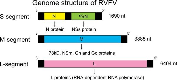 Figure 1.
Genome structure of RVFV
RVFV has a tripartite negative-sense or ambisense RNA genome named S-, M-, and L-segment. S-segment encodes N and NSs genes in an ambisense manner. N mRNA is synthesized from viral-sense (negative-sense) S-segment, while NSs mRNA is synthesized from anti-viral-sense (positive-sense) S-segment. M-segment encodes a single M mRNA and synthesizes the78kD, NSm, Gn or Gc proteins by leaky scanning of several AUGs at 5'region of M mRNA, followed by their co-translational cleavage22,23. L-segment encodes the L protein. Both N and L proteins are essential for viral transcription and replication, while Gn and Gc are the viral envelope proteins. NSs and NSm proteins are nonstructural proteins, which are not incorporated into virus particles.
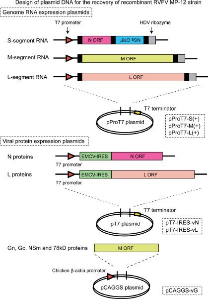 Figure 2.
. Design of plasmid DNA for the recovery of recombinant RVFV MP-12 strain
The cDNA encoding full-length anti-viral-sense S-, M-, or L-segment are cloneddownstream of T7 promoter and upstream of hepatitis delta virus (HDV) ribozyme sequences, designated as pProT7-S(+), pProT7-M(+), or pProT7-L(+), respectively2. T7 RNA polymerase expressed in BHK/T7-9 cells transcribes the RNA encoding full-length S-, M-, or L-segment with precise genome 3’ end. The open reading frame (ORF) of N or L proteins are cloned under encephalomyocarditis virus (EMCV) internal ribosome entry site (IRES), which are designated as pT7-IRES-vN or pT7-IRES-vL, respectively, to allow the uncapped T7 RNA transcript to be recognized by ribosomes in cap-independent manner. The M ORF is cloned under chicken b-actin promoter of pCAGGS plasmid24, which is designated as pCAGGS-vG, to allow the synthesis of 78kD, NSm, Gn and Gc proteins, which are generated from different AUGs by leaky scanning23. Both N and L proteins are required for initiating transcription or RNA replication, while the pT7-IRES-vN and pT7-IRES-vL are not essential for the recovery of recombinant MP-122, probably due to the presumable leaky expression of Pol-II-driven capped RNA transcripts encoding N-ORF and L-ORF from pProT7-S(+) and pProT7-L(+), respectively.
 Figure 3.
Recovery of RVFV MP-12 from plasmid DNA
Transfection of BHK/T7-9 cells with pProT7-S(+), pProT7-M(+), pProT7-L(+), pT7-IRES-vN, pT7-IRES-vL and pCAGGS-vG plasmids (Fig.1) generates infectious recombinant RVFV MP-12 strain in culture supernatants. The supernatant at 5 day post transfection is collected, and passaged into fresh Vero E6 cells for viral amplification. Typically, more than 1 x 10⁶ pfu/ml of virus can be recovered at 3 to 4 days post infection. The amplified virus (E6P1 virus) is titrated by using plaque assay with Vero E6 cells and used for phenotype analysis and immunogenicity studies.
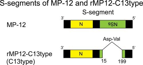 Figure 4.
S-segment of MP-12 and rMP12-C13type
The comparison of MP-12 and rMP12-C13type (C13type) S-segments. Compared to NSs of MP-12 strain, the NSs ORF of C13type is truncated by 69%, and is identical to that of naturally isolated clone 13 strain2,25.
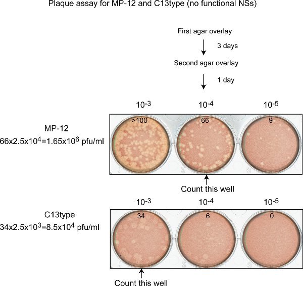 Figure 5.
Plaque assay for MP-12 (functional NSs) and C13type (non-functional NSs)
The monolayer of Vero E6 cells in a 6-well plate is used for plaque assay. After 3 days incubation with 0.6% agar overlay, the second agar overlay containing neutral red solution is added. Then, plaques are counted at day 4 post infection. MP-12 forms clear plaques of varied sizes, while C13type forms large turbid plaques (or foci). The well with 10 to 100 plaques should be used for counting.
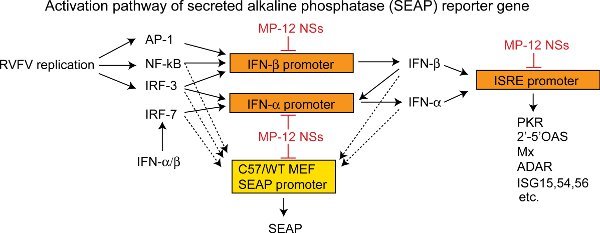 Figure 6. Activation pathway of secreted alkaline phosphatase (SEAP) reporter gene in C57/WT MEF cells
Interferon-beta promoter includes the binding sequences of AP-1, NF-kB and IRF-3. RVFV replication activates AP-1, NF-kB and IRF-37,11. However, NSs inhibit the release of repressor complex from the IFN-beta promoter even after the binding of those transcription factors, thus suppressing the synthesis of IFN-beta mRNA26. Furthermore, NSs sequesters TFIIH p44 subunits8 and also promotes degradation of TFIIH p62 subunits27, thus inducing a general host transcription suppression including IFN-alpha gene and genes under the ISRE promoter. C13type or other NSs mutants lacking IFN-beta suppression function induce IFN-beta synthesis, which in turn activates the IFN-alpha promoter and the interferon-sensitive response element (ISRE) promoter. IRF-7 is then transcriptionally upregulated by IFN-alpha/beta stimulation and further upregulated by IFN-alpha in support of IRF-328,29. In this assay, C57/WT MEF cells encode secreted alkaline phosphatase (SEAP) at the downstream of an artificial binding sequence of NF-kB, IRF-3 and IRF-7. Thus, the NSs mutants lacking IFN-beta suppression function upregulate SEAP whose secretion is then measured.
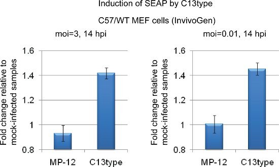 Figure 7.
Induction of SEAP by C13type
C57/WT MEF cells (InvivoGen) were mock-infected or infected with MP-12 or rMP12-C13type (C13type) at moi of 3 (left panel) or 0.01 (right panel). Culture supernatants (50 μl) at 14 hpi were mixed with 200 μl of QUANTI-Blue (InvivoGen) substrate in 96 well plate and the OD values at 650 nm were measured after 1 h incubation at 37°C by a plate reader. The relative increases of SEAP to mock-infected cells are shown. The data represent the mean +/- standard deviation of three independent experiments. The culture supernatant from C13type-infected cells shows increased SEAP, suggesting the lack of host transcription suppression by NSs.
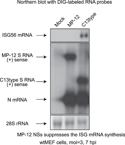 Figure 8. Northern blot with DIG-labeled RNA probe
Wild-type mouse embryonic fibroblast (MEF) cells were mock-infected or infected with MP-12 or rMP12-C13type (C13type) at moi of 3. Total RNA was collected at 7 hpi by using Trizol (Invitrogen), and Northern blot was performed by using digoxigenin-labeled RNA probe specific to mouse endogenous ISG56 mRNA or MP-12 N mRNA/anti-viral-sense S-segment2,30. For making the probes for mouse endogenous ISG56 mRNA or RVFV N mRNA/anti-viral-sense S-segment, the PCR fragments amplified by the primer set of KpnmISG56F (GGG TGG TAC CGC TCC ACT TTC AGA GCC TTC GCA AAG CAG) and HindmISG56 (TAC AAA GCT TAT GGG AGA GAA TGC TGA TGG TGA CCA GG) for ISG56 mRNA or KpnNF (AGT TGG TAC CAT GGA CAA CTA TCA AGA GCT TGC G) and HindNR (GGG CAA GCT TTT AGG CTG CTG TCT TGT AAG) for RVFV N mRNA/anti-viral-sense S-segment were digested with KpnI and HindIII, and ligated into pSPT18 plasmid (Roche. Then RNA probes labeled with digoxigenin were synthesized by using DIG RNA Labeling Kit (SP6/T7) (Roche, Cat#1 175 025). The 28S rRNA level of each sample is also shown as loading control. Wild-type MEF cells infected with C13type induced ISG56 mRNA synthesis, while those infected with MP-12 did not induce it, suggesting the lack of host transcription suppression in C13type-infected cells. The data is consistent with those obtained by SEAP assay in Figure 6.
Discussion
Reverse genetics systems for RVFV have been developed by several groups by utilizing T7 promoter2,4,5 or mouse3 or human4 pol-I promoter. In this manuscript, we describe a protocol to generate recombinant RVFV MP-12 strains by using BHK/T7-9 cells15 that stably express T7 RNA polymerase. The efficiency of viral recovery varied depending on the condition of BHK/T7-9 cells, the amount of plasmids, the number of transfected cells and so on. We always amplify the P0 virus in Vero E6 cells to obtain high titer virus stocks for experiments. Type-I IFN-competent cells such as human lung diploid (MRC-5) cells could be used for viral amplification only when the NSs function supported an efficient viral replication by inhibiting antiviral activities including type-I IFN or PKR.
A plaque assay is a quite useful method for titrating MP-12 and the NSs mutants. As a general precaution for the plaque assay, cells should be immediately added with overlay at the appropriate temperature after the removal of viral inocula. Crystal violet staining is not suitable for detecting C13type virus plaques, because C13type virus does not disrupt the monolayer of Vero E6 cells2. On the other hand, neutral red staining could successfully detect plaques formed by C13type virus. The strength of cellular staining with neutral red is determined by the relative incorporation of neutral red dye into live cells. Thus, it is likely that C13type-infected cells have a reduced metabolic activity which leads to decreased incorporation of neutral red dye compared to that of uninfected cells, and which forms a weak staining focus. The required amount of neutral red varies by the lot of neutral red solution. Long-term storage of neutral red solution often generates precipitates. This could be prevented by heat-dissolving of neutral red solution at 55°C for 10 min; however, it affects the staining of cells.
It is important to use viruses with accurate viral titer in experiments. The titer in a viral stock could be decreased by media with pH below 6.531 which is indicated by yellowish color of phenol red in the viral stock, long-term storage, repeated freeze-thaw cycles and so on. Thus, it is important to use fresh viral stock for animal vaccination or titrate the stock again just before experiment.
RVFV encode two characterized nonstructural proteins; NSs and NSm. The RVFV lacking NSs is significantly attenuated in animals25,32, while the RVFV lacking NSm is still highly virulent in rats33. Thus, NSs plays a major role as virulence factor in the pathogenesis. NSs is a multifunctional protein which induces 1) suppression of host general transcription8, 2) suppression of IFN-beta mRNA synthesis26 and 3) degradation of dsRNA-dependent protein kinase (PKR)9,10. The parental MP-12 strain could be further attenuated by introducing truncation or mutations into NSs. On the other hand, it is also important to maintain the immunogenicity of such mutant MP-12. Type-I IFN plays a role for the maturation of dendritic cells, which is important for the differentiations of T cells and B cells12-14. Various NSs mutants lacking IFN-beta suppression function, which maintain full or partial NSs functions, are useful to analyze the impact of the NSs-mediated IFN-beta suppression function on the immunogenicity and safety of vaccine candidates. It is known that cells infected with RVFV wt ZH548 strain activate NF-kB, IRF-3, and AP-1 (Figure 6), while IFN-beta mRNA synthesis is suppressed by NSs at transcriptional level7. Thus, MP-12, which expresses fully functional NSs3, inhibits the synthesis of NF-kB/IRF-3/7-inducible SEAP reporter gene mRNA at transcriptional level, while recombinant MP-12 lacking NSs functions such as C13type could induce the SEAP mRNA synthesis (Figure 7). To correctly understand the result of immunogenicity of such mutants, the phenotypes should be further characterized in cell culture system. For example, the viral replication kinetics should be tested in type-I IFN competent cells such as human lung diploid MRC-5 cells or mouse embryonic fibroblast (MEF) cells. The PKR degradation function can be tested by Western blot with antibodies specific to human or mouse PKR. IFN-beta mRNA induction by NSs mutation should be confirmed by Northern blot with digoxigenin-labeled RNA probe specific to human or mouse IFN-beta mRNA. Host general transcription suppression can be tested by analyzing the incorporation of a uridine analog into nascent mRNA. Eventually, the immunogenicity and safety of candidate MP-12 NSs mutants should be tested in mice. Typically, 1 x 10⁵ pfu of the candidate vaccine diluted in PBS is given subcutaneously and sera collected from the retro-orbital sinus at day 30 (or 42) and day 180 are tested for the presence of neutralizing antibodies by plaque reduction neutralizing test (PRNT80) and total IgG against RVFV N and Gn/Gc by IgG ELISA coated with purified recombinant viral proteins. The efficacy of vaccine candidates can be tested by challenging mice at 42 days post immunization with wild-type ZH501 (e.g.; i.p., 1 x 10³ pfu) at animal biosafety level (BSL)4 facility or enhanced BSL3+ facility in the U.S.A. The reverse genetics approach is a powerful tool to design and generate safe and immunogenic live-attenuated RVFV vaccines.
Disclosures
We have nothing to disclose.
Acknowledgments
This work was funded by Grant Number 5 U54 AI057156-07 through Western Regional Center of Excellence (WRCE), 1 R01 AI08764301-A1 from National Institute of Allergy and Infectious Diseases, and an internal funding from Sealy Center for Vaccine Development at the University of Texas Medical Branch.
References
- Bird BH, Ksiazek TG, Nichol ST, Maclachlan NJ. Rift Valley fever virus. J. Am. Vet. Med. Assoc. 2009;234:883–893. doi: 10.2460/javma.234.7.883. [DOI] [PubMed] [Google Scholar]
- Ikegami T, Won S, Peters CJ, Makino S. Rescue of infectious rift valley fever virus entirely from cDNA, analysis of virus lacking the NSs gene, and expression of a foreign gene. J. Virol. 2006;80:2933–2940. doi: 10.1128/JVI.80.6.2933-2940.2006. [DOI] [PMC free article] [PubMed] [Google Scholar]
- Billecocq A. RNA polymerase I-mediated expression of viral RNA for the rescue of infectious virulent and avirulent Rift Valley fever viruses. Virology. 2008;378:377–384. doi: 10.1016/j.virol.2008.05.033. [DOI] [PMC free article] [PubMed] [Google Scholar]
- Habjan M, Penski N, Spiegel M, Weber F. T7 RNA polymerase-dependent and -independent systems for cDNA-based rescue of Rift Valley fever virus. J. Gen. Virol. 2008;89:2157–2166. doi: 10.1099/vir.0.2008/002097-0. [DOI] [PubMed] [Google Scholar]
- Gerrard SR, Bird BH, Albarino CG, Nichol ST. The NSm proteins of Rift Valley fever virus are dispensable for maturation, replication and infection. Virology. 2007;359:459–465. doi: 10.1016/j.virol.2006.09.035. [DOI] [PMC free article] [PubMed] [Google Scholar]
- Billecocq A. NSs protein of Rift Valley fever virus blocks interferon production by inhibiting host gene transcription. J. Virol. 2004;78:9798–9806. doi: 10.1128/JVI.78.18.9798-9806.2004. [DOI] [PMC free article] [PubMed] [Google Scholar]
- May NLe. TFIIH transcription factor, a target for the Rift Valley hemorrhagic fever virus. Cell. 2004;116:541–550. doi: 10.1016/s0092-8674(04)00132-1. [DOI] [PubMed] [Google Scholar]
- Ikegami T. Rift Valley fever virus NSs protein promotes post-transcriptional downregulation of protein kinase PKR and inhibits eIF2alpha phosphorylation. PLoS Pathog. 2009;5:e1000287–e1000287. doi: 10.1371/journal.ppat.1000287. [DOI] [PMC free article] [PubMed] [Google Scholar]
- Habjan M. NSs protein of Rift valley fever virus induces the specific degradation of the double-stranded RNA-dependent protein kinase. J. Virol. 2009;83:4365–4375. doi: 10.1128/JVI.02148-08. [DOI] [PMC free article] [PubMed] [Google Scholar]
- Garcia-Sastre A, Biron CA. Type 1 interferons and the virus-host relationship: a lesson in detente. Science. 2006;312:879–882. doi: 10.1126/science.1125676. [DOI] [PubMed] [Google Scholar]
- Bon ALe. Type i interferons potently enhance humoral immunity and can promote isotype switching by stimulating dendritic cells in vivo. Immunity. 2001;14:461–470. doi: 10.1016/s1074-7613(01)00126-1. [DOI] [PubMed] [Google Scholar]
- Le Bon A, Tough DF. Links between innate and adaptive immunity via type I interferon. Curr. Opin. Immunol. 2002;14:432–436. doi: 10.1016/s0952-7915(02)00354-0. [DOI] [PubMed] [Google Scholar]
- Tough DF. Type I interferon as a link between innate and adaptive immunity through dendritic cell stimulation. Leuk. Lymphoma. 2004;45:257–264. doi: 10.1080/1042819031000149368. [DOI] [PubMed] [Google Scholar]
- Ito N. Improved recovery of rabies virus from cloned cDNA using a vaccinia virus-free reverse genetics system. Microbiol. Immunol. 2003;47:613–617. doi: 10.1111/j.1348-0421.2003.tb03424.x. [DOI] [PubMed] [Google Scholar]
- Terasaki K, Murakami S, Lokugamage KG, Makino S. Mechanism of tripartite RNA genome packaging in Rift Valley fever virus. Proc. Natl. Acad. Sci. U.S.A. 2010;108:804–809. doi: 10.1073/pnas.1013155108. [DOI] [PMC free article] [PubMed] [Google Scholar]
- Buchholz UJ, Finke S, Conzelmann KK. Generation of bovine respiratory syncytial virus (BRSV) from cDNA: BRSV NS2 is not essential for virus replication in tissue culture, and the human RSV leader region acts as a functional BRSV genome promoter. J. Virol. 1999;73:251–259. doi: 10.1128/jvi.73.1.251-259.1999. [DOI] [PMC free article] [PubMed] [Google Scholar]
- Diaz MO. Homozygous deletion of the alpha- and beta 1-interferon genes in human leukemia and derived cell lines. Proc. Natl. Acad. Sci. U.S.A. 1988;85:5259–5263. doi: 10.1073/pnas.85.14.5259. [DOI] [PMC free article] [PubMed] [Google Scholar]
- Mosca JD, Pitha PM. Transcriptional and posttranscriptional regulation of exogenous human beta interferon gene in simian cells defective in interferon synthesis. Mol. Cell. Biol. 1986;6:2279–2283. doi: 10.1128/mcb.6.6.2279. [DOI] [PMC free article] [PubMed] [Google Scholar]
- Constantinescu SN. Expression and signaling specificity of the IFNAR chain of the type I interferon receptor complex. Proc. Natl. Acad. Sci. U.S.A. 1995;92:10487–10491. doi: 10.1073/pnas.92.23.10487. [DOI] [PMC free article] [PubMed] [Google Scholar]
- Kumar KG, Tang W, Ravindranath AK, Clark WA, Croze E, Fuchs SY. SCF(HOS) ubiquitin ligase mediates the ligand-induced down-regulation of the interferon-alpha receptor. EMBO J. 2003;22:5480–5490. doi: 10.1093/emboj/cdg524. [DOI] [PMC free article] [PubMed] [Google Scholar]
- Kakach LT, Suzich JA, Collett MS. Rift Valley fever virus M segment: phlebovirus expression strategy and protein glycosylation. Virology. 1989;170:505–510. doi: 10.1016/0042-6822(89)90442-x. [DOI] [PubMed] [Google Scholar]
- Kakach LT, Wasmoen TL, Collett MS. Rift Valley fever virus M segment: use of recombinant vaccinia viruses to study Phlebovirus gene expression. J. Virol. 1988;62:826–833. doi: 10.1128/jvi.62.3.826-833.1988. [DOI] [PMC free article] [PubMed] [Google Scholar]
- Niwa H, Yamamura K, Miyazaki J. Efficient selection for high-expression transfectants with a novel eukaryotic vector. Gene. 1991;108:193–199. doi: 10.1016/0378-1119(91)90434-d. [DOI] [PubMed] [Google Scholar]
- Muller R. Characterization of clone 13, a naturally attenuated avirulent isolate of Rift Valley fever virus, which is altered in the small segment. Am. J. Trop. Med. Hyg. 1995;53:405–411. doi: 10.4269/ajtmh.1995.53.405. [DOI] [PubMed] [Google Scholar]
- Le May N. A SAP30 complex inhibits IFN-beta expression in Rift Valley fever virus infected cells. PLoS Pathog. 2008;4:e13–e13. doi: 10.1371/journal.ppat.0040013. [DOI] [PMC free article] [PubMed] [Google Scholar]
- Kalveram B, Lihoradova O, Ikegami T. NSs Protein of Rift Valley Fever Virus Promotes Post-Translational Downregulation of the TFIIH Subunit p62. J. Virol. 2011;85:6234–6243. doi: 10.1128/JVI.02255-10. [DOI] [PMC free article] [PubMed] [Google Scholar]
- Taniguchi T, Ogasawara K, Takaoka A, Tanaka N. IRF family of transcription factors as regulators of host defense. Annu. Rev. Immunol. 2001;19:623–655. doi: 10.1146/annurev.immunol.19.1.623. [DOI] [PubMed] [Google Scholar]
- Marie I, Durbin JE, Levy DE. Differential viral induction of distinct interferon-alpha genes by positive feedback through interferon regulatory factor-7. EMBO J. 1998;17:6660–6669. doi: 10.1093/emboj/17.22.6660. [DOI] [PMC free article] [PubMed] [Google Scholar]
- Ikegami T, Won S, Peters CJ, Makino S. Rift Valley fever virus NSs mRNA is transcribed from an incoming anti-viral-sense S RNA segment. J. Virol. 2005;79:12106–12111. doi: 10.1128/JVI.79.18.12106-12111.2005. [DOI] [PMC free article] [PubMed] [Google Scholar]
- Mims CA. Rift Valley Fever virus in mice. I. General features of the infection. Br. J. Exp. Pathol. 1956;37:99–109. [PMC free article] [PubMed] [Google Scholar]
- Bouloy M. Genetic evidence for an interferon-antagonistic function of rift valley fever virus nonstructural protein NSs. J. Virol. 2001;75:1371–1377. doi: 10.1128/JVI.75.3.1371-1377.2001. [DOI] [PMC free article] [PubMed] [Google Scholar]
- Bird BH, Albarino CG, Nichol ST. Rift Valley fever virus lacking NSm proteins retains high virulence in vivo and may provide a model of human delayed onset neurologic disease. Virology. 2007;362:10–15. doi: 10.1016/j.virol.2007.01.046. [DOI] [PubMed] [Google Scholar]


