Figure 7.
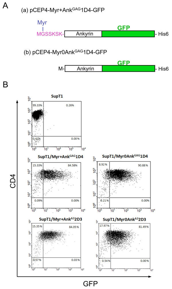
Characterization of SupT1 cells stably expressing artificial ankyrins. (A), Ankyrin constructs. Schematic representation of the artificial ankyrin constructs designed for stable expression in SupT1 cells, using pCEP4-based vector. Histidine-tag and green fluorescence protein (GFP; green box) were inserted at the C-terminus and in-phase with the ankyrin sequence (white box). Addition of a N-myristoylation signal (in purple red letters) to the Myr+AnkGAG1D4-GFP clone resulted in the removal of the N-terminal methionine (M) and covalent linkage of myristic acid chain (Myr; in blue letters) to glycine-2 (G). (B), Flow cytometry. Expression of CD4 molecules at the surface of control SupT1 cells, SupT1/Myr+AnkGAG1D4-GFP and SupT1/Myr0AnkGAG1D4-GFP cells. Flow cytometry analysis was performed on nonpermeabilized cells, using monoclonal antibody against CD4, followed by PE-conjugated goat anti-mouse IgG.

