Abstract
Pseudomyxoma peritonei is rare being characterized by intraperitoneal accumulation of mucinous ascites produced by neoplastic cells, mostly originating from a perforated appendiceal adenoma. The clinical presentatation of the disease is variable, and preoperative diagnosis is often difficult. We describe the clinical case of a 60-year-old patient who presentated predominantly with urological symptoms. CECT revealed an appendiceal lesion infiltrating and projecting into the urinary bladder. Surgical cytoreduction was performed and patient remains symptomatically better on follow up.
Keywords: pseudomyxoma
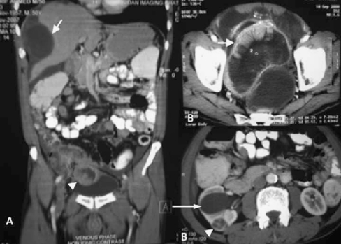
A 60 year old non diabetic and non hypertensive gentleman, reported with passing of jelly like material in urine with occasional haematuria, abdominal pain, distension, fever and burning micturation of 8 months duration. He also had loss of appetite with significant weight loss of 20 kg. Clinically he was malnourished and had generalized abdominal distension with dilated veins over the flanks. His hematological and biochemical parameters were normal expect urine routine examination, which revealed significant pus cells with positive urine culture .He was evaluated with ultrasound of abdomen which showed heterogenous mass in suprapubic region, thickening of urinary bladder and multiple lesions in the liver. Contrast enhanced computed tomography of abdomen revealed large irregular thick walled peripherally enhancing polypoidal lesion in right iliac fossa abutting the caecum and terminal ileum with solid and cystic components. The lesion was seen infiltrating the urinary bladder and projecting into its lumen (Fig. 1, Panel A). It also showed perihepatic high density fluid collections and marked scalloping of the liver (Fig. 1, Panel A). Pelvic scans showed large cystic peripherally enhancing lesion abutting the rectum (Fig. 1, Panel B). There was also dilatation of renal pelvis of the right kidney with a small round well defined hypodense peripherally enhancing lesion with spoke wheel central scar in the lower pole (Fig. 1, Panel B).
Fig. 1.
Panel A—CECT abdomen showing irregular thick walled peripherally enhancing polypoidal lesion abutting the caecum and terminal ileum with solid-cystic components and infiltration and projection into the urinary bladder (arrow head). Perihepatic high density fluid collections and marked scalloping of the liver (long arrow). Panel B (upper panel)—Pelvic scans showing large cystic peripherally enhancing lesion abutting the rectum (white arrow). Panel B (lower panel)—Dilatation of renal pelvis of the right kidney (long arrow) with a small round well defined hypodense peripherally enhancing lesion with spoke wheel central scar in the lower pole (arrow head)
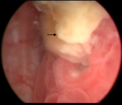
These findings were suggestive of tumor arising probably from the appendix with pseudomyxoma peritonea and hydronephrosis with oncocytoma in the right kidney. Cystoscopy showed proliferative growth with jelly like material extruding from the dome of urinary bladder (Fig. 2). Biopsy from this lesion was reported as mucinous adenocarcinoma of appendix. In the view of extensive disease debulking of the peritoneal lesions was done and the patient is symptomatically better on follow up.
Fig. 2.


