Endometrial cancer is the most common invasive cancer of the female genital tract, with an increasing incidence rate. Abnormal uterine bleeding is the presenting symptom in 75-90% of cases [1, 2]. The majority of patients with endometrial cancer are diagnosed with no evidence of extra-uterine spread (70-80% stage I), which gives patients better prognosis [1, 3]. In more advanced disease the sites commonly affected outside the uterus are pelvic and para-aortic lymph nodes and the ovaries [4]. Similarly to uterine sarcomas, distant metastases in advanced or recurrent endometrial cancer most commonly involve the lungs, liver, central nervous system and skin [4–6].
Metastases to bones have been described in 2-15% of patients with metastatic disease, and the most common site of osseous metastases are vertebrae, with pelvic bones, ribs and sternum [3, 7]. Isolated metastases to bone extremities are extremely rare, and thought to result from the haematological spread of cancer cells [5]. The review of the Medline database by searching the items endometrial cancer and metastases to extremities showed twenty such cases, with only nine cases when metastatic tumour of the foot was the first manifestation of endometrial cancer (Table I) [3, 5, 7–13].
Table I.
Isolated metastasis to the bones of the extremities as the first manifestation of endometrial cancer – review of the literature
| Author/year (references) | Age [years] | Histology | Grade | Bone involved | Other metastasis | Treatment | Survival [months] |
|---|---|---|---|---|---|---|---|
| Vanecko RM, et al./1967 [10] | 54 | – | – | Fibula | – | – | – |
| Onuba O/1983 [13] | 57 | Endometrioid adenocarcinoma | – | Tibia | Lung, kidney | – | – |
| Cooper JK, et al./1994 [7] | 59 | Endometrioid adenocarcinoma | 2 | Calcaneus | No | Chemotherapy, radiotherapy | > 60 |
| Petru E, et al./1995 [11] | 61 | Endometrioid adenocarcinoma | 1 | Tarsus | No | Chemotherapy, hormone therapy | 10 |
| Malicky ES, et al./1997 [12] | 44 | Endometrioid adenocarcinoma | 2 | Femur | No | Radiotherapy, hormone therapy | > 12 |
| Manolitsas TP, et al./2002 [3] | 76 | Endometrioid adenocarcinoma | 3 | Calcaneus | No | Radiotherapy | 20 |
| Uharcek P, et al./2006 [8] | 67 | Endometrioid adenocarcinoma | 1 | Calcaneus, talus and metatarsal bones | No | Surgery, chemotherapy, hormone therapy | > 20 |
| Loizzi V, et al. 2006 [5] | 73 | Endometrioid adenocarcinoma | 3 | Tibia | No | Chemotherapy | 9 |
| Kaya A, et al./2007 [9] | 70 | Endometrioid adenoca rcinoma | 1 | Tibia | No | Radiotherapy | > 47 |
| This case, 2010 | 74 | Endometrioid adenocarcinoma | 2 | Calcaneus, talus and metatarsal bones | No | Radiotherapy | > 40 |
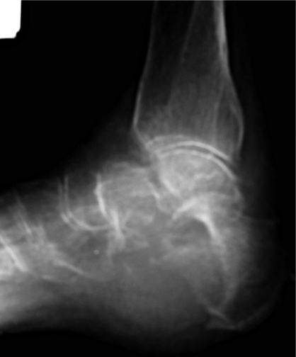
A 74-year-old female suffered from pain and swelling of the right foot from October 2006. In the X-ray picture and bone scintigraphy, suspected lesion of the right calcaneus, talus and metatarsal bones was detected (Figure 1). Afterwards it was histologically verified in the biopsy as metastatic cancer (Figure 2). The patient had no vaginal bleeding or other gynaecological symptoms, but subsequent computed tomography scans showed an enlarged uterus. Uterine curettage confirmed the diagnosis of endometrial cancer. The patient was treated with total abdominal hysterectomy, bilateral salpingo-oophorectomy without pelvic lymph node dissection in December 2006. The histological diagnosis was a moderate differentiated (G2) endometrioid endometrial cancer invading the outer half of the myometrium (> 1/2) with invasion of cervical stroma. The fallopian tubes and the ovaries showed no signs of metastases. No evidence of macroscopic abdominopelvic metastases was found at surgery. Her disease was classified as clinical stage IVB according to FIGO 2009 staging, but in the TNM classification it was pT2 Nx M2.
Figure 1.
Radiograph of the right foot demonstrating metastases of the endometrial cancer
Figure 2.
Metastatic endometrial cancer cells in the fine needle aspiration of the tumour of the foot (H + E, 200× magnification)
In January 2007 the patient was admitted to the department of palliative radiotherapy. She received irradiation by Co 60 until a total dose of 20 Gy to the tumour of the right foot, with complete resolution of symptoms. The patient was discharged from hospital in March 2007. During the observation from surgery in December 2006 until June 2010 no other metastases were detected. The patient remains alive and asymptomatic 43 months after the diagnosis of metastatic endometrial cancer to the foot.
Our report presented in the previous section has three main peculiar features: 1) it demonstrates endometrial cancer that presented with osseous metastasis, which is a rare occurrence, 2) the metastatic tumour was located in the foot, which is extremely rare, 3) the bone lesion was a single bone metastasis, which is unusual in such cases. Additionally, the case in which isolated metastatic tumour of the extremity was the first manifestation of endometrial cancer is the tenth such case reported in the literature (Table I), and the second one located in the calcaneus and involving talus and metatarsal bones [8].
Despite the rarity of metastases to the upper and lower extremities, there is a need to have a high index of suspicion for metastasis in patients with a history of endometrial cancer who present with swelling or bony tenderness. The initial diagnosis can be challenging, as the symptoms of pain and swelling are often attributed to other more common benign conditions such as soft tissue inflammation, trauma, arthritis, and osteomyelitis [7]. It is important to consider bone metastasis as a possible diagnosis also in patients without history of cancer, but with osseous pain not responding to conservative treatment [5, 9–13]. Appropriate imaging may include plain X-ray picture and radionuclide bone scans [3]. Technetium diphosphonate bone scans can be positive up to 18 months before a lesion is detectable on plain X-ray [14]. Therefore, a biopsy should be performed in patients with suspected lesions, and who demonstrate evidence of bony destruction [4].
The treatment strategy in patients with confirmed isolated metastatic lesion in the bone extremity still remains a topic of controversy, because of the few descriptions available in the literature and the different bone sites involved. For these reasons, a common suitable treatment regimen cannot be established and treatment should be tailor suited to each patient. The treatment of irradiation with or without surgery, hormone therapy and chemotherapy is reported as effective in most cases and may be curative [4, 5, 7, 9, 11, 12]. The main goal of treatment should be to eliminate or palliate pain and prolong survival.
References
- 1.Bokhman JV. Two pathogenetic types of endometrial carcinoma. Gynecol Oncol. 1983;15:10–7. doi: 10.1016/0090-8258(83)90111-7. [DOI] [PubMed] [Google Scholar]
- 2.Seebacher V, Schmid M, Polterauer S, et al. The presence of postmenopausal bleeding as prognostic parameter in patients with endometrial cancer: a retrospective multi-center study. BMC Cancer. 2009;9:460. doi: 10.1186/1471-2407-9-460. [DOI] [PMC free article] [PubMed] [Google Scholar]
- 3.Manolitsas TP, Fowler JM, Gahbauer RA, Gupta N. Pain in the foot: calcaneal metastasis as the presenting feature of endometrial cancer. Obstet Gynecol. 2002;100:1067–9. doi: 10.1016/s0029-7844(02)02015-x. [DOI] [PubMed] [Google Scholar]
- 4.Amiot RA, Wilson SE, Reznicek MJ, Webb BS. Endometrial carcinoma metastasis to the distal phalanx of the hallux: a case report. J Foot Ankle Surg. 2005;44:462–5. doi: 10.1053/j.jfas.2005.07.014. [DOI] [PubMed] [Google Scholar]
- 5.Loizzi V, Cormio G, Cuccovillo A, Fattizzi N, Selvaggi L. Two cases of endometrial cancer diagnosis associated with bone metastasis. Gynecol Obstet Invest. 2006;61:49–52. doi: 10.1159/000088530. [DOI] [PubMed] [Google Scholar]
- 6.Karolewski K, Glinski B, Jakubowicz J, et al. Uterine sarcomas – an evaluation of treatment results and prognostic factors. Arch Med Sci. 2009;5:215–21. [Google Scholar]
- 7.Cooper JK, Wong FL, Swenerton KD. Endometrial adenocarcinoma presenting as an isolated calcaneal metastasis. A rare entity with good prognosis. Cancer. 1994;73:2779–81. doi: 10.1002/1097-0142(19940601)73:11<2779::aid-cncr2820731121>3.0.co;2-u. [DOI] [PubMed] [Google Scholar]
- 8.Uharcek P, Mlyncek M, Ravinger J. Endometrial adenocarcinoma presenting with an osseous metastasis. Gynecol Obstet Invest. 2006;61:200–2. doi: 10.1159/000091402. [DOI] [PubMed] [Google Scholar]
- 9.Kaya A, Olmezoglu A, Eren CS, et al. Solitary bone metastasis in the tibia as a presenting sign of endometrial adenocarcinoma: a case report and the review of the literature. Clin Exp Metastasis. 2007;24:87–92. doi: 10.1007/s10585-007-9061-2. [DOI] [PubMed] [Google Scholar]
- 10.Vanecko RM, Yao ST, Schmitz RL. Metastasis to the fibula from endometrial carcinoma. Report of 2 cases. Obstet Gynecol. 1967;29:803–5. [PubMed] [Google Scholar]
- 11.Petru E, Malleier M, Lax S, et al. Solitary metastasis in the tarsus preceding the diagnosis of primary endometrial cancer. A case report. Eur J Gynaecol Oncol. 1995;16:387–91. [PubMed] [Google Scholar]
- 12.Malicky ES, Kostic KJ, Jacob JH, Allen WC. Endometrial carcinoma presenting with an isolated osseous metastasis: a case report and review of the literature. Eur J Gynaecol Oncol. 1997;18:492–4. [PubMed] [Google Scholar]
- 13.Onuba O. Pathological fracture of the right tibia, an unusual presentation of endometrial carcinoma: a case report. Injury. 1983;14:541–5. doi: 10.1016/0020-1383(83)90058-x. [DOI] [PubMed] [Google Scholar]
- 14.Litton GJ, Ward JH, Abbott TM, Williams HJ., Jr Isolated calcaneal metastasis in a patient with endometrial adenocarcinoma. Cancer. 1991;67:1979–83. doi: 10.1002/1097-0142(19910401)67:7<1979::aid-cncr2820670726>3.0.co;2-6. [DOI] [PubMed] [Google Scholar]


