Abstract
A total of 1569 patients with chronic myeloid leukemia (CML) referred to our institution within 1 month of diagnosis since 1965 were reviewed: 1148 chronic phase (CP), 175 accelerated phase (AP), and 246 blastic phase (BP). The median survival was 8.9 years in CP, 4.8 years in AP, and 6 months in BP. In CP, the 8-year survival was ≤ 15% before 1983, 42%-65% from 1983-2000, and 87% since 2001. Survival was worse in older patients (P = .004), but this was less significant since 2001 (P = .07). Survival by Sokal risk was significantly different before 2001 (P < .001), but not since 2001 (P = .4). In AP, survival improved over time (P < .001); the 8-year survival in patients treated since 2001 was 75%. Survival by age was not different in years < 2001 (P = .09), but was better since 2001 in patients ≤ 70 years of age (P = .004). In BP, the median survival improved over time (P < .001), although it has been only 7 months since 2001. In summary, survival in CML has significantly improved since 2001, particularly so in CP-AML and AP-CML. Imatinib therapy minimized the impact of known prognostic factors and Sokal risk in CP-CML and accentuated the impact of age in AP- and BP-CML.
Introduction
The International Randomized Study of Interferon and STI571 (IRIS) compared imatinib mesylate, a selective BCR-ABL tyrosine kinase inhibitor (TKI), with the standard of care, IFN-α and low-dose cytarabine, in patients with newly diagnosed Philadelphia chromosome–positive chronic myeloid leukemia (CML) in the chronic phase (CP). This study showed that imatinib therapy resulted in higher rates of complete cytogenetic response, major molecular response, long-term event-free survival, and transformation-free survival.1 The IRIS study did not show an improvement in the survival of patients treated with imatinib because of the crossover design, which allowed 90% of patients to change therapy from IFN-α to imatinib within 9 months from the start of therapy. Therefore, the improved survival of patients with CML relied on comparisons of the outcome of patients with CML receiving imatinib to those historically treated with other modalities.2–4
At our institution, a CML database updates the characteristics and outcomes of all patients with CML referred since 1965. In the present study, we evaluated the outcome of patients with CML referred to our institution within 1 month from diagnosis and included patients presenting in CP, accelerated phase (AP), or blastic phase (BP). The aim of the study was to analyze outcome by year of therapy in different phases of CML and to define the prognostic factors associated with outcome.
Methods
A total of 3548 patients with CML were referred to our institution from 1965 until 2010 with different durations of prior therapy before referral. A diagnosis of Philadelphia chromosome–positive CML documented by cytogenetic analysis was required for a patient to be included in the analysis. The group included 2465 patients referred in CP, 640 patients referred in AP, and 443 patients referred in BP. For the present study, we restricted the analysis to patients referred to our institution within 1 month from diagnosis. Therefore, the analysis of CML transformation applies only to patients who presented with de novo AP- or BP-CML and does not apply to patients who evolved from CP-AML and developed AP- or BP-CML later. A total of 1569 such patients were referred, including 1148 patients in CP, 175 in AP, and 246 in BP. The diagnostic criteria for CML phases were as described previously.5,6 The characteristics of patients are detailed in Table 1.
Table 1.
Characteristics of study group
| Parameter | Category | CP (n = 1148) | AP (n = 175) | BP (n = 246) |
|---|---|---|---|---|
| Age, y, n (%) | < 50 | 667 (58) | 118 (67) | 111 (45) |
| 50-60 | 273 (24) | 30 (17) | 62 (25) | |
| 61-70 | 160 (14) | 17 (10) | 59 (24) | |
| > 70 | 48 (4) | 10 (6) | 14 (6) | |
| Median (range) | 46 (4-86) | 42 (7-85) | 51 (16-81) | |
| Sex | Female | 454 (40) | 54 (31) | 97 (39) |
| Splenomegaly | Yes | 520/1142 (46) | 122/172 (71) | 86/191 (45) |
| Hemoglobin, g/dL | Median (range) | 11.9 (5-16.7) | 10.5 (5.1-16.3) | 9.7 (2-15.1) |
| WBC, × 109/L) | Median (range) | 78 (1.5-725) | 98.2 (0.6-813) | 37 (0-574) |
| Platelets, × 109/L) | Median (range) | 371 (21-3940) | 365 (22-1610) | 73 (2-2275) |
| Peripheral basophils, % | Median (range) | 3 (0-19) | 5 (0-43) | 1 (0.75) |
| Peripheral blasts, % | Median (range) | 1 (0-15) | 2 (0-29) | 35 (0-100) |
| Marrow basophils, % | Median (range) | 2 (0-19) | 5 (0-46) | 1 (0-44) |
| Marrow blasts, % | Median (range) | 1 (0-14) | 2 (0-26) | 46 (0-98) |
| Sokal risk, n (%) | Low | 549 (48) | ||
| Intermediate | 349 (30) | |||
| High | 228 (20) | |||
| NA | 22 (2) | |||
| Year of treatment, n (%) | < 1975 | 106 (9) | 21 (12) | 13 (5) |
| 1975-1982 | 110 (10) | 25 (14) | 29 (12) | |
| 1983-1990 | 165 (14) | 24 (14) | 41 (17) | |
| 1991-2000 | 352 (31) | 59 (34) | 80 (32) | |
| > 2000 | 415 (36) | 46 (26) | 83 (34) |
Before 1983, patients were treated with busulfan and hydroxyurea. From 1983 until 2000, most patients received IFN-α–based therapy if they presented in CP and combinations of IFN-α and chemotherapy if they presented in transformation. Since 2001, most patients presenting in CP were treated with a TKI: either imatinib or a second-generation TKI such as nilotinib or dasatinib. Allogeneic stem cell transplantation (SCT) was not a frequent frontline CP-CML therapy at our institution, but was offered to patients who were candidates for allogeneic SCT and had available donors and a reasonable risk of allogeneic SCT–associated outcome (usually after failure of the frontline therapy of the time). The frontline treatments of patients by different CML phases and by time period are detailed in Table 2. The numbers of patients receiving allogeneic SCT as front- or second-line therapy for each CML phase are also shown in Table 2.
Table 2.
Characteristics of therapy
| Treatment | CP (n = 1148) | AP (n = 175) | BP (n = 246) | < 1975 (n = 140) | 1975-1982 (n = 164) | 1983-1990 (n = 230) | 1991-2000 (n = 492) | > 2000 (n = 543) |
|---|---|---|---|---|---|---|---|---|
| Hydroxyurea or busulfan, n (%) | 138 (12) | 31 (17) | 2 (< 1) | 100 (71) | 37 (22) | 5 (2) | 23 (5) | 4 (< 1) |
| IFN-α based, n (%) | 430 (38) | 47 (27) | 8 (3) | 0 | 16 (10) | 152 (66) | 316 (64) | 2 (< 1) |
| Single agents, n (%) | 8 (< 1%) | 9 (5) | 34 (14) | 10 (7) | 2 (1) | 4 (2) | 29 (6) | 6 (1) |
| Cytarabine ± other, n (%) | 97 (8) | 27 (15) | 55 (22) | 26 (19) | 94 (57) | 38 (16) | 18 (4) | 3 (< 1) |
| Combination chemotherapy, n (%) | 1 (< 1) | 2 (1) | 42 (17) | 1 (< 1) | 10 (6) | 17 (7) | 16 (3) | 1 (< 1) |
| Allogeneic stem cell transplantation, n (%) | 16 (1) | 5 (3) | 1 (< 1) | 0 | 0 | 1 (< 1) | 20 (4) | 1 (< 1) |
| Tyrosine kinase inhibitor, n (%) | 420 (37) | 50 (29) | 65 (26) | 0 | 0 | 0 | 36 (7) | 499 (92) |
| No therapy, n (%) | 1 (< 1) | 7 (3) | 0 | 0 | 1 | 1 | 6 (1) | |
| Unknown/not available, n (%) | 37 (3) | 3 (2) | 32 (13) | 2 (1) | 4 (2) | 12 (5) | 33 (7) | 21 (4) |
| Stem cell transplantation salvage by CML phase at time of transplantation, n (%)* | 74 (39) | 42 (22) | 75 (39) | 0 | 2 (1) | 28 (15) | 114 (60) | 47 (25) |
191 patients received allogeneic stem cell transplantation as salvage therapy.
The details of therapy in different time periods have been described previously.7–10 All patients consented to therapy as per institutional guidelines and in compliance with the Declaration of Helsinki.
Prognostic factors for survival used standard statistical methods.11,12 Patients in CP-CML were categorized by Sokal risk group.13
Results
The overall median survival was 8.9 years for patients presenting in CP, 4.8 years for those presenting in AP, and 6 months for those presenting in BP. Details on survival for each phase are detailed in the following sections.
CP-CML
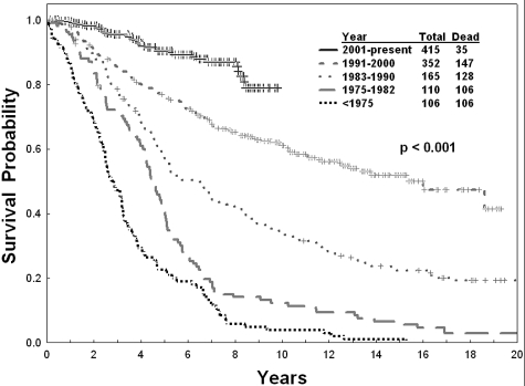
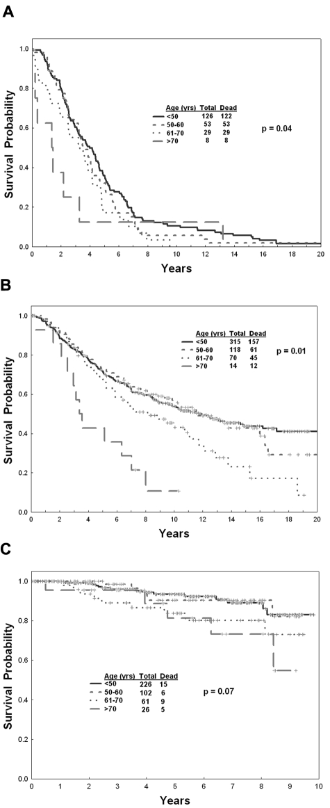
The overall survival of newly diagnosed CML has improved significantly since the introduction of imatinib therapy. The estimated 8-year survival rate has increased from 6% before 1975 up to 87% since 2001 (Figure 1). Survival was significantly different by age overall (P = .004), particularly in years ≤ 2000 (P < .001), but has differed less significantly since 2001 (P = .07; Figure 2A-C).
Figure 1.
Survival in newly diagnosed CP-CML by year of therapy.
Figure 2.
Survival in newly diagnosed CP-CML. (A) By age before 1983 overall, (B) from 1983-2000, and (C) since 2001.
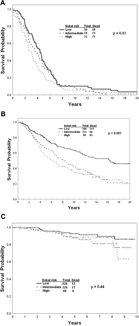
Survival was statistically significantly different by Sokal risk (P < .001). However, the survival by Sokal risk groups was significantly different only in years ≤ 2000 (P = .03 in years < 1983; P < .001 for years 1983-2000). Since 2001, the significance of the Sokal risk model in defining different risk groups has disappeared (P = .44; Figure 3A-C).
Figure 3.
Survival in newly diagnosed CP-CML. (A) By Sokal risk factor before 1983, (B) from 1983-2000, and (C) since 2001.
Prognostic factors associated with differences in survival in CP-CML are shown in Table 3. By multivariate analysis, the independent risk factors for survival were: year of therapy, older age, anemia, percent BM basophils, percent BM blasts, and male sex. Among patients treated since 2001, the independent adverse prognostic factors for survival were: year of therapy (hazard ratio [HR], 0.71; P = .01) and older age (HR, 1.02; P = .055). Among the 35 of 415 patients who died since 2001, 34 deaths were among patients referred during the years 2001-2004, and 1 death was in patients referred in 2005; no deaths occurred in patients referred in the years 2006-2010. Death was attributed to CML consequences in 16 patients, and attributed to non-CML causes in 18 patients: non-CML causes included 5 other cancers, 4 complications of allogeneic SCT–related GVHD, 1 car accident, 1 suicide, 2 postsurgical deaths/sepsis, 3 cardiac events, and 2 other causes. Therefore, with TKI therapy, the significance of most established prognostic factors, including Sokal risk, were eliminated except for a marginal effect of older age.
Table 3.
Univariate and multivariate analyses for survival in chronic phase
| Univariate |
Multivariate |
||||
|---|---|---|---|---|---|
| Adverse | HR | P | HR | P | |
| Age, y | Older; continuous | 1.01 | .002 | 1.01 | < .001 |
| Sex | Male vs female | 1.08 | .38 | 1.25 | .03 |
| Splenomegaly | Yes vs no | 1.85 | < .001 | 1.09 | .51 |
| Hemoglobin, g/dL | Lower; continuous | 0.88 | < .001 | 0.94 | .02 |
| WBC count, × 109/L) | Higher; continuous | 1.003 | < .001 | 0.999 | .31 |
| Platelet count, × 109/L) | Lower; continuous | 1.004 | < .001 | 1.00 | .41 |
| Peripheral blasts, % | Higher; continuous | 1.14 | < .001 | 0.995 | .83 |
| Peripheral basophils, % | Higher; continuous | 0.98 | .09 | 0.98 | .17 |
| BM blasts, % | Higher continuous | 1.07 | < .001 | 1.06 | .02 |
| BM basophils, % | Higher; continuous | 1.05 | .004 | 1.08 | < .001 |
| Sokal | Intermediate vs low | 1.84 | < .001 | 1.28 | .07 |
| High vs low | 2.23 | < .001 | 1.10 | .62 | |
| Year of therapy | Continuous | 0.93 | < .001 | 0.925 | < .001 |
AP-CML
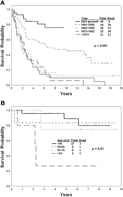
Among patients presenting in AP, survival has improved significantly by year of therapy (Figure 4A). The estimated 8-year survival rate has increased from less than 20% before 1990, to 45% in the years 1991-2000, to 75% since 2001. This suggests the strong impact of imatinib therapy in AP-CML and the need to develop new “AP” criteria that predict for a very short survival.
Figure 4.
Survival in AP-CML. (A) By year of therapy and (B) by age since 2001.
The effect of older age on survival was influenced by year of therapy. Overall, there was a trend for older age to be associated with a shorter survival (P = .08). Before 2001, the outcome of patients was so poor that different age groups did not significantly distinguish different prognostic groups (P = .07-.32). Since 2001, the prognosis of patients improved significantly, and older age was then associated with worse survival (P = .03), considering the small numbers involved (Figure 4B).
Prognostic factors for survival in AP by univariate analysis are shown in Table 4. By multivariate analysis, the independent adverse factors were: year of therapy, older age, an increased percentage of BM blasts, an increased percentage of BM basophils, and male sex. Among patients treated since 2001, the independent adverse prognostic factors for survival by multivariate analysis were older age (HR, 1.07; P = .049) and an increased percentage of BM blasts (HR, 1.10; P = .03).
Table 4.
Univariate and multivariate analyses for AP-CML and BP-CML
| AP |
BP |
|||||||
|---|---|---|---|---|---|---|---|---|
| Univariate | Multivariate | Univariate | Multivariate | |||||
| HR | P | HR | P | HR | P | HR | P | |
| Age, y | 1.02 | .002 | 1.02 | .02 | 1.01 | .22 | 1.01 | .04 |
| Male sex | 1.10 | .65 | 1.97 | .004 | 0.89 | .39 | 0.79 | .18 |
| Splenomegaly | 1.86 | .01 | 1.51 | .17 | 1.04 | .82 | 1.004 | .98 |
| Hemoglobin, g/dL | 1.04 | .38 | 1.09 | .26 | 1.03 | .31 | 1.06 | .12 |
| White blood cell counts × 109/L | 1.01 | .35 | 0.999 | .32 | 0.999 | .96 | 1.000 | .94 |
| Platelet count × 109/L | 1.002 | .51 | 1.000 | .55 | 0.999 | .005 | 0.999 | .006 |
| Peripheral blasts, % | 1.03 | .009 | 0.98 | .16 | 1.01 | < .001 | 1.005 | .15 |
| Peripheral basophils, % | 0.98 | .10 | 0.98 | .17 | 0.99 | .19 | 0.99 | .20 |
| BM blasts, % | 1.05 | < .001 | 1.07 | < .001 | 1.003 | .16 | 0.997 | .46 |
| BM basophils, % | 0.99 | .56 | 1.06 | .04 | 0.99 | .36 | 1.02 | .2 |
| Y of therapy | 0.947 | < .001 | 0.93 | < .001 | 0.97 | < .001 | 0.956 | < .001 |
BP-CML
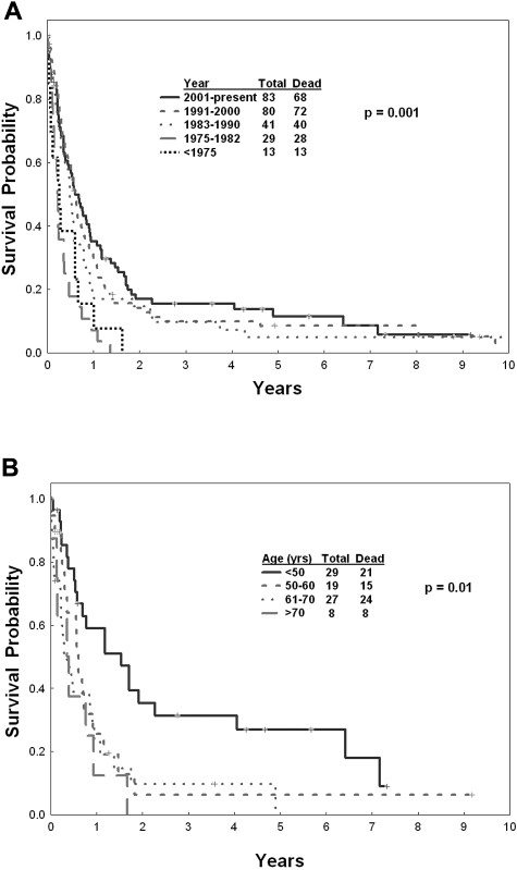
The median survival of patients presenting in BP was 6 months. The median survival in BP has improved (Figure 5A; P < .001), but the improvement is clinically modest: the median survival among patients treated since 2001 is only 7 months. Survival by age has not improved in this poor-prognosis population, particularly among patients treated until 2000. Since 2001, younger patients (age ≤ 50 years) have had a significant improvement in survival (Figure 5B; P = .01).
Figure 5.
Survival in BP-CML. (A) By year of therapy and (B) by age since 2001.
Prognostic factors for survival in BP by univariate analysis are shown in Table 4. By multivariate analysis, older age, thrombocytopenia, and year of therapy remained prognostically significant for survival. Among patients treated since 2001, older age was identified as an independent significant factor for survival (HR, 1.03; P = .001), as was the percentage of peripheral blasts (HR, 1.012; P = .01).
Impact of postfrontline therapy on prognosis
Postfrontline therapy may influence outcome. In particular, outcome in CML may be influenced by subsequent allogeneic SCT among patients with inadequate response to frontline therapy (eg, IFN-α) and by the availability of imatinib and other TKI therapies in patients who failed IFN-α but were able to access the new agents in their later courses of TKI therapy.
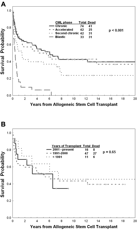
Overall, 191 patients underwent postfrontline allogeneic SCT from related (n = 131) or unrelated donors (n = 60), in CP (n = 74) or in transformed (n = 117) phases. The outcome of patients from the date of allogeneic SCT by CML phase is shown in Figure 6A. Outcome of allogeneic SCT in CP has not improved significantly by year of therapy at our institution (P = .65; Figure 6B). Interestingly, there is no difference in the median time from diagnosis to allogeneic SCT by year of therapy. In addition, only 14% of the total patients underwent allogeneic SCT.
Figure 6.
Survival after allogeneic SCT. (A) By phase of CML at the time of SCT and (B) by year of therapy in CP.
To assess the benefit of allogeneic SCT in BP, we evaluated the outcome of 2 groups of patients who underwent allogeneic SCT in connection with BP-CML. The first group of patients includes 32 of 246 patients (13%) who presented with BP-CML and underwent allogeneic SCT subsequent to their BP treatment. These included 20 of 163 patients (12%) who presented before 2001 and 12 of 83 patients who presented since 2001. The estimated 5-year survival rate from date of allogeneic SCT was 10% for patients before 2001 and 30% for patients since 2001 (P = .03). The estimated 5-year survival of these 32 patients dated from the BP presentation was 10% for patients before 2001 and 38% for patients since 2001 (P = not significant). The second group was 75 patients (Tables 1–2) who underwent allogeneic SCT as salvage therapy (not frontline therapy) either in BP (n = 33) or in second CP (n = 42). This group of 75 patients may have presented in any phase of CML (CP, AP, or BP), but underwent allogeneic SCT as postfrontline therapy in either BP or second CP. Fifty patients had allogeneic SCT before 2001 and 25 since 2001. The estimated 5-year survival was 18% for patients treated before 2001 and 40% for patients treated since 2001 (P = .038). This suggests that allogeneic SCT in general, and since 2001 (availability of TKIs that could reduce safely and effectively the CML burden before stem cell transplant) has improved the outcome of a subset of patients (10%-15%) who were able to undergo allogeneic SCT in postblastic transformation.
The survival of patients treated from 1991-2000 is significantly better than those treated from 1983-1990 (Figure 1), even though IFN-α–based therapy was essentially the frontline therapy in both decades. However, 44 of 165 patients (27%) treated from 1983-1990 had access to TKI therapy in their later course, in contrast to 277 of 352 patients (79%) treated from 1991-2000. This explains the similarity of the survival curve in the first 3 years, but the significant diversions beyond that period, a time that allowed many patients diagnosed in the years 1991-2000 to have the opportunity to receive the new TKIs.
Discussion
This single-institution experience shows the survival improvement in patients with CML presenting in different phases over 5 decades. It confirms the major prognostic effect of the introduction of imatinib therapy into the treatment of CP-CML (Figure 1). In these patients, the estimated 8-year survival has improved from a historical rate of less than 20% up to 87% in the imatinib era, accounting for all deaths whether CML related or not.
The analysis demonstrates how a new, highly effective therapy such as imatinib minimizes the prognostic impact of other previously established prognostic factors in CML, such as older age, thrombocytosis, splenomegaly, basophilia, and others. It also shows how established risk models such as the Sokal risk model in CP-CML (Figure 3A-C) become less relevant once new effective therapies are introduced. Since 2001, only age had a persistent but marginal significance. Year of therapy since 2001 also remained prognostic. This may be because of increasing experience with and penetration of imatinib therapy, access of patients after imatinib failure to second-generation TKIs, and the use of second-generation TKIs as frontline therapy.
In patients presenting in AP, the estimated 8-year survival with imatinib therapy is now 75%. This is extremely encouraging and suggests that a TKI, rather than allogeneic SCT, is a reasonable frontline therapy, based on pretreatment of characteristics and initial response to TKI therapy. It also highlights the need to develop new criteria for “AP” that would define survival times shorter than a median of 2 years.6 In this regard, the multivariate analysis in patients in AP presenting since 2001 identified older age and percentage of BM blasts as independently adverse.
It was interesting to analyze the time-dependent effect of different prognostic factors. For example, older age, an established prognostic factor in CP-CML, was highly significant in patients treated before 2001, but had only marginal significance—and only among very old patients—with imatinib therapy. Conversely, in AP- and BP-CML patients, in whom prognosis is so poor, older age was not identified as an adverse factor before 2001. With the introduction of effective imatinib therapy that prolonged survival, older age (in contrast to the situation in CP), became significant in defining survival (Figures 4B and 5B).
Effective imatinib therapy has minimized the prognostic effect of most well-known prognostic factors, eliminating the significance of most. In fact, in other studies incorporating treatment-associated factors such as achievement of complete cytogenetic response or major molecular response with imatinib at different time points, imatinib therapy–associated prognostic factors became predominant over pretreatment prognostic factors.2–4 This is similar to the findings in other tumors, for which highly effective therapies eliminated the previously established prognostic factors (eg, testicular cancer, hairy cell leukemia, and acute promyelocytic leukemia) and identified at times newer, usually treatment-associated factors (eg, minimal residual disease at different times on therapy).
The findings of the present study are consistent with other studies investigating in a large number of patients the evolution of prognosis in CML by year of therapy.14,15 In a Swedish study of 3173 patients diagnosed with CML treated over 5 decades, the investigators demonstrated a significant improvement in survival.15 In that study, frontline allogeneic SCT was offered more commonly in the years before 2000 and was reported by the investigators to account for the survival improvement from 1987-2000. Allogeneic SCT was performed in 18% of patients from 1987-1993 and in 42% from 1994-2000. As shown in our study, this survival benefit might also be related to the access to subsequent therapy with TKI. In the Swedish study, older age was identified as adverse even in the era of imatinib therapy, particularly among patients 80 years or older. The investigators attributed this to the poor penetration of imatinib therapy among this very old age group. In our study, the number of patients older than 80 years was small. However, we also showed older age (≥ 70 years) to be associated with worse outcome in CP-CML, but the association became less significant in the imatinib era (P = .07). In contrast, in the CML-transformed phases, the prognostic effect of age became more prominent once the survival of patients improved from poor or extremely poor to significantly better (as in AP) or to modestly better (as in BP).
In summary, the results of the present study demonstrate the significant survival improvement of patients with CML, not only in CP, but also in newly diagnosed AP or BP. It emphasizes the need to develop new prognostic models for CP-CML, new definitions for AP-CML, and newer strategies, including combined modality therapies in patients with BP-CML, in whom the median survival remains very poor.
Acknowledgements
This study was funded in part by the National Institutes of Health (grant P01 CA049639).
Footnotes
The publication costs of this article were defrayed in part by page charge payment. Therefore, and solely to indicate this fact, this article is hereby marked “advertisement” in accordance with 18 USC section 1734.
Authorship
Contribution: H.K. and J.C. designed and performed the research, contributed analytical tools, analyzed the data, and wrote the manuscript; S.O., E.J., G.G.-M., A.Q.-C., F.R., S.F., T.K., G.B., R.C., and M.T. performed the research, contributed analytical tools, analyzed the data, and wrote the manuscript; and J.S., M.B.R., and X.H. contributed analytical tools, analyzed the data, and wrote the manuscript.
Conflict-of-interest disclosure: H.K. is a consultant for Novartis and receives research funding from Novartis, Pfizer, and BMS. E.J. and A.Q.-C. receive honoraria from Novartis for consultancy. G.B. is on the speakers' bureau for Novartis. M.T. is a consultant for and on the speakers' bureau for Novartis. J.C. has research funding from Novartis, BMS, Pfizer, Ariad, Chemgenex, and Deciphera, and is a consultant for BMS, Novartis, Ariad, and Chemgenex. The remaining authors declare no competing financial interests.
Correspondence: Hagop Kantarjian, MD, The M. D. Anderson Cancer Center, 1515 Holcombe Blvd, Unit 428, Leukemia Department, Houston, TX 77030; e-mail: hkantarj@mdanderson.org.
References
- 1.Druker B, Guilhot F, O'Brien S, Gathman I, Kantarjian H, et al. Five-year follow-up of patients receiving imatinib for chronic myeloid leukemia. N Engl J Med. 2006;355(23):2408–17. doi: 10.1056/NEJMoa062867. [DOI] [PubMed] [Google Scholar]
- 2.Kantarjian H, Talpaz M, O'Brien S, et al. Survival benefit with imatinib mesylate versus interferon alpha-based regimens in newly diagnosed chronic-phase chronic myelogenous leukemia. Blood. 2006;108(6):1835–1840. doi: 10.1182/blood-2006-02-004325. [DOI] [PubMed] [Google Scholar]
- 3.Roy L, Guilhot J, Krahnke T, et al. Survival advantage from imatinib compared with the combination interferon alpha plus cytarabine in chronic-phase chronic myelogenous leukemia: historical comparison between two phase 3 trials. Blood. 2006;108(5):1478–1484. doi: 10.1182/blood-2006-02-001495. [DOI] [PubMed] [Google Scholar]
- 4.Kantarjian H, O'Brien S, Cortes J, et al. Analysis of the impact of imatinib mesylate therapy on the prognosis of patients with Philadelphia chromosome-positive chronic myelogenous leukemia treated with interferon alpha regimens for early chronic phase. Cancer. 2003;98(7):1430–7. doi: 10.1002/cncr.11665. [DOI] [PubMed] [Google Scholar]
- 5.Walters R, Kantarjian H, Keating M, et al. Therapy of lymphoid and undifferentiated chronic myelogenous leukemia in blast crisis with continuous vincristine and adriamycin infusions plus high-dose decadron. Cancer. 1987;60(8):1708–1712. doi: 10.1002/1097-0142(19871015)60:8<1708::aid-cncr2820600803>3.0.co;2-1. [DOI] [PubMed] [Google Scholar]
- 6.Kantarjian HM, Dixon D, Keating MJ, et al. Characteristics of accelerated disease in chronic myelogenous leukemia. Cancer. 1988;61(7):1441–1446. doi: 10.1002/1097-0142(19880401)61:7<1441::aid-cncr2820610727>3.0.co;2-c. [DOI] [PubMed] [Google Scholar]
- 7.Kantarjian H, O'Brien S, Cortes J, et al. Complete cytogenetic and molecular responses to interferon alpha-based therapy for chronic myelogenous leukemia are associated with excellent long-term prognosis. Cancer. 2003;97(4):1033–41. doi: 10.1002/cncr.11223. [DOI] [PubMed] [Google Scholar]
- 8.Kantarjian H, O'Brien S, Shan J, et al. Cytogenetic and molecular responses and outcome in chronic myelogenous leukemia. Cancer. 2008;112(4):837–45. doi: 10.1002/cncr.23238. [DOI] [PubMed] [Google Scholar]
- 9.Cortes J, Jones D, O'Brien S, et al. Nilotinib as frontline treatment for patients with chronic myeloid leukemia in early chronic phase. J Clin Oncol. 2010;28(3):392–397. doi: 10.1200/JCO.2009.25.4896. [DOI] [PMC free article] [PubMed] [Google Scholar]
- 10.Cortes J, Jones D, O'Brien S, et al. Results of dasatinib therapy in patients with early chronic-phase chronic myeloid leukemia. J Clin Oncol. 2010;28(3):398–404. doi: 10.1200/JCO.2009.25.4920. [DOI] [PMC free article] [PubMed] [Google Scholar]
- 11.Kaplan EL, Maier P. Non-parametric estimation from incomplete observations. J Am Stat Assoc. 1965;53:457–81. [Google Scholar]
- 12.Cox Regression models and life tables. J R Stat Soc B. 1972;34:187–220. [Google Scholar]
- 13.Sokal JE, Cox EB, Baccarani M, Tura S, Gomez GA, Robertson JE, et al. Prognostic discrimination in “good risk” chronic granulocytic leukemia. Blood. 1984;63(4):789–799. [PubMed] [Google Scholar]
- 14.Brenner H, Gondos A, Pulte D. Recent trends in long-term survival of patients with chronic myelocytic leukemia: disclosing the impact of advances in therapy on the population level. Haematologica. 2008;93(10):1544–9. doi: 10.3324/haematol.13045. [DOI] [PubMed] [Google Scholar]
- 15.Bjorkholm M, Ohm L, Eloranta S, Derolf A, Hultcrantz M, et al. The success story of targeted therapy in chronic myeloid leukemia: a population based study of 3173 patients diagnosed in Sweden 1973-2008. J Clin Oncol. 2011;28:2514–2525. doi: 10.1200/JCO.2011.34.7146. [DOI] [PMC free article] [PubMed] [Google Scholar]


