Abstract
Benign smooth muscle proliferations are relatively rare in the oral cavity. Most are classified as angioleiomyomas, some as hamartomatous growths and a few as cutaneous-type leiomyomas. We present two cases of benign smooth muscle proliferations in the tongue, provide a review, briefly discuss histogenesis and offer a clinico-pathological differential diagnosis.
Keywords: Tongue, Smooth muscle lesions
Introduction
Benign smooth muscle lesions and tumours (leiomyomas) are relatively rare in the oral tissues. They are much more common in the skin and gastrointestinal tract [1, 2]. These tumours represent only 0.4% of all soft tissue lesions in the oral cavity [3]. The most common sites in the mouth are the lips, tongue, palate and buccal mucosa [4]. Four groups of benign tumours of smooth muscle are described: cutaneous, angioleiomyoma (or angiomyoma), leiomyomas of deep soft tissue, and a final group linked in name to the myofibroblast (the myofibroblastomas) [1].
Most oral smooth muscle lesions are angioleiomyomas (in our biopsy service, approximately 95%), arising from the smooth muscle of blood vessels [4–6]. They are often painful and typically enlarge slowly, sometimes reaching several centimetres in diameter, but are usually small at the time of biopsy. Solid lesions are characteristically in colour to normal mucosa but the vascular lesions are red or blue [6]. Leiomyomas of the posterior dorsal tongue are generally accepted to arise from the smooth muscle in the region of the circumvallate papillae [7]. Although rare, it is important that they be considered in a differential diagnosis of a variety of soft tissue lesions which may arise in the posterior tongue. Such lesions could include lingual thyroid, rhabdomyoma, granular cell tumour, schwannoma, and neurofibroma.
Leiomyomatous hamartomas of the midline of the tongue have been described [8]. Hamartomas are masses of disorganized mature specialised cells or tissue indigenous to a particular affected site, and are composed of a mixture of different relatively mature tissues although one particular type of tissue may dominate [9]. When smooth muscle dominates in a hamartoma, the distinction between a hamartoma and leiomyoma may be difficult.
In this report, we present and discuss two unusual cases of benign smooth muscle proliferations arising in the tongue.
Case Reports
Case 1
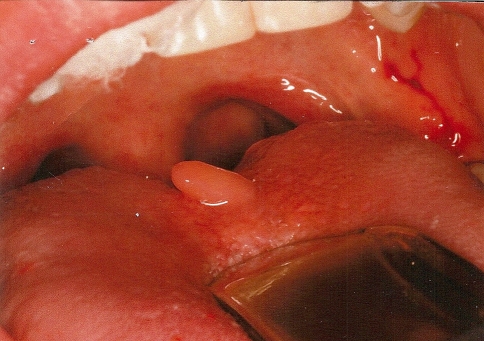
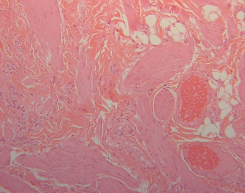
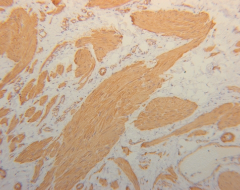
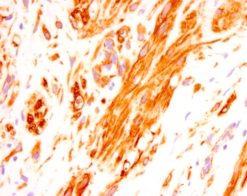
An otherwise healthy 14 year-old male presented with a mass of the midline dorsum of the tongue (Fig. 1). His medical history was non-contributory, and there was no history of trauma. A clinical diagnosis of papilloma or elongated papilla was submitted. The lesion was completely excised, and on gross examination consisted of a nodule of grey brown soft tissue 11 × 5 × 5 mm. Microscopically, the specimen consisted of a nodule of vascular fibrous connective tissue which was dominated by an unencapsulated proliferation of benign smooth muscle fibre bundles peripheral to a core of thick walled blood vessels (Fig. 2) There were occasional areas of interspersed fibro-adipose tissue. The smooth muscle bundles exhibited bland cellular morphology but mild nuclear pleomorphism. Routine immunoperoxidase stains were positive for smooth muscle actin (Fig. 3), desmin and h-caldesmon, but negative for CD31, CD34 and S100 in the tumour cells. The histological features were interpreted to represent those of a leiomyoma.
Fig. 1.
Polypoid mass of the mid-posterior dorsum of the tongue
Fig. 2.
Smooth muscle fascicles as seen in Case 1, with interspersed collagen fibre bundles and occasional fat cells (H&E stain, original magnification ×100)
Fig. 3.
Smooth muscle actin stain showing smooth muscle fascicles in case 1 (original magnification ×100)
Case 2
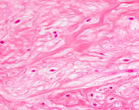
A healthy 42 year-old male patient reported to his dental surgeon, presenting with a smooth 5 × 4 mm, round, white nodule on the left ventral anterior surface of the tongue. His past medical history was uneventful: he weighed 150 lb, and reported no history of smoking, drug abuse, and/or alcoholic intake. The mass in his tongue was soft on palpation, mobile and asymptomatic. The nodule was removed under local anesthetic. A clinical diagnosis of papilloma was submitted. A subsequent post-operative examination was revealed no evidence of recurrence. The area was asymptomatic and had healed completely. Palpation of the area displayed no scar tissue and no palpable mass. The gross pathological specimen consisted of a smooth, round, white nodule 3 × 2 × 2 mm with a small base. Microscopically, it was a nodule of pale, spindle and round cells exhibiting small oval bland nuclei, often with clear cytoplasm and arranged in clusters (Fig. 4). The nodule was well circumscribed but unencapsulated. This tumour nodule was surrounded by a vascular loose fibrous connective tissue which was surfaced by a para-keratinizing thin stratified squamous epithelium. Immunohistochemical stains for desmin, muscle specific actin, smooth muscle actin (Fig. 5), h-caldesmon and vimentin were positive while stains for CD68, CD34, S100 and cytokeratin were all negative. The histological features were interpreted to be those of a leiomyoma, with some clear cell change.
Fig. 4.
Smooth muscle bundles with clear cell change (Case 2; H&E stain, original magnification ×400)
Fig. 5.
H-caldesmon showing strong positive staining (Case 2; original magnification ×400)
Discussion
Although angiomyomas are fairly common, non-vascular leiomyomas are extremely rare in the oral cavity. An extensive review published in 2000 revealed only 28 cases of leiomyoma in the tongue, of which only eight were non-vascular leiomyomas [10]. Subsequently, we found that only two other possible non-vascular leiomyomas of the tongue have been described of which only one could be absolutely confirmed [11, 12].
Leiomyomas arising in the posterior tongue appear to be non-vascular, and histologically resemble the cutaneous-type which develop from the pilar arrector muscles. It has been proposed that non-vascular leiomyomas arising in the posterior tongue develop from smooth muscle in the circumvallate papilla or in the lingual duct [7, 13]. The cutaneous type lesions may be solitary or multiple, developing during adolescence or adult life although occasionally appearing at birth or during early childhood [1]. Mature lesions are nodular, often associated with significant pain and may reach a size of 2 cm. Histologically, non-vascular leiomyomas of the tongue are like their cutaneous counterparts: they are less well defined than the angiomyoma, unencapsulated and blend with the surrounding connective tissues. They form bundles of smooth muscle fibres, which usually intersect in an orderly fashion and resemble normal smooth muscle cells. They are positive for desmins, smooth muscle actin and caldesmon [1] as was seen in the two cases described here.
Distinction from lesions reported as smooth muscle hamartomas on a histological basis is not so clear cut, and would be mostly related to differences in clinical presentation. Most smooth muscle hamartomas are lesions which appear as large lesions, usually in the lumbar region during childhood or early adult life, and are sometimes associated with hyperpigmentation [1]. Histologically, oral smooth muscle hamartomas of the tongue have fibrous connective tissue, adipose tissue, vascular and neural tissue interspersed between well-formed smooth muscle fascicles [8, 14]. Smooth muscle hamartomas may have a similar histological appearance to leiomyomas of pilar-arrector origin, but usually are represented by a single lesion which is several centimetres in diameter [1]. In our case 1, an argument could be made for the diagnosis of a hamartoma as it showed a central large blood vessel, with interspersed small areas of adipose tissue. However, the proliferation of smooth muscle fascicles around this central zone was non vascular in nature, and it was relatively tightly packed although unencapsulated. In addition, adipose tissue has been described within smooth muscle tumours of all types [1].
In case 2 described above, clear cell change was seen, a phenomenon which has been described in somatic leiomyomas of deep soft tissue [1]. In this case, all muscle stains were positive, and S100 protein staining was negative, leading to a conclusive diagnosis of leiomyoma. Many of the fascicles were cut in cross-section leading to an appearance of smaller rounder nuclei in some areas, a phenomenon also shown by Enzinger and Weiss [1]. Because of the presence of clear cells, benign mesenchymoma, a controversial entity, would have to be considered in the histologic differential diagnosis. The mesenchymoma, by definition, contains fibrous tissue and 2 or more mesenchymal tissues that are not normally associated with each other [15]. Several benign mesenchymomas have been described in the oral regions [15–17]. In case 2, the clear cells did not have the morphology of fat cells (Fig. 4), and because of the predominance of the muscle staining, the possibility of mesenchymoma was discarded.
Myofibromas would probably form the most important group in a histological differential diagnosis, although the distinction would be of little clinical significance [18]. Microscopically, myofibromas are typically well-circumscribed, having a central hemangiopericytoma-like appearance and paucicellular areas dominating the periphery of the lesions [1, 18, 19]. Myofibromas are typically desmin negative [1].
Other soft tissue lesions to consider in a clinical differential diagnosis would include lingual thyroid, rhabdomyoma, granular cell tumour, schwannoma, and neurofibroma, but these would be relatively easy to differentiate microscopically using a Masson-Trichrome stain, and immunohistochemical markers such as alpha smooth muscle actin and S100 protein.
In conclusion, pathologists should be aware of the range of benign smooth muscle lesions which may be encountered in this region. The general dental practitioner should be aware that lesions of smooth muscle could arise in the posterior tongue, even though distinction from other benign lesions such as hamartomas and myofibromas may not be clinically possible and surgical excision is usually curative. The two cases reported herein demonstrate some of this diversity, ranging from hamartoma-like proliferation, to clear cell change.
References
- 1.Weiss SW, Goldblum JR, Enzinger FM. Enzinger and weiss’s soft tissue tumours. 5. Philadelphia, PA: Mosby Elsevier; 2008. pp. 695–726. [Google Scholar]
- 2.Damm DD, Neville BW. Oral leiomyomas. Oral Surg Oral Med Oral Pathol. 1979;47:343–348. doi: 10.1016/0030-4220(79)90257-3. [DOI] [PubMed] [Google Scholar]
- 3.Baden E, Doyle JL, Lederman DA. Leiomyoma of the oral cavity: a light microscopic and immunohistochemical study with review of the literature from 1884 to 1992. Eur J Cancer B Oral Oncol. 1994;30B:1–7. doi: 10.1016/0964-1955(94)90043-4. [DOI] [PubMed] [Google Scholar]
- 4.Neville BW, Damm DD, Allen CM, Bouquot JE. Oral and maxillofacial pathology. 3. St. Louis, Mo.: Saunders/Elsevier; 2009. [Google Scholar]
- 5.Epivatianos A, Trigonidis G, Papanayotou P. Vascular leiomyoma of the oral cavity. J Oral Maxillofac Surg. 1985;43:377–382. doi: 10.1016/0278-2391(85)90261-7. [DOI] [PubMed] [Google Scholar]
- 6.Gnepp DR. Diagnostic surgical pathology of the head and neck. 2. Philadelphia, PA: Saunders/Elsevier; 2009. [Google Scholar]
- 7.Luaces Rey R, Lorenzo Franco F, Gomez Oliveira G, Patino Seijas B, Guitian D, Lopez-Cedrun Cembranos JL. Oral leiomyoma in retromolar trigone. A case report. Med Oral Patol Oral Cir Bucal. 2007;12:E53–E55. [PubMed] [Google Scholar]
- 8.Nava-Villalba M, Ocampo-Acosta F, Seamanduras-Pacheco A, Aldape-Barrios BC. Leiomyomatous hamartoma: report of two cases and review of the literature. Oral Surg Oral Med Oral Pathol Oral Radiol Endod. 2008;105:e39–e45. doi: 10.1016/j.tripleo.2007.12.021. [DOI] [PubMed] [Google Scholar]
- 9.Kumar V, Abbas AK, Fausto N, Robbins SL, Cotran RS. Robbins and Cotran pathologic basis of disease. 7. Philadelphia, Pa: Elsevier Saunders; 2005. [Google Scholar]
- 10.Natiella JR, Neiders ME, Greene GW. Oral leiomyoma, report of six cases and a review of the literature. J Oral Pathol. 1982;11:353–365. doi: 10.1111/j.1600-0714.1982.tb00177.x. [DOI] [PubMed] [Google Scholar]
- 11.Kotler HS, Gould NS, Gruber B. Leiomyoma of the tongue presenting as congenital airway obstruction. Int J Pediatr Otorhinolaryngol. 1994;29:139–145. doi: 10.1016/0165-5876(94)90093-0. [DOI] [PubMed] [Google Scholar]
- 12.Felix F, Gomes GA, Tomita S, Fonseca Junior A, El Hadj Miranda LA, Arruda AM. Painful tongue leiomyoma. Braz J Otorhinolaryngol. 2006;72:715. doi: 10.1016/S1808-8694(15)31032-6. [DOI] [PMC free article] [PubMed] [Google Scholar]
- 13.Wertheimer-Hatch L, Hatch GF, III, HatchB SKF, Davis GB, Blanchard DK, Foster RS, Jr, et al. Tumors of the oral cavity and pharynx. World J Surg. 2000;24:395–400. doi: 10.1007/s002689910064. [DOI] [PubMed] [Google Scholar]
- 14.Faria PR, Batista JD, Duriguetto AF, Jr, Souza KC, Candelori I, Cardoso SV, et al. Giant leiomyomatous hamartoma of the tongue. J Oral Maxillofac Surg. 2008;66:1476–1480. doi: 10.1016/j.joms.2007.06.679. [DOI] [PubMed] [Google Scholar]
- 15.Kessler DA, Kademani D, Feldman RS, Howlett P. Mesenchymoma: an unusual tumour of the lip. Br J Oral Maxillofac Surg. 2000;42(4):348–350. doi: 10.1016/j.bjoms.2004.03.002. [DOI] [PubMed] [Google Scholar]
- 16.Jones AC, Trochesset D, Freedman PD. Intraoral benign mesenchymoma: a report of 10 cases and review of the literature. Oral Surg Oral Med Oral Pathol Oral Radiol Endod. 2003;95(1):67–76. doi: 10.1067/moe.2003.52. [DOI] [PubMed] [Google Scholar]
- 17.Quiñónez RE, Astor FC, Romaguera RL. Benign mesenchymoma of the tongue. Otolaryngol Head Neck Surg. 2002;127(4):357–358. doi: 10.1067/mhn.2002.127887. [DOI] [PubMed] [Google Scholar]
- 18.Montgomery E, Speight PM, Fisher C. Myofibromas presenting in the oral cavity: a series of 9 cases. Oral Surg Oral Med Oral Pathol Oral Radiol Endod. 2000;89:343–348. doi: 10.1016/S1079-2104(00)70100-4. [DOI] [PubMed] [Google Scholar]
- 19.Azevedo Rde S, Pires FR, Della Coletta R, Almeida OP, Kowalski LP, Lopes MA. Oral myofibromas: report of two cases and review of clinical and histopathologic differential diagnosis. Oral Surg Oral Med Oral Pathol Oral Radiol Endod. 2008;105:e35–e40. doi: 10.1016/j.tripleo.2008.02.022. [DOI] [PubMed] [Google Scholar]


