Abstract
Background. We determined the consistency of positive interferon-gamma (IFN-γ) release assays (IGRAs) to detect latent TB infection (LTBI) over one-year postpartum in HIV-1-infected women. Methods. Women with positive IGRAs during pregnancy had four 3-monthly postpartum IGRAs. Postpartum change in magnitude of IFN-γ response was determined using linear mixed models. Results. Among 18 women with positive pregnancy IGRA, 15 (83%) had a subsequent positive IGRA; 9 (50%) were always positive, 3 (17%) were always negative, and 6 (33%) fluctuated between positive and negative IGRAs. Women with pregnancy IGRA IFN-γ>8 spot forming cells (SFCs)/well were more likely to have consistent postpartum IGRA response (odds ratio: 10.0; 95% confidence interval (CI): 0.9–117.0). Change in IFN-γ response over postpartum was 10.2 SFCs/well (95% CI: −1.5–21.8 SFCs/well). Conclusion. Pregnancy positive IGRAs were often maintained postpartum with increased consistency in women with higher baseline responses. There were modest increases in magnitude of IGRA responses postpartum.
1. Background
Tuberculosis (TB) and human immunodeficiency virus type 1 (HIV-1) infection are major health problems in women, particularly during their reproductive years (15–49) [1]. In a recent analysis, we observed that HIV-1-infected women with latent TB infection (LTBI) as detected by a positive interferon-gamma (IFN-γ) release assay (IGRA) during pregnancy are at increased risk of active TB during the postpartum period [2]. Postpartum active TB is associated with increased risk of mortality in HIV-1-infected women and their infants and is also associated with an increased risk of HIV-1 transmission to the infants [3, 4]. Thus, IGRAs during or after pregnancy may be useful in identifying women at increased risk for future active TB who in turn may expose their infants.
LTBI has traditionally been detected using the tuberculin skin test (TST), which has limitations in specificity due to cross-reactivity with bacille Calmette-Guerin (BCG) vaccine and in sensitivity due to anergy in immunocompromised and malnourished individuals [5]. In contrast, IGRAs are not confounded by prior BCG, correlate better with exposure to active TB than TST, and are not prone to boosting on repeat testing [5–8]. IGRAs measure immune responses to MTB antigens: early secretory antigenic target 6-kD protein (ESAT-6) and culture filtrate protein 10 (CFP-10).
While there are data on IGRA performance following known recent TB exposure or in presence of active TB, little is known regarding variability of IGRA responses among individuals without these risk factors [9–12]. In high TB prevalence settings, 30–50% of individuals have evidence of latent TB infection (LTBI), and specific TB exposure may not be known or defined. Individuals with LTBI would be expected to have positive IGRA responses; however, these may vary due to changes in antigenic burden, T-cell response variability, or test performance. The pregnancy/postpartum period is associated with hormonal and immunologic changes during which time it is plausible that MTB-specific IGRA responses might fluctuate. Performance of IGRAs through pregnancy and postpartum has not been described, overall or in presence of HIV-1 infection.
Using a historical repository from a peripartum HIV-1 infected cohort, we conducted a study to determine the magnitude and consistency of IGRA-positive responses over 1 year postpartum in HIV-1-infected women with positive IGRA responses during pregnancy.
2. Materials and Methods
2.1. Ethics Statement
Written informed consent for the parent study was obtained from the women. Human Subjects Division at University of Washington and Ethical Review Committee at University of Nairobi approved the parent and current studies.
2.2. Study Design and Population
This study used a specimen repository from a historical cohort of HIV-1-infected women who were enrolled during pregnancy at 32 weeks gestation and followed for at least 1-year postpartum [13, 14]. As previously reported, we tested 361 women at ~32-week gestation with T-SPOT.TB IGRA using cryopreserved PBMCs; 135 (37.4%) and 170 (47.1%) were positive and negative, respectively, and 56 (15.5%) had indeterminate responses [2]. We selected 18 (13.3%) women from the 135 with positive IGRAs who had further specimens available at postpartum months 3, 6, 9, and 12. Because this study aimed at defining consistency of IGRAs in absence of active TB, we excluded women who developed active TB during followup to avoid changes in IGRAs due to active TB.
2.3. Laboratory Methods
Cryopreserved peripheral blood mononuclear cells (PBMCs) were tested for IFN-γ responses using T-SPOT.TB, following the manufacturers' instructions on assay procedure and interpretation of results which have been previously described [2]. The methods used for PBMC isolation in this cohort have been previously described [15]. Cells were isolated within 8 hours and were preserved in freezing medium containing 90% fetal calf serum and 10% dimethyl sulfoxide (DMSO) using a temperature rate controlled freezing unit overnight at −80°C and transferred to liquid nitrogen storage tank within 3 days for long-term storage. IGRA tests for the previously published study [2] and the current serial study were conducted by the same technician in the same laboratory in Nairobi, Kenya.
2.4. Statistical Analysis
The 18 women selected for this serial assay study were compared with the remaining 117 women with positive IGRA responses at 32-week gestation using Student t-tests for means and z-test based on bootstrapped standard errors to detect differences in medians for continuous variables, χ2 test (or Fisher's exact when cell counts were ≤5) for categorical variables.
Women were classified as consistent responders if they had positive IGRAs at all four postpartum visits at months 3, 6, 9, and 12, excluding visits with indeterminate responses. Logistic regression was used to estimate the odds ratio (OR) of having 100% positive postpartum IGRA responses and having >50% positive IGRA responses. Women with combined IFN-γ response (maximum of ESAT-6 or CFP-10-specific response above background) during pregnancy of >8 SFCs/well were compared to those with ≤8 SFCs/well. The cut-point of >8 SFCs/well in the magnitude of pregnancy IFN-γ response was identified because this cut-point falls above the grey zone of 5–7 spots identified by T-SPOT.TB manufacturer and by the United States Food and Drug Administration (FDA) as an indication for retesting and is the 25th percentile of the magnitude of IFN-γ response during pregnancy in our data [16, 17].
We used continuous spot count data to estimate the rate of change in the magnitude of the combined and antigen-specific response between postpartum months 3 and 12, using linear mixed models (LMMs) with random intercepts. Using the LMMs, we estimated the intraclass correlation coefficient (ICC), expressed as the within-person variability in responses as a proportion of the overall variability. Rate of change in mean postpartum CD4 counts were assessed using LMM with random intercepts.
Analyses were done using Stata Intercooled v11.1 [18].
3. Results
3.1. Enrollment and Followup
Baseline characteristics (age, education, and medical history) of the 18 HIV-1-infected IGRA positive women selected for serial assessment were comparable to IGRA positive women (n = 117) from the cohort who were not included in this serial assessment study. The baseline median CD4 count (518 versus 469 cells/uL, P = 0.59) and median HIV-1 plasma viral load (4.2 versus 4.7 log10 copies/mL, P = 0.11), baseline median ESAT-6 (16.0 versus 23.5; P = 0.22), and CFP-10 (16.0 versus 23.0; P = 0.70) were similar between women selected and not selected for this study, respectively. Among the 18 selected women, 1 reported having had TB approximately one year prior to being enrolled in the cohort. At 3, 6, 9, and 12 months, 61%, 44%, 33%, and 33% of women reported breastfeeding, respectively. None of the women were hospitalized or initiated antiretroviral therapy and 5 (28%) were diagnosed with pneumonia during followup. None of the women received LTBI treatment during followup because there was no LTBI testing during the period of cohort followup and it was not recommended as standard of care.
3.2. Consistency of Postpartum IGRA
Individual positive, negative, and indeterminate responses at each postpartum visit are shown in Table 1. Of the 72 (18 women × 4 postpartum time points) tests performed, 9 (12.5%) were indeterminate. Excluding visits with indeterminate responses, 83.3% of women had a postpartum positive IGRA, 50% (9/18) had positive IGRA response at all postpartum visits, 33.3% (6/18) had responses fluctuating between positive and negative, and 16.7% (3/18) had negative IGRAs at all postpartum visits.
Table 1.
Interferon-γ release assay results during postpartum in HIV-1-infected women who were interferon-γ release assay positive during pregnancy.
| Postpartum IGRA responses at months† | |||||||
|---|---|---|---|---|---|---|---|
| ID | Interferon-γ response* during pregnancy, SFCs/well | 3 | 6 | 9 | 12 | Consistent‡ positive postpartum IGRAs | Percent of positive IGRA visits of all subsequent visits with valid responses |
| 307 | 160.5 | + | + | Yes | 100.0 | ||
| 237 | 144 | + | + | + | + | Yes | 100.0 |
| 379 | 119.5 | + | + | + | + | Yes | 100.0 |
| 268 | 79 | + | + | + | Yes | 100.0 | |
| 310 | 42.5 | + | + | + | Yes | 100.0 | |
| 330 | 40 | + | + | + | + | Yes | 100.0 |
| 277 | 24 | + | + | + | + | Yes | 100.0 |
| 362 | 15.5 | + | + | + | Yes | 100.0 | |
| 410 | 8 | + | + | + | + | Yes | 100.0 |
| 249 | 97.5 | + | − | + | + | No | 75.0 |
| 300 | 96.5 | + | − | + | + | No | 75.0 |
| 228 | 8 | + | + | − | + | No | 75.0 |
| 381 | 8 | − | + | + | No | 66.6 | |
| 377 | 64 | − | + | − | + | No | 50.0 |
| 287 | 6 | − | − | + | No | 33.0 | |
| 376 | 120 | − | − | − | No | 0.0 | |
| 265 | 7.5 | − | − | − | − | No | 0.0 |
| 233 | 7 | − | − | − | No | 0.0 | |
Note: IGRA interferon-gamma release assay.
This table is sorted by consistency of positive postpartum IGRAs. The first nine rows display IGRA responses in women who had all positive postpartum IGRAs. The next 6 rows display IGRA responses in women who fluctuated between positive and negative IGRAs postpartum. The last 3 rows display IGRA responses in women who had all negative postpartum IGRA.
*Maximum of ESAT-6/CPF-10 minus negative control.
†Blank cells represent visits with indeterminate responses.
‡Consistent positive IGRAs is defined as all postpartum visits with positive IGRAs, excluding visits with indeterminate responses.
3.3. Pregnancy IFN-γ Response and Consistency of Postpartum IGRAs
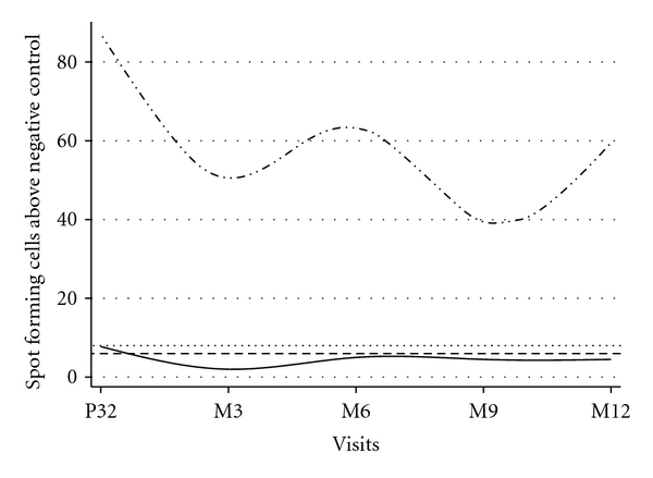
Women with combined IFN-γ response >8 SFCs/well during pregnancy were 10 times more likely to have consistently positive IGRAs postpartum compared to women with ≤8 SFCs/well (OR: 10.0; 95% confidence interval (CI): 0.85–117.0; P = 0.07) (Table 2(a)) and 5 times more likely to have >50% of postpartum visits with positive IGRAs (OR: 5.0; 95% CI: 0.55–45.39; P = 0.15) (Table 2(b)). Median magnitude of a combined IFN-γ response during postpartum in women with baseline IFN-γ response of >8 versus ≤8 SFCs/well is displayed in Figure 1.
Table 2.
(a) Odds of consistently positive interferon-γ release assays in women associated with baseline magnitude of interferon-γ response >8 compared to ≤8 SFCs/well. (b) Odds of >50% postpartum visits with positive interferon-γ release assays in women with baseline magnitude of interferon-γ response >8 compared to ≤8 SFCs/well.
(a)
| Postpartum consistency of IGRAs | |||
|---|---|---|---|
| Interferon-γ response* (SFCs/well) during pregnancy | Yes | No | Odds ratio (95% confidence interval); P value |
| >8 | 8 | 4 | 10.0 (0.85–117.0); 0.07 |
| ≤8 | 1 | 5 | |
(b)
| Greater than 50% postpartum visits with positive IGRAs | |||
| Interferon-γ response* (SFCs/well) during pregnancy | Yes | No | Odds ratio (95% confidence interval); P value |
|
| |||
| >8 | 10 | 2 | 5.0 (0.55–45.39); 0.15 |
| ≤8 | 3 | 3 | |
Note: IGRA interferon-gamma release assay.
*Maximum of ESAT-6/CPF-10 minus negative control.
Figure 1.

Change in magnitude of postpartum interferon-γ response by baseline interferon-γ response. The dash and dotted line represents women with baseline interferon-γ response of >8 SFCs/well, and the solid line represents women with baseline interferon-γ response of ≤8 SFCs/well. The horizontal dashed line corresponds to the manufacturer defined cut-point at 6 SFCs/well above background for a positive response and the dotted horizontal line represents 8 SFCs/well. Interferon-γ response is defined as the maximum of ESAT-6/CFP-10 minus spot count in negative control.
3.4. Change in Magnitude of Postpartum IFN-γ Response
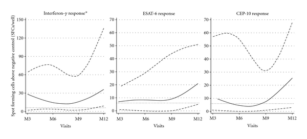
Median magnitude of the combined IFN-γ and antigen-specific responses over postpartum are shown in Figure 2. Using a LMM with random intercepts, the average rate of change in magnitude (SFCs/well) per 3 monthly visits was estimated to be 10.2 (95% CI: −1.5–21.8; P = 0.09) for the combined postpartum IFN-γ response and 5.0 (95% CI: −3.2–13.1; P = 0.23) and 7.2 (95% CI: −3.0–17.2; P = 0.17) for ESAT-6 and CFP-10 responses, respectively. Using this model, we estimated an ICC of 0.51, 0.61, and 0.41 for combined, ESAT-6 and CFP-10 responses, respectively, suggesting substantial within-person variability in IGRA responses.
Figure 2.

Change in magnitude of interferon-γ and antigen-specific response during postpartum. The solid lines represent the median response, and the two dashed lines represent the 25th and 75th percentiles of the response. The horizontal dotted line represents the manufacturer defined cut-point at 6 SFCs/well above background for a positive response. Interferon-γ response* is defined as the maximum of ESAT-6/CFP-10 minus spot count in negative control.
The average rate of decline in CD4 count between months 3 and 12 was −24.0 cells/mm3 per 3 monthly visit (95% CI: −57.6–9.6; P = 0.16) using a LMM with random intercepts. Change in CD4 count was plotted against the change in magnitude of the combined IFN-γ response and ESAT-6 and CFP-10 responses. Adjusted analyses incorporating CD4 did not alter estimates for rate of change in IFN-γ response.
4. Discussion
In this study, we serially tested 18 HIV-1-infected women who were IGRA-positive at baseline during pregnancy, at 3 monthly intervals during the first-year postpartum to determine long-term within-person consistency of IGRA responses and to describe the effect of the peripartum period on IGRA responses. We observed that 83% of women had a subsequent positive response and 50% retained a positive IGRA response at all subsequent assays throughout the postpartum period. Women with higher magnitude of combined response at baseline were more likely to have consistently positive responses at subsequent time points. Women with weak positive response close to the cut-off value at baseline had subsequently fluctuating IGRA responses. The magnitude of the combined IFN-γ response and antigen-specific responses increased slightly during the 1-year postpartum period.
Our study was conducted in a setting with high TB incidence in HIV-1-infected women with high probability of TB exposure but no specific known TB exposure. We found that HIV-1-infected pregnant women with latent TB infection had generally reproducible positive IGRA responses. Although 2011 WHO guidelines recommend universal isoniazid preventive therapy (IPT) in HIV-1-infected individuals, IGRA-targeted IPT may be a strategy to consider for pregnant women [19]. Consistent detection of latent TB by IGRAs despite physiologic changes during/after pregnancy would strengthen specificity of this approach.
While most women had repeated positive IGRAs in our study, we observed some reversions and noted within-person variability of responses, which could be due to changes in TB exposure, TB immune responses, or test reproducibility. Our observation of fluctuation in responses is consistent with previous serial IGRA studies, in which conversions and reversions were observed more frequently in individuals with responses close to the predefined cut-point for a positive response [20–22]. One South African study with close repeat testing (2 days apart) of 15 health care workers (HCWs) noted 100% concordance in results [23]. However, in TB-exposed HCWs in India with serial IGRAs, 2/14 (14%) had a change in QFT responses with magnitude of IFN-γ declining 12 days after baseline assessment [21]. Another study in HCWs in South Africa using both QFT and T-SPOT.TB at 4 times over 21 days observed that T-SPOT responses were more likely to change (revert/convert) than QFT and noted considerable within-person variability of responses [20]. Finally, among HCWs in Germany, a low TB incidence country, there were more reversions than conversions during serial testing and age and prior positive TST predicted consistent QFT positivity [22].
In our study, women with baseline response of >8 SFCs/well were 10 times more likely to have consistently positive IGRAs postpartum while those with lower levels had less consistent responses. These data are consistent with previous studies noting that responses close to the cut-point are more likely to fluctuate [20–22]. The US FDA and Oxford Immunotec recommend a borderline “grey zone” of 5–7 spots above negative control for the T-SPOT.TB and suggest that results be considered in conjunction with clinical information or to retest [16, 17]. We also observed a substantial within person variability in the quantitative responses, which probably accounts for much of the fluctuation in the qualitative responses between positive and negative. These findings underscore the importance of considering quantitative IFN-γ response data in addition to the dichotomous (positive/negative) results in serial testing and in clinical decisions.
Our study has important strengths and limitations. Our study is the first to describe long-term within-person reproducibility of T-SPOT.TB IGRA in HIV-1-infected women during the pregnancy/postpartum period in a TB-endemic setting in the absence of any specific exposure to TB. The women included in this study had longitudinally measured clinical and immunological outcomes, enabling us to take these factors into account since they could potentially alter IGRA responses. We were also able to repeat the IGRAs for these women at relatively close intervals and at critical time points such as early postpartum and during breastfeeding. Our study involves women without a defined time of TB exposure or LTBI treatment. This enabled us to describe the natural variation in IFN-γ responses during the pregnancy/postpartum period. In contrast to previous studies on serial IGRAs, which have been typically shorter, our period of serial evaluation was longer (over 1 year). A limitation of our study was the use of cryopreserved PBMCs as opposed to fresh samples, which might have contributed to test variability [24]. In a previous study of T-SPOT.TB results comparing fresh and frozen PBMCs, cryopreserved samples had lower sensitivity [25]. In our study, lower sensitivity due to cryopreserved PBMCs may have contributed to greater fluctuation of results between positive and negative. However, a new serial assessment study using fresh assays would not be feasible because a positive test would indicate LTBI treatment based on new guidelines. In addition, a few published studies have noted adequacy of cryopreserved samples for T.SPOT.TB and other ELISpot assays [26, 27]. A design constraint of studying LTBI responses in this setting is that women are frequently exposed to TB. Thus, the alternative design including women who were IGRA negative at baseline and followed for consistency of “negative” responses would be confounded by women with new positive IGRAs who newly acquired LTBI. We therefore restricted the study to those with baseline positive IGRAs to assess consistency. Selective inclusion of baseline positives would be expected to bias estimates of rate of change in IFN-γ response. To circumvent this potential bias, we evaluated changes after the baseline visit. Our sample size, though small, was comparable or larger than previous serial studies on IGRAs.
In conclusion, our study demonstrates consistency of IGRAs during pregnancy and postpartum in HIV-1-infected women, particularly among those with higher magnitude responses. Fluctuation in responses between positive and negative was seen among women with weak positive responses at baseline. Despite hormonal and immune perturbations during the postpartum period, the magnitude of response did not change markedly over the postpartum period, as shown by the results from the linear mixed models. The slight increase in levels over time during the postpartum period may reflect some impact of immunosuppression during pregnancy, which may explain increased susceptibility to active TB during this period. The burden of TB in HIV-1-infected women in the childbearing years and the consequent risk of TB morbidity and mortality in their infants is well established [1–4, 28–31]. Further serial studies in this setting will be useful to define optimal timing for IGRA testing during or after pregnancy and to understand biologic determinants of IGRA responses and magnitude.
Acknowledgments
We thank everyone at the Paediatrics Research Laboratory, Kenyatta National Hospital for laboratory support for this study and the study staff and participants of this perinatal cohort. This work is supported by the US National Institutes Health (NIH) through grant no. R21 HD058477-01 and Firland Foundation Grant no. 200910. B. L. Payne and D. Wamalwa were scholars in the International AIDS Training and Research Program, NIH Research Grant D43 TW000007, funded by the Fogarty International Center and the Office of Research on Women's Health. K. Tapia is supported by the University of Washington Center for AIDS Research (CFAR), an NIH-funded program (P30 AI027757), which is supported by the following NIH Institutes and Centers (NIAID, NCI, NIMH, NIDA, NICHD, NHLBI, and NCCAM). G. C. John-Stewart is supported by NIH Research Grant K24 HD054314-04. The funders had no role in study design, data collection and analysis, decision to publish, or preparation of the paper.
References
- 1.Mofenson LM, Laughon BE. Human immunodeficiency virus, mycobacterium tuberculosis, and pregnancy: a deadly combination. Clinical Infectious Diseases. 2007;45(2):250–253. doi: 10.1086/518975. [DOI] [PubMed] [Google Scholar]
- 2.Jonnalagadda S, Payne BL, Brown E, et al. Latent tuberculosis detection by interferon γ release assay during pregnancy predicts active tuberculosis and mortality in human immunodeficiency virus type 1-infected women and their children. Journal of Infectious Diseases. 2010;202(12):1826–1835. doi: 10.1086/657411. [DOI] [PMC free article] [PubMed] [Google Scholar]
- 3.Gupta A, Nayak U, Ram M, et al. Postpartum tuberculosis incidence and mortality among HIV-infected women and their infants in Pune, India, 2002–2005. Clinical Infectious Diseases. 2007;45(2):241–249. doi: 10.1086/518974. [DOI] [PubMed] [Google Scholar]
- 4.Gupta A, Bhosale R, Kinikar A, et al. Maternal tuberculosis: a risk factor for mother-to-child transmission of human immunodeficiency virus. Journal of Infectious Diseases. 2011;203(3):358–362. doi: 10.1093/jinfdis/jiq064. [DOI] [PMC free article] [PubMed] [Google Scholar]
- 5.Menzies D, Pai M, Comstock G. Meta-analysis: new tests for the diagnosis of latent tuberculosis infection: areas of uncertainty and recommendations for research. Annals of Internal Medicine. 2007;146(5):340–354. doi: 10.7326/0003-4819-146-5-200703060-00006. [DOI] [PubMed] [Google Scholar]
- 6.Shams H, Weis SE, Klucar P, et al. Enzyme-linked immunospot and tuberculin skin testing to detect latent tuberculosis infection. American Journal of Respiratory and Critical Care Medicine. 2005;172(9):1161–1168. doi: 10.1164/rccm.200505-748OC. [DOI] [PMC free article] [PubMed] [Google Scholar]
- 7.Ewer K, Deeks J, Alvarez L, et al. Comparison of T-cell-based assay with tuberculin skin test for diagnosis of Mycobacterium tuberculosis infection in a school tuberculosis outbreak. The Lancet. 2003;361(9364):1168–1173. doi: 10.1016/S0140-6736(03)12950-9. [DOI] [PubMed] [Google Scholar]
- 8.Pai M, Zwerling A, Menzies D. Systematic review: T-cell-based assays for the diagnosis of latent tuberculosis infection: an update. Annals of Internal Medicine. 2008;149(3):177–184. doi: 10.7326/0003-4819-149-3-200808050-00241. [DOI] [PMC free article] [PubMed] [Google Scholar]
- 9.Pai M, Joshi R, Dogra S, et al. Serial testing of health care workers for tuberculosis using interferon-γ assay. American Journal of Respiratory and Critical Care Medicine. 2006;174(3):349–355. doi: 10.1164/rccm.200604-472OC. [DOI] [PMC free article] [PubMed] [Google Scholar]
- 10.Pai M, Joshi R, Dogra S, et al. Persistently elevated T cell interferon-γ responses after treatment for latent tuberculosis infection among health care workers in India: a preliminary report. Journal of Occupational Medicine and Toxicology. 2006;1(1, article 7) doi: 10.1186/1745-6673-1-7. [DOI] [PMC free article] [PubMed] [Google Scholar]
- 11.Hill PC, Brookes RH, Fox A, et al. Longitudinal assessment of an ELISPOT test for Mycobacterium tuberculosis infection. Plos Medicine. 2007;4(6, article e192) doi: 10.1371/journal.pmed.0040192. [DOI] [PMC free article] [PubMed] [Google Scholar]
- 12.Pai M, O’Brien R. Serial testing for tuberculosis: can we make sense of T cell assay conversions and reversions? Plos Medicine. 2007;4(6, article e208) doi: 10.1371/journal.pmed.0040208. [DOI] [PMC free article] [PubMed] [Google Scholar]
- 13.Walson JL, Brown ER, Otieno PA, et al. Morbidity among HIV-1-infected mothers in Kenya: prevalence and correlates of illness during 2-year postpartum follow-up. Journal of Acquired Immune Deficiency Syndromes. 2007;46(2):208–215. doi: 10.1097/QAI.0b013e318141fcc0. [DOI] [PMC free article] [PubMed] [Google Scholar]
- 14.Otieno PA, Brown ER, Mbori-Ngacha DA, et al. HIV-1 disease progression in breast-feeding and formula-feeding mothers: a prospective 2-year comparison of T cell subsets, HIV-1 RNA levels, and mortality. Journal of Infectious Diseases. 2007;195(2):220–229. doi: 10.1086/510245. [DOI] [PMC free article] [PubMed] [Google Scholar]
- 15.Lohman BL, Slyker JA, Richardson BA, et al. Longitudinal assessment of human immunodeficiency virus type 1 (HIV-1)-specific gamma interferon responses during the first year of life in HIV-1-infected infants. Journal of Virology. 2005;79(13):8121–8130. doi: 10.1128/JVI.79.13.8121-8130.2005. [DOI] [PMC free article] [PubMed] [Google Scholar]
- 16. T-SPOT.TB, 2011, http://www.Oxfordimmunotec.Com/t-spot_international/
- 17.U.S Food and Drug Administration. T-spot.Tb—p070006, 2008, http://www.accessdata.fda.gov/scripts/cdrh/cfdocs/cftopic/pma/pma.cfm?num=p070006.
- 18.StataCorp. Stata Statistical Software: Release 11. College Station TSL, 2009.
- 19.Guidelines for intensified tuberculosis case-finding and isoniazid preventive therapy for people living with HIV in resource-constrained settings. World Health Organization, 2011.
- 20.van Zyl-Smit RN, Pai M, Peprah K, et al. Within-subject variability and boosting of t-cell interferon-γ responses after tuberculin skin testing. American Journal of Respiratory and Critical Care Medicine. 2009;180(1):49–58. doi: 10.1164/rccm.200811-1704OC. [DOI] [PubMed] [Google Scholar]
- 21.Veerapathran A, Joshi R, Goswami K, et al. T-cell assays for tuberculosis infection: deriving cut-offs for conversions using reproducibility data. Plos ONE. 2008;3(3) doi: 10.1371/journal.pone.0001850. Article ID e1850. [DOI] [PMC free article] [PubMed] [Google Scholar]
- 22.Ringshausen FC, Nienhaus A, Schablon A, Schlösser S, Schultze-Werninghaus G, Rohde G. Predictors of persistently positive Mycobacterium-tuberculosis-specific interferon-gamma responses in the serial testing of health care workers. BMC Infectious Diseases. 2010;10, article 220 doi: 10.1186/1471-2334-10-220. [DOI] [PMC free article] [PubMed] [Google Scholar]
- 23.Detjen AK, Loebenberg L, Grewal HM, et al. Short-term reproducibility of a commercial interferon gamma release assay. Clinical and Vaccine Immunology. 2009;16(8):1170–1175. doi: 10.1128/CVI.00168-09. [DOI] [PMC free article] [PubMed] [Google Scholar]
- 24.Tuuminen T, Sorva S, Liippo K, et al. Feasibility of commercial interferon-γ-based methods for the diagnosis of latent Mycobacterium tuberculosis infection in Finland, a country of low incidence and high bacille Calmette-Guérin vaccination coverage. Clinical Microbiology and Infection. 2007;13(8):836–838. doi: 10.1111/j.1469-0691.2007.01750.x. [DOI] [PubMed] [Google Scholar]
- 25.Meier T, Eulenbruch HP, Wrighton-Smith P, Enders G, Regnath T. Sensitivity of a new commercial enzyme-linked immunospot assay (T SPOT-TB) for diagnosis of tuberculosis in clinical practice. European Journal of Clinical Microbiology and Infectious Diseases. 2005;24(8):529–536. doi: 10.1007/s10096-005-1377-8. [DOI] [PubMed] [Google Scholar]
- 26.Smith JG, Liu X, Kaufhold RM, Clair J, Caulfield MJ. Development and validation of a gamma interferon ELISPOT assay for quantitation of cellular immune responses to varicella-zoster virus. Clinical and Diagnostic Laboratory Immunology. 2001;8(5):871–879. doi: 10.1128/CDLI.8.5.871-879.2001. [DOI] [PMC free article] [PubMed] [Google Scholar]
- 27.Russell ND, Hudgens MG, Ha R, Havenar-Daughton C, McElrath MJ. Moving to human immunodeficiency virus type 1 vaccine efficacy trials: defining T cell responses as potential correlates of immunity. Journal of Infectious Diseases. 2003;187(2):226–242. doi: 10.1086/367702. [DOI] [PubMed] [Google Scholar]
- 28.Marais BJ, Gupta A, Starke JR, El Sony A. Tuberculosis in women and children. The Lancet. 2010;375(9731):2057–2059. doi: 10.1016/S0140-6736(10)60579-X. [DOI] [PubMed] [Google Scholar]
- 29.Khan M, Pillay T, Moodley JM, Connolly C. Maternal mortality associated with tuberculosis-HIV-1 co-infection in Durban, South Africa. AIDS. 2001;15(14):1857–1863. doi: 10.1097/00002030-200109280-00016. [DOI] [PubMed] [Google Scholar]
- 30.Pillay T, Khan M, Moodley J, et al. The increasing burden of tuberculosis in pregnant women, newborns and infants under 6 months of age in Durban, KwaZulu-Natal. South African Medical Journal. 2001;91(11):983–987. [PubMed] [Google Scholar]
- 31.Pillay T, Khan M, Moodley J, Adhikari M, Coovadia H. Perinatal tuberculosis and HIV-1: considerations for resource-limited settings. Lancet Infectious Diseases. 2004;4(3):155–165. doi: 10.1016/S1473-3099(04)00939-9. [DOI] [PubMed] [Google Scholar]


