Abstract
We have evaluated the potential use of various polyamidoamine (PAMAM) dendrimer [dendrimer, generation (G) 2-4] conjugates with cyclodextrins (CyDs) as novel DNA and RNA carriers. Among the various dendrimer conjugates with CyDs, the dendrimer (G3) conjugate with α-CyD having an average degree of substitution (DS) of 2.4 [α-CDE (G3, DS2)] displayed remarkable properties as DNA, shRNA and siRNA delivery carriers through the sensor function of α-CDEs toward nucleic acid drugs, cell surface and endosomal membranes. In an attempt to develop cell-specific gene transfer carriers, we prepared sugar-appended α-CDEs. Of the various sugar-appended α-CDEs prepared, galactose- or mannose-appended α-CDEs provided superior gene transfer activity to α-CDE in various cells, but not cell-specific gene delivery ability. However, lactose-appended α-CDE [Lac-α-CDE (G2)] was found to possess asialoglycoprotein receptor (AgpR)-mediated hepatocyte-selective gene transfer activity, both in vitro and in vivo. Most recently, we prepared folate-poly(ethylene glycol)-appended α-CDE [Fol-PαC (G3)] and revealed that Fol-PαC (G3) imparted folate receptor (FR)-mediated cancer cell-selective gene transfer activity. Consequently, α-CDEs bearing integrated, multifunctional molecules may possess the potential to be novel carriers for DNA, shRNA and siRNA.
Keywords: cyclodextrin, PAMAM dendrimer, carrier, DNA, siRNA, shRNA
1. Introduction
Gene therapy is emerging as a potential strategy for the treatment of genetic diseases, cancers, cardiovascular diseases and infectious diseases [1]. Clinical trials employing over 1,500 gene therapy protocols have been carried out for various diseases [2–4]. Recently, gene silencing induced by small interfering RNA (siRNA), RNA interference (RNAi), became a powerful tool of gene analysis and gene therapy [5–7]. Likewise, vector-based short-hairpin RNAs (shRNA) expression systems have been developed in order to prolong the RNAi effect [8]. However, standard therapeutic use of DNA (gene) and siRNA in clinical settings in humans has been hampered by the lack of effective methods to deliver these nucleic acid drugs into the diseased organs and cells [9–12]. For these reasons, the improvement in transfer activity of a non-viral vector (carrier) is of utmost importance [13–15].
The two gene delivery methods are well known: the viral method and the non-viral method [16,17]. In general, viral vectors have, however, safety risks such as immunogenicity, oncogenicity and potential viral recombination to be solved [18–21]. Hence, more attention is given to the applications of non-viral vectors, because the non-viral method has the profound advantage of being non-pathogenic and non-immunogenic. The non-viral method is further subdivided into two methods, i.e. physical delivery and chemical delivery methods, and the latter method includes three types: lipofection, polyfection and lipopolyfection methods [22,23]. Recently, numerous polycations and polymer micelle have been used for formulating gene, shRNA and siRNA into complexes now termed “polyplexes”. Polycations include histons, polylysine, cationic oligopeptides, polyethyleneimine (PEI), polypropyleneimine (PPI), dendrimers, poly(2-(dimethylamino)ethyl methacrylate and chitosan [13]. The potential use of polyelectrolyte complex micelles for delivering nucleic acid drugs has also been reported [23–27].
2. Polyamidoamine (PAMAM) Starburst™ Dendrimers (Dendrimers) as DNA, shRNA and siRNA Carriers
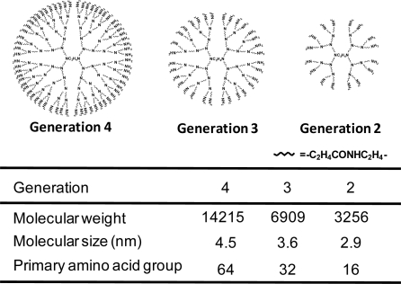
Dendrimers, which were developed by Tomalia et al., are biocompatible, non-immunogenic and water-soluble, and possess terminal modifiable amine functional groups as the sensor for binding various targeting or guest molecules [28,29]. Unlike classical polymers, dendrimers have a high degree of molecular uniformity, narrow molecular weight distribution, specific size and shape characteristics, and a highly-functionalized terminal surface [30]. The family of cationic dendrimers with low generations is shown in Figure 1. Dendrimers can form complexes with nucleic acid drugs such as plasmid DNA (pDNA), shRNA and siRNA through electrostatic interactions and bind to glycosaminoglycans (heparan sulfate, hyaluronic acid and chondroitin sulfate) on cell surface [31,32], and have been shown to be more efficient and safer than either cationic liposomes or other cationic polymers for in vitro gene transfer [33,34]. In addition, the high transfection efficiency of dendrimers can not only be due to their well-defined shape but also be caused by the low pKa of the amines (3.9 and 6.9). The low pKa allows the dendrimer to buffer the pH changes in the endosomal compartment [35], i.e., the enhanced transfection has been attributed to the dendrimer acting as a proton sponge, similar to polyethyleneimine (PEI) in the acidic endosomes, leading to osmotic swelling and lysis of endosomes/lysosomes [36]. It is evident that the nature of dendrimers as non-viral vectors depends significantly on their generation (G). Gene transfer activity of dendrimers with high generations is likely to be superior to that of low generation [32,37]. Furthermore, maximal transfection efficiency using dendrimer (G6) was reported to be obtained, compared to higher generation’s dendrimers, possibly due to rigid structure and cytotoxicity of the dendrimers with higher (>G7) generation [38]. In fact, the cytotoxicity of dendrimers augmented as the generation increased.
Figure 1.
Chemical structures of PAMAM Dendrimers (G4, G3, G2).
Therefore, there has been a growing interest in developing low generation dendrimers (<G4) because of their extremely low cytotoxicity [39]. It should be noted that PAMAM dendrimers developed by Szoka et al. represents a new class of transfection regents based on activated-dendrimer technology, removing some of the branches [40]. Indeed, Superfect™ has been reported with enhanced transfection activities due to the increased flexibility of the fractured dendrimers that enable them to be compact when forming complexes with DNA and to swell when released from DNA [41].
Surprisingly, anti-inflammatory effects and apoptotic activity of dendrimers were recently reported, although the detailed mechanisms are still unknown [42,43]. Hereafter, these pharmacological and physiological properties of dendrimers should be considered, and some improvement of the unexpected activity of dendrimers through conjugation with functional moieties may be required.
3. α-Cyclodextrin Conjugates with Dendrimers (α-CDE) as DNA, shRNA and siRNA Carriers
Cyclodextrins (CyDs) were first isolated approximately 100 years ago and were characterized as cyclic oligosaccharides [44–46]. The α-, β-, and γ-CyDs, consisting of six, seven, and eight glucose units, respectively, are the most common natural CyDs. CyDs can improve the solubility, dissolution rate and bioavailability of the drugs, so the widespread use of CyDs is well known in the pharmaceutical field [47,48]. CyDs have been reported to interact with cell membrane constituents such as cholesterol and phospholipids, resulting in the induction of hemolysis of human and rabbit red blood cells (RRBC) [49–51]. Additionally, we have reported that CyDs induced hemolysis at high concentration: the magnitude of hemolytic activity of CyDs in human erythrocytes increased in the order of γ-CyD < α-CyD < 2-hydroxypropyl-β-CyD (HP-β-CyD) < β-CyD < 2,3,6-tri-O-methyl-β-CyD (TM-β-CyD) < 2,6-di-O-methyl-β-CyD (DM-β-CyD) [52]. The CyD-induced hemolysis is probably a secondary event resulting from the membrane disruption which elicited the removal of membrane components from erythrocytes [53]. The species and amounts of released components are dependent upon the cavity size of CyDs. The removal of cholesterol and proteins from the biomembranes is significant for β-CyD, which α-CyD releases phospholipids selectively. In addition, the hemolytic activity of methylated CyDs is well known to be rather high, compared to natural CyDs. Recently, we reported that DM-β-CyD and methyl-β-CyD (M-β-CyD) induced morphological changes in RRBC from discocyte to echinocyte through the extraction of cholesterol from cholesterol-rich lipid rafts [54], while 2,6-di-O-methyl-α-CyD (DM-α-CyD) induced morphological changes from discocyte to stomatocyte by the extraction of sphingomyelin from sphingolipid-rich lipid rafts, but not extraction of cholesterol [54]. Moreover, we demonstrated that DM-β-CyD induces apoptosis through the PI3K-Akt-Bad pathway, resulting from cholesterol depletion in cholesterol-rich lipid rafts, whereas DM-α-CyD induces necrosis, resulting from sphingolipids depletion in sphingolipid-rich lipid rafts [55]. Therefore, CyDs surely have the novel sensing function to release membrane components from biomembranes.
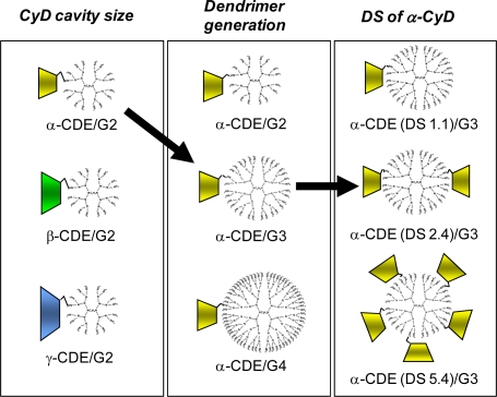
Regarding the delivery of nucleic acid drugs using CyDs, it is acknowledged that CyDs interact with nucleic acids only very slightly [56]. Therefore, the potential of CyDs as carriers for nucleic acids on the basis of their direct interaction would not be expected. In view of this idea, the alternative use of CyDs for carriers of nucleic acids has been required. In addition, the use of CyD and its derivatives for increased transformation efficiency of competent bacterial cells through the interaction between CyDs and bacterial cell wall, not DNA [57]. Meanwhile, Davis and co-workers have reported a number of uses of β-CyD-containing polycations (CDP) with adamantine-PEG or adamantine-PEG-transferrin for gene, DNAzyme and siRNA transfer [46,58–61]. On the other hand, the widespread use of various CyD-appended polymers and polyrotaxanes as gene carriers has been reported, e.g., cationic star polymers consisting of α-CyD core and oligoethylenimine arms [62], PPI dendrimer graft β-CyD [63], low molecular weight PEI cross-linked by HP-β-CyD or HP-γ-CyD [64], low molecular weight PEIs linked by β-CyD [65], linear PEI through γ-CyD and biocleavable polyrotaxane [66], cationic supramolecules consisting of oligoethylenimine-grafted α-CyDs [67] and chitosan/CyD nanoparticles for the airway epithelium [68]. On the other hand, we have reported that CyD-conjugated dendrimers would have a significant impact as non-viral vectors (Figure 2), e.g., we prepared dendrimers (G2, G3, G4) conjugates (CDE) with CyDs [69–71]. Here the reasons why we used dendrimers with low generation and CyDs were their low cytotoxicity and endosome-disrupting effects through the release of membrane components from endosomal membranes after endocytosis, respectively. Of three CDE (G2) with α-, β- or γ-CyD at the molar ratio of 1:1 (dendrimer:CyD), dendrimers (G2) functionalized with α-CyD [α-CDE (G2)] showed luciferase gene expression about 100 times higher than for unfunctionalized PAMAM or for non-covalent mixtures of dendrimer and α-CyD, when pDNA encoding luciferase gene was used [69]. Of various α-CDEs, α-CDE (G3) with the degree of substitution (DS) of 2.4 [α-CDE (G3, DS2)] was revealed to have best transfection efficiency with low cytotoxicity, i.e., the gene transfer activity of α-CDE (G3, DS2) was found to be superior to commercially-available transfection reagents such as TransFast™ (TF) and Lipofectin™ (LF) [70,71]. Moreover, α-CDE (G3, DS2) was found to induce gene expression in spleen after intravenous injection of the pDNA complexes-containing suspension [71]. The enhanced gene transfer activity through the conjugation of α-CyD with dendrimer (G3) could be ascribed to the improved endosomal-escaping ability, i.e., the additive or synergetic effects of the proton sponge effects of dendrimers and the endosomal membrane-disrupting effects of α-CyD, based on the sensing function of α-CyD towards endosomal membranes [72]. However, the transfection efficiency of the pDNA complexes with α-CDEs seems to be still low, probably due to the lack of the translocation ability of the carriers into nucleus. Furthermore, we recently prepared dendrimer conjugates (G2) with glucuronylglucosyl-β-CyD [GUG-β-CDE (G2)] as a gene carrier and clarified the findings that gene transfer activity of GUG-β-CDE (G2) was superior to that of α-CDE (G2) and β-CDE (G2), with negligible cytotoxicity.
Figure 2.
Optimization study of CDE as non-viral carriers.
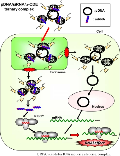
We have revealed that α-CDE (G3, DS2) have potential as siRNA carriers (Figure 3) [73,74]. The luciferase reporter gene system has been widely used to evaluate the efficiency of the siRNA carrier. Firstly, we used the cotransfection system: the ternary complex of luciferase reporter plasmids with siRNA duplexes and a carrier is transfected and it is acknowledged to be useful for simple evaluation of the RNAi effect at the early phase [73]. Then, we examined the sequence specific gene silencing effects using α-CDE (G3, DS2) as a siRNA carrier. Here we evaluated by measuring of luciferase activity after transfected with ternary complexes of DNA/siRNA/α-CDE (G3, DS2) and compared its RNAi effect with the other commercial transfection reagents, i.e. Lipofectamine™ 2000 (L2000), TF and LF. The ternary complex of α-CDE (G3, DS2) induced sequence-specific gene silencing without the off-target effect and its luciferase activity was reduced to half of a control. Meanwhile, all of the commercial transfection reagents used also displayed pGL2 siRNA specific inhibition, but L2000 and TF had non-specific effects on pGL3 siRNA and gave the unstable gene expression effect, compared with α-CDE (G3, DS2). Secondly, we used the transfection system: the binary complex of siRNA/α-CDE (G3, DS2) was transfected to cells transiently and stably expressing luciferase reporter genes. In these systems, α-CDE (G3, DS2) was found to have the potent RNAi effects, compared to L2000 and TF [73]. Thus, α-CDE (G3, DS2) may be a new candidate for a potential therapeutic agent for a siRNA carrier. Thirdly, we examined the in vivo RNAi effect in mice inoculated Colon-26 tumor cells stably expressing luciferase reporter gene [75]. When siRNA complex with α-CDE (G3, DS2) was intratumorally injected, luciferase activity was significantly decreased, but siRNA complexes with L2000 provided the off-target effects. Thus, α-CDE (G3, DS2) has the potential as a siRNA carrier in vitro and in vivo.
Figure 3.
Proposed scheme of RNAi effects of the ternary complexes of pDNA/siRNA/α-CDE.
Recently, shRNA has been developed in order to improve duration of the RNAi effect [8]. Therefore, the shRNA transfer activity of α-CDE (G3, DS2) was compared with that of dendrimer (G3). α-CDE (G3, DS2) formed a stable and condensed complex with shRNA and induced a conformational transition of shRNA in solution even in the low charge ratios. In addition, α-CDE (G3, DS2) markedly inhibited the enzymatic degradation of shRNA by DNase I. The shRNA complex with α-CDE (G3, DS2) at the charge ratio of 20/1 (carrier/shRNA) elicited the most potent RNAi effects in cells transiently and stably expressing the GL3 and GL2 luciferase genes without cytotoxicity. Besides, the RNAi effects were strikingly enhanced by the further addition of the adequate amounts of siRNA to the shRNA complex with α-CDE (G3, DS2). Taken together, the prominent RNAi effects of the shRNA complex with α-CDE could be attributed to its stabilizing effect on enzymatic degradation of shRNA and negligible cytotoxicity. These results suggest that α-CDE (G3, DS2) has the potential to be a novel carrier for shRNA as well as siRNA.
4. Sugar-appended α-CDEs as DNA Carriers
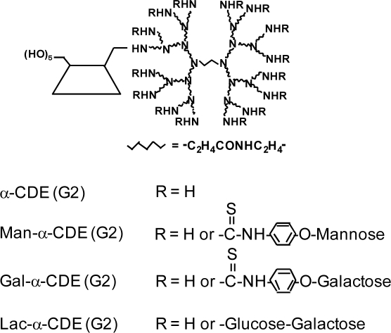
α-CDE (G3, DS2) possesses the potential to be a novel carrier for nucleic acid drugs, but the lack of cell-specific gene transfer activity of α-CDEs has been shown. A carrier system needs to fulfill the following requirements to be a promising candidate for in vivo gene delivery. The carrier should be able to efficiently accumulate in specific target tissues with the lack of toxicity and immunogenicity, and deliver the intact gene into the nucleus of target cell to get high levels of gene expression. Instead of viral vectors, synthetic carriers such as polymers have become an attractive alternative due to their relative safety and their lack of restraints on the size of the pDNA to be delivered. Among the non-viral methods, the glycofection method has recently come to attention [76]. Glycosylated polymers are used for transfection and interact with pDNA to give a glycoplex [77]. In general, glycoplexes are used for delivery to the specific cells and/or to augment gene transfer activity [78]. For example, a mannosylated PEI has high transfection efficiency to macrophages and dendritic cells, which were mediated by the mannose receptor and DEC-205, respectively [79]. Additionally, galactosylated PEI has high transfection efficiency to hepatocytes expressing an asialoglycoprotein receptor (AgpR) [80]. Furthermore, some findings showing glycosyl residues to be very promising candidates of a nuclear targeting signal have been reported [78]. Thus, glycosylation of polymers is an effective method to deliver gene to target cells and/or to enhance gene transfer activity. To possess the cell-specific gene transfer activity of α-CDE (G3, DS2), we prepared the three types of sugar-appended α-CDEs: mannosylated α-CDEs [Man-α-CDEs (G2, G3)] [67,81], galactosylated α-CDEs [Gal-α-CDEs (G2)] [82] and lactosylated α-CDEs [Lac-α-CDE (G2)] [75] with the various degree of substitution (DS) of these sugar moieties (Figure 4).
Figure 4.
Chemical structures of α-CDE (G2) and sugar-appended α-CDEs (G2).
Firstly, to achieve antigen presenting cells (APC)-specific gene delivery of α-CDE (G2), we prepared Man-α-CDE (G2) with the various DS of the mannose moiety (DSM) and evaluated their gene transfer activity in a variety of cells [83], because APC express mannose receptors. Man-α-CDEs (G2, DSM3, 5) were found to have much higher gene transfer activity than dendrimer, α-CDE (G2) and Man-α-CDE (G2, DSM1, 8) in various cells, which are independent of the expression of cell surface mannose receptors. The surface plasmon resonance (SPR) study demonstrated that the specific binding activity of Man-α-CDE (G2, DSM3) to concanavalin A, a mannose lectin, was not very strong. It should be noted that Man-α-CDE (G2, DSM3) provided gene transfer activity higher than dendrimer and α-CDE (G2) in kidney 12 h after intravenous injection in mice. These results suggest the potential use of Man-α-CDE (G2, DSM3) as a non-viral vector, although Man-α-CDE (G2, DSM3) did not show cell-specific gene delivery.
Secondly, to improve APC-specific gene transfer activity of Man-α-CDE (G2), we prepared Man-α-CDEs (G3) with various DSM (5, 10, 13, 20) and compared their cytotoxicity and gene transfer activity, and elucidated the enhancing mechanism for the activity [81]. Of the various carriers used here, Man-α-CDE (G3, DSM10) provided the highest gene transfer activity in NR8383, A549, NIH3T3 and HepG2 cells and the activity of Man-α-CDE (G3, DSM10) was not decreased by the addition of 10% serum in A549 cells. Additionally, no cytotoxicity of the polyplex with Man-α-CDE (G3, DSM10) was observed in A549 and NIH3T3 cells up to the charge ratio of 200:1 (carrier:pDNA). However, the gene transfer activity of Man-α-CDE (G3, DSM10) was independent of the expression of mannose receptors. Interestingly, Alexa-pDNA complex with TRITC-Man-α-CDE (G3, DSM10), but not the complex with TRITC-α-CDE (G3), was found to translocate to the nucleus at 24 h after incubation in A549 cells. HVJ-E vector including mannan, but neither the vector alone nor the vector including dextran, suppressed the nuclear localization of TRITC-Man-α-CDE (G3, DSM10) to a striking degree after 24 h incubation in A549 cells. These results suggest that Man-α-CDE (G3, DSM10) has less cytotoxicity and prominent gene transfer activity through not only its serum resistant and endosome-escaping abilities but also nuclear localization ability, although Man-α-CDE (G3, DSM10) did not elicit cell-specific gene delivery.
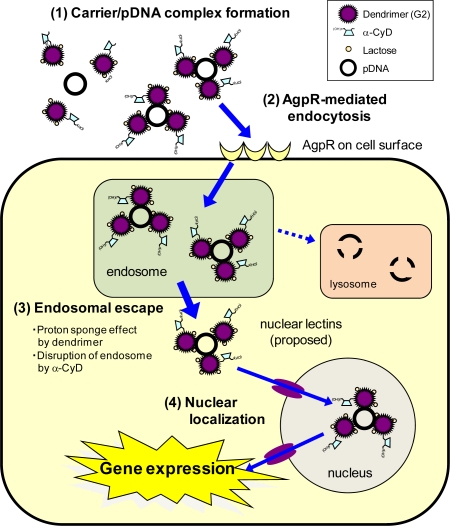
Thirdly, to improve gene transfer efficiency and/or achieve cell-specific gene delivery of α-CDE (G2), we prepared α-CDE bearing galactose [Gal-α-CDE (G2)] with the various DS of the galactose moiety (DSG) as a novel non-viral vector [82]. Gal-α-CDE (G2, DSG4) was found to have much higher gene transfer activity than dendrimer, α-CDE (G2) and Gal-α-CDEs (G2, DSG8, 15) in HepG2, NIH3T3 and A549 cells, which are independent of AgpR expression. Gene transfer activity of Gal-α-CDE (G2, DSG4) was insensitive to the existence of competitors (asialofetuin and galactose) and serum. These results suggest the potential use of Gal-α-CDE (G2, DSG4) as a non-viral vector in various cells, although Gal-α-CDE (G2, DSG4) did not have a cell-specific gene transfer activity. Here we envisaged that α-D-mannopyranosylphenyl isothiocyanate and α-D-galactopyranosylphenyl isothio-cyanate as a space between sugar moiety and dendrimer in the Man-α-CDEs (G2, G3) and Gal-α-CDE (G2) may be involved in the lack of cell-specific gene delivery of these sugar-appended α-CDEs owing to the short length of the spacer. Thereby, we prepared, therefore, α-CDEs bearing α-lactose, a disaccharide formed from α-glucose and α-galactose (Lac-α-CDE) without using the spacer [75]. Of Lac-α-CDEs (G2) having various DS of lactose moiety (DLS1 ,3, 5, 6, 10), Lac-α-CDE (G2, DSL3) was found to have the highest gene transfer activity than dendrimer, α-CDE and the other Lac-α-CDEs in HepG2 cells, AgpR-positive cells, but not in A549 cells, AgpR-negative cells. In addition, the luciferase gene transfer activity of Lac-α-CDE (G2, DSL3) was markedly suppressed in HepG2 cells by adding asialofetuin, a competitor against AgpR, but not bovine serum albumin (BSA). Furthermore, the flow cytometric study showed that cellular association of polyplex with Lac-α-CDE (G2, DSL3) was also suppressed by addition of asialofetuin, not BSA, in HepG2 cells. Thus, it should be noted that Lac-α-CDE (G2, DSL3) provided hepatocyte-selective gene transfer activity through the binding of the carrier to AgpR on HepG2 cells. In fact, the SPR study clearly demonstrated that the association constant of Lac-α-CDE (G2, DSL3) to peanut lectin, a galactose lectin, was approximately 100-fold higher than that of α-CDE (G2). Moreover, Alexa-pDNA complex with TRITC-Lac-α-CDE (G2, DSL3), but not the complex with TRITC-α-CDE (G2), was found to translocate to nucleus in HepG2 cells, suggesting the lactose-mediated nuclear translocation. These proposed hepatocyte-selective gene transfer behaviors of Lac-α-CDE (G2, DSL3) are shown in Figure 5. Importantly, the AgpR-dependent gene delivery of Lac-α-CDE (G2, DSL3) was observed in vivo: Lac-α-CDE (G2, DSL3) provided gene transfer activity much higher than α-CDE (G2) in parenchymal cells and much lower than in spleen 12 h after intravenous injection in mice. In addition, neither cytotoxicity nor change in serum chemistry value was observed, when the complex of pDNA with Lac-α-CDE (G2, DSL3) was applied to culture cells and mice, respectively. Hence, these results hold promise for the potential use of Lac-α-CDE (G2, DSL3) as a hepatocyte-selective non-viral vector with negligible cytotoxicity.
Figure 5.
Proposed scheme for improved effects of gene transfer activity by Lac-α-CDE (G2, DSL3).
5. Folate-appended α-CDEs as DNA Carriers
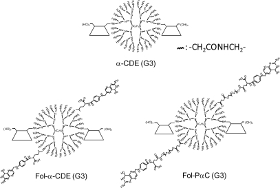
Targeting of the folate receptor (FR) had received much attention in recent years, since the FR has been shown to be over expressed in human cancer cells [84]. Additionally, folic acid is a relatively small molecule (MW 441 Da) which consequently has only limited effects on the dimensions of the carrier system. Some papers regarding folate-appended dendrimers have been published so far. For example, Konda et al. reported the folate-dendrimer MRI contrast agents to the high affinity folate receptor expressed in ovarian tumor xenografts [85]. Shukla et al. demonstrated that folate receptor-targeted boronated PAMAM dendrimers are potential agents for neutron capture therapy [86]. In addition, Singh et al. reported that folate-PEG-dendrimer conjugate was significantly safe and effective in tumor targeting for 5-fluorouracil, an anticancer drug, compared to a non-PEGylated formulation [87]. In an attempt to develop FR-overexpressing cancer cell-specific gene transfer carriers, we prepared folate-appended α-CDEs [Fol-α-CDE (G3)] and folate-PEG-appended α-CDEs [Fol-PαC (G3)] (Figure 6) and evaluated their potential as a novel cell-specific gene transfer carrier. Gene transfer activity of Fol-α-CDEs (DS of folate; DSF2, 5, 7) was lower than that of α-CDE (G3) in KB cells, FR-overexpressing cells. Of the three Fol-PαC (G3, DSF2, 5, 7), Fol-PαC (G3, DSF5) had the highest gene transfer activity in KB cells. This activity was significantly higher than that of α-CDE (G3) in KB cells, but not in A549 cells, FR-negative cells. The cellular uptake of the pDNA complexes with Fol-PαC (G3, DSF5) was inhibited by adding folic acid as a competitor of FR, suggesting the FR-mediated endocytosis. In fact, the SPR data indicated that the association constant of Fol-PαC (G3, DSF5) with folate binding protein (FBP) was approximately 320-fold higher than that of α-CDE (G3). No cytotoxicity of the DNA complex with Fol-PαC (G3, DSF5) was observed in KB cells or A549 cells up to the charge ratio of 100:1 (carrier:DNA), although the DNA complexes with PEI (10 kDa, 25 kDa) showed cytotoxicity even at a charge ratio of 10:1 (carrier:DNA). Most recently, we revealed that DNA complex with Fol-PαC (G3, DSF5) elicited in-vivo gene transfer activity in tumor tissues in mice. In conclusion, potentially, Fol-PαC (G3, DSF5) could be used as a FR-overexpressing cancer cell-selective gene transfer carrier because of FR-mediated gene delivery and the extremely low cytotoxicity.
Figure 6.
Chemical structures of α-CDE (G3), Fol-α-CDE (G3) and Fol-PαC (G3).
6. Conclusions
Many attempts have been made to design and evaluate CyD conjugates with polymers for DNA, shRNA and siRNA carriers. In this review, we have demonstrated the potential of α-CDEs as DNA, shRNA and siRNA carriers. However, their clinical use may be still very limited, so we have sought to extend the function of α-CDEs. Moreover, the development of the sustained release systems of polyplexes with α-CDEs would be required. Elaborate studies are further required to develop novel carriers for various nucleic acid drugs such as gene, shRNA, siRNA, decoy DNA, antisense DNA, ribozyme and aptamers. The future should see certain clinical use products using CyD-containing carriers for DNA and RNA.
Acknowledgments
We would like to express sincere thanks to Dr. K. Uekama and Dr. F. Hirayama, Faculty of Pharmaceutical Sciences, Sojo University, for their valuable advice, warm support and kind help. We thanks Dr. H. Kihara, Dr. K. Wada, Dr. T. Tsutsumi, Ms. Y. Chihara, Mr. S. Yamashita, Ms. M. Arizono, Mr. T. Anno, Mr. M. Mori, Ms. A. Yoshimatsu, Ms. H. Ikeda and K. Hayashida, Graduate School of Pharmaceutical Sciences, Kumamoto University, for their excellent contribution to this study. We appreciate Dr. Hattori and Dr. Takeuchi, Tokyo Polytechnic University, for measurement of SPR. This work was partially supported by Grant-in-Aid for Scientific Research (C) from Japan Society for the Promotion of Science (16590114, 18590144, 20590037).
References and Notes
- 1.Pfeifer A., Verma I.M. Gene therapy: promises and problems. Annu. Rev. Genomics Hum. Genet. 2001;2:177–211. doi: 10.1146/annurev.genom.2.1.177. [DOI] [PubMed] [Google Scholar]
- 2.Lowenstein P.R. Clinical trials in gene therapy: ethics of informed consent and the future of experimental medicine. Curr. Opin. Mol. Ther. 2008;10:428–430. [PMC free article] [PubMed] [Google Scholar]
- 3.Fischer A., Cavazzana-Calvo M. Gene therapy of inherited diseases. Lancet. 2008;371:2044–2047. doi: 10.1016/S0140-6736(08)60874-0. [DOI] [PubMed] [Google Scholar]
- 4.Raty J.K., Lesch H.P., Wirth T., Yla-Herttuala S. Improving safety of gene therapy. Curr. Drug Saf. 2008;3:46–53. doi: 10.2174/157488608783333925. [DOI] [PubMed] [Google Scholar]
- 5.Shen Y. Advances in the development of siRNA-based therapeutics for cancer. IDrugs. 2008;11:572–578. [PubMed] [Google Scholar]
- 6.Ghildiyal M., Zamore P.D. Small silencing RNAs: an expanding universe. Nat. Rev. Genet. 2009;10:94–108. doi: 10.1038/nrg2504. [DOI] [PMC free article] [PubMed] [Google Scholar]
- 7.Castanotto D., Rossi J.J. The promises and pitfalls of RNA-interference-based therapeutics. Nature. 2009;457:426–433. doi: 10.1038/nature07758. [DOI] [PMC free article] [PubMed] [Google Scholar]
- 8.Rao D.D., Vorhies J.S., Senzer N., Nemunaitis J. siRNA vs. shRNA: similarities and differences. Adv. Drug Deliv. Rev. 2009;61:746–759. doi: 10.1016/j.addr.2009.04.004. [DOI] [PubMed] [Google Scholar]
- 9.Blau H.M., Springer M.L. Gene therapy--a novel form of drug delivery. N. Engl. J. Med. 1995;333:1204–1207. doi: 10.1056/NEJM199511023331808. [DOI] [PubMed] [Google Scholar]
- 10.Afione S.A., Conrad C.K., Flotte T.R. Gene therapy vectors as drug delivery systems. Clin. Pharmacokinet. 1995;28:181–189. doi: 10.2165/00003088-199528030-00001. [DOI] [PubMed] [Google Scholar]
- 11.Grimm D. Small silencing RNAs: state-of-the-art. Adv. Drug Deliv. Rev. 2009;61:672–703. doi: 10.1016/j.addr.2009.05.002. [DOI] [PubMed] [Google Scholar]
- 12.Takeda K. Delivery of magic bullets: on the still rocky road to gene therapy. Br. J. Pharmacol. 2009;157:151–152. doi: 10.1111/j.1476-5381.2009.00289.x. [DOI] [PMC free article] [PubMed] [Google Scholar]
- 13.Li S.D., Huang L. Gene therapy progress and prospects: non-viral gene therapy by systemic delivery. Gene Ther. 2006;13:1313–1319. doi: 10.1038/sj.gt.3302838. [DOI] [PubMed] [Google Scholar]
- 14.Akhtar S. Non-viral cancer gene therapy: beyond delivery. Gene Ther. 2006;13:739–740. doi: 10.1038/sj.gt.3302692. [DOI] [PubMed] [Google Scholar]
- 15.Rettig G.R., Rice K.G. Non-viral gene delivery: from the needle to the nucleus. Expert Opin. Biol. Ther. 2007;7:799–808. doi: 10.1517/14712598.7.6.799. [DOI] [PubMed] [Google Scholar]
- 16.Cristiano R.J., Xu B., Nguyen D., Schumacher G., Kataoka M., Spitz F.R., Roth J.A. Viral and nonviral gene delivery vectors for cancer gene therapy. Cancer Detect. Prev. 1998;22:445–454. doi: 10.1046/j.1525-1500.1998.00051.x. [DOI] [PubMed] [Google Scholar]
- 17.Ma H., Diamond S.L. Nonviral gene therapy and its delivery systems. Curr. Pharm. Biotechnol. 2001;2:1–17. doi: 10.2174/1389201013378770. [DOI] [PubMed] [Google Scholar]
- 18.Smaglik P. Tighter watch urged on adenoviral vectors.with proposal to report all ‘adverse events’. Nature. 1999;402:707. doi: 10.1038/45321. [DOI] [PubMed] [Google Scholar]
- 19.Yi Y., Hahm S.H., Lee K.H. Retroviral gene therapy: safety issues and possible solutions. Curr. Gene Ther. 2005;5:25–35. doi: 10.2174/1566523052997514. [DOI] [PubMed] [Google Scholar]
- 20.Boyce N. Trial halted after gene shows up in semen. Nature. 2001;414:677. doi: 10.1038/414677a. [DOI] [PubMed] [Google Scholar]
- 21.Hacein-Bey-Abina S., Von Kalle C., Schmidt M., McCormack M.P., Wulffraat N., Leboulch P., Lim A., Osborne C.S., Pawliuk R., Morillon E., Sorensen R., Forster A., Fraser P., Cohen J.I., de Saint Basile G., Alexander I., Wintergerst U., Frebourg T., Aurias A., Stoppa-Lyonnet D., Romana S., Radford-Weiss I., Gross F., Valensi F., Delabesse E., Macintyre E., Sigaux F., Soulier J., Leiva L.E., Wissler M., Prinz C., Rabbitts T.H., Le Deist F., Fischer A., Cavazzana-Calvo M. LMO2-associated clonal T cell proliferation in two patients after gene therapy for SCID-X1. Science. 2003;302:415–419. doi: 10.1126/science.1088547. [DOI] [PubMed] [Google Scholar]
- 22.Thomas M., Klibanov A.M. Non-viral gene therapy: polycation-mediated DNA delivery. Appl. Microbiol. Biotechnol. 2003;62:27–34. doi: 10.1007/s00253-003-1321-8. [DOI] [PubMed] [Google Scholar]
- 23.Eliyahu H., Barenholz Y., Domb A.J. Polymers for DNA delivery. Molecules. 2005;10:34–64. doi: 10.3390/10010034. [DOI] [PMC free article] [PubMed] [Google Scholar]
- 24.Park J.S., Akiyama Y., Yamasaki Y., Kataoka K. Preparation and characterization of polyion complex micelles with a novel thermosensitive poly(2-isopropyl-2-oxazoline) shell via the complexation of oppositely charged block ionomers. Langmuir. 2007;23:138–146. doi: 10.1021/la061431j. [DOI] [PubMed] [Google Scholar]
- 25.Akagi D., Oba M., Koyama H., Nishiyama N., Fukushima S., Miyata T., Nagawa H., Kataoka K. Biocompatible micellar nanovectors achieve efficient gene transfer to vascular lesions without cytotoxicity and thrombus formation. Gene Ther. 2007;14:1029–1038. doi: 10.1038/sj.gt.3302945. [DOI] [PubMed] [Google Scholar]
- 26.Gao K., Huang L. Nonviral Methods for siRNA Delivery. Mol. Pharm. 2009;6:651–658. doi: 10.1021/mp800134q. [DOI] [PMC free article] [PubMed] [Google Scholar]
- 27.Reischl D., Zimmer A. Drug delivery of siRNA therapeutics: potentials and limits of nanosystems. Nanomedicine. 2009;5:8–20. doi: 10.1016/j.nano.2008.06.001. [DOI] [PubMed] [Google Scholar]
- 28.Tomalia D., Baker H., Dewald J., Hall M., Kallos G., Martin S., Roek J., Ryder J., Smith P. A new class of polymers: starburt-dendritic macromolecules. Polymer J. 1985;17:117–132. [Google Scholar]
- 29.Tomalia D.A. Dendrimer Research. Science. 1991;252:1231. doi: 10.1126/science.252.5010.1231-b. [DOI] [PubMed] [Google Scholar]
- 30.Esfand R., Tomalia D.A. Poly(amidoamine) (PAMAM) dendrimers: from biomimicry to drug delivery and biomedical applications. Drug Discov. Today. 2001;6:427–436. doi: 10.1016/s1359-6446(01)01757-3. [DOI] [PubMed] [Google Scholar]
- 31.Fant K., Esbjorner E.K., Lincoln P., Norden B. DNA condensation by PAMAM dendrimers: self-assembly characteristics and effect on transcription. Biochemistry. 2008;47:1732–1740. doi: 10.1021/bi7017199. [DOI] [PubMed] [Google Scholar]
- 32.Braun C.S., Vetro J.A., Tomalia D.A., Koe G.S., Koe J.G., Middaugh C.R. Structure/function relationships of polyamidoamine/DNA dendrimers as gene delivery vehicles. J. Pharm. Sci. 2005;94:423–436. doi: 10.1002/jps.20251. [DOI] [PubMed] [Google Scholar]
- 33.Dutta T., Jain N.K., McMillan N.A., Parekh H.S. Dendrimer Nanocarriers as Versatile Vectors in Gene Delivery. Nanomedicine. 2009 doi: 10.1016/j.nano.2009.05.005. Epub ahead of print. [DOI] [PubMed] [Google Scholar]
- 34.Hui Z., He Z.G., Zheng L., Li G.Y., Shen S.R., Li X.L. Studies on polyamidoamine dendrimers as efficient gene delivery vector. J. Biomater. Appl. 2008;22:527–544. doi: 10.1177/0885328207080005. [DOI] [PubMed] [Google Scholar]
- 35.Klajnert B., Bryszewska M. Dendrimers: properties and applications. Acta. Biochim. Pol. 2001;48:199–208. [PubMed] [Google Scholar]
- 36.Boussif O., Lezoualc’h F., Zanta M.A., Mergny M.D., Scherman D., Demeneix B., Behr J.P. A versatile vector for gene and oligonucleotide transfer into cells in culture and in vivo: polyethylenimine. Proc. Natl. Acad. Sci. USA. 1995;92:7297–7301. doi: 10.1073/pnas.92.16.7297. [DOI] [PMC free article] [PubMed] [Google Scholar]
- 37.Kukowska-Latallo J.F., Bielinska A.U., Johnson J., Spindler R., Tomalia D.A., Baker J.R., Jr. Efficient transfer of genetic material into mammalian cells using Starburst polyamidoamine dendrimers. Proc. Natl. Acad. Sci. USA. 1996;93:4897–4902. doi: 10.1073/pnas.93.10.4897. [DOI] [PMC free article] [PubMed] [Google Scholar]
- 38.Haensler J., Szoka F.C., Jr. Polyamidoamine cascade polymers mediate efficient transfection of cells in culture. Bioconjug. Chem. 1993;4:372–379. doi: 10.1021/bc00023a012. [DOI] [PubMed] [Google Scholar]
- 39.Morgan D.M., Larvin V.L., Pearson J.D. Biochemical characterisation of polycation-induced cytotoxicity to human vascular endothelial cells. J. Cell Sci. 1989;94(Pt 3):553–559. doi: 10.1242/jcs.94.3.553. [DOI] [PubMed] [Google Scholar]
- 40.Tang M.X., Redemann C.T., Szoka F.C., Jr. In vitro gene delivery by degraded polyamidoamine dendrimers. Bioconjug. Chem. 1996;7:703–714. doi: 10.1021/bc9600630. [DOI] [PubMed] [Google Scholar]
- 41.Hudde T., Rayner S.A., Comer R.M., Weber M., Isaacs J.D., Waldmann H., Larkin D.F., George A.J. Activated polyamidoamine dendrimers, a non-viral vector for gene transfer to the corneal endothelium. Gene Ther. 1999;6:939–943. doi: 10.1038/sj.gt.3300886. [DOI] [PubMed] [Google Scholar]
- 42.Chauhan A.S., Diwan P.V., Jain N.K., Tomalia D.A. Unexpected in vivo anti-inflammatory activity observed for simple, surface functionalized poly(amidoamine) dendrimers. Biomacromolecules. 2009;10:1195–1202. doi: 10.1021/bm9000298. [DOI] [PubMed] [Google Scholar]
- 43.Kuo J.H., Jan M.S., Chiu H.W. Mechanism of cell death induced by cationic dendrimers in RAW 264.7 murine macrophage-like cells. J. Pharm. Pharmacol. 2005;57:489–495. doi: 10.1211/0022357055803. [DOI] [PubMed] [Google Scholar]
- 44.Szejtli J. Medicinal applications of cyclodextrins. Med. Res. Rev. 1994;14:353–386. doi: 10.1002/med.2610140304. [DOI] [PubMed] [Google Scholar]
- 45.Uekama K. Pharmaceutical application of cyclodextrins as multi-functional drug carriers. Yakugaku Zasshi. 2004;124:909–935. doi: 10.1248/yakushi.124.909. [DOI] [PubMed] [Google Scholar]
- 46.Davis M.E., Brewster M.E. Cyclodextrin-based pharmaceutics: past, present and future. Nat. Rev. Drug Discov. 2004;3:1023–1035. doi: 10.1038/nrd1576. [DOI] [PubMed] [Google Scholar]
- 47.Uekama K., Hirayama F., Irie T. Cyclodextrin Drug Carrier Systems. Chem. Rev. 1998;98:2045–2076. doi: 10.1021/cr970025p. [DOI] [PubMed] [Google Scholar]
- 48.Szente L., Szejtli J. Highly soluble cyclodextrin derivatives: chemistry, properties, and trends in development. Adv. Drug Deliv. Rev. 1999;36:17–28. doi: 10.1016/s0169-409x(98)00092-1. [DOI] [PubMed] [Google Scholar]
- 49.Irie T., Otagiri M., Sunada M., Uekama K., Ohtani Y., Yamada Y., Sugiyama Y. Cyclodextrin-induced hemolysis and shape changes of human erythrocytes in vitro. J. Pharmacobiodyn. 1982;5:741–744. doi: 10.1248/bpb1978.5.741. [DOI] [PubMed] [Google Scholar]
- 50.Fauvelle F., Debouzy J.C., Crouzy S., Goschl M., Chapron Y. Mechanism of α-cyclodextrin-induced hemolysis. 1. The two-step extraction of phosphatidylinositol from the membrane. J. Pharm. Sci. 1997;86:935–943. doi: 10.1021/js9602453. [DOI] [PubMed] [Google Scholar]
- 51.Ohtani Y., Irie T., Uekama K., Fukunaga K., Pitha J. Differential effects of α-, β- and γ-cyclodextrins on human erythrocytes. Eur. J. Biochem. 1989;186:17–22. doi: 10.1111/j.1432-1033.1989.tb15171.x. [DOI] [PubMed] [Google Scholar]
- 52.Irie T., Uekama K. Pharmaceutical applications of cyclodextrins. III. Toxicological issues and safety evaluation. J. Pharm. Sci. 1997;86:147–162. doi: 10.1021/js960213f. [DOI] [PubMed] [Google Scholar]
- 53.Uekama K., Otagiri M. Cyclodextrins in drug carrier systems. Crit. Rev. Ther. Drug Carrier Syst. 1987;3:1–40. [PubMed] [Google Scholar]
- 54.Motoyama K., Toyodome H., Onodera R., Irie T., Hirayama F., Uekama K., Arima H. Involvement of lipid rafts of rabbit red blood cells in morphological changes induced by methylated beta-cyclodextrins. Biol. Pharm. Bull. 2009;32:700–705. doi: 10.1248/bpb.32.700. [DOI] [PubMed] [Google Scholar]
- 55.Motoyama K., Kameyama K., Onodera R., Araki N., Hirayama F., Uekama K., Arima H. Involvement of PI3K-Akt-Bad pathway in apoptosis induced by 2,6-di-O-methyl-β-cyclodextrin, not 2,6-di-O-methyl-α-cyclodextrin, through cholesterol depletion from lipid rafts on plasma membranes in cells. Eur. J. Pharm. Sci. 2009 doi: 10.1016/j.ejps.2009.07.010. in press. [DOI] [PubMed] [Google Scholar]
- 56.Arima H. Polyfection as nonviral gene transfer method - design of novel nonviral vector using α-cyclodextrin. Yakugaku Zasshi. 2004;124:451–464. doi: 10.1248/yakushi.124.451. [DOI] [PubMed] [Google Scholar]
- 57.Aachmann F.L., Aune T.E. Use of cyclodextrin and its derivatives for increased transformation efficiency of competent bacterial cells. Appl. Microbiol. Biotechnol. 2009;83:589–596. doi: 10.1007/s00253-009-1907-x. [DOI] [PubMed] [Google Scholar]
- 58.Gonzalez H., Hwang S.J., Davis M.E. New class of polymers for the delivery of macromolecular therapeutics. Bioconjug. Chem. 1999;10:1068–1074. doi: 10.1021/bc990072j. [DOI] [PubMed] [Google Scholar]
- 59.Hwang S.J., Bellocq N.C., Davis M.E. Effects of structure of β-cyclodextrin-containing polymers on gene delivery. Bioconjug. Chem. 2001;12:280–290. doi: 10.1021/bc0001084. [DOI] [PubMed] [Google Scholar]
- 60.Pun S.H., Tack F., Bellocq N.C., Cheng J., Grubbs B.H., Jensen G.S., Davis M.E., Brewster M., Janicot M., Janssens B., Floren W., Bakker A. Targeted delivery of RNA-cleaving DNA enzyme (DNAzyme) to tumor tissue by transferrin-modified, cyclodextrin-based particles. Cancer Biol. Ther. 2004;3:641–650. doi: 10.4161/cbt.3.7.918. [DOI] [PubMed] [Google Scholar]
- 61.Bartlett D.W., Davis M.E. Impact of tumor-specific targeting and dosing schedule on tumor growth inhibition after intravenous administration of siRNA-containing nanoparticles. Biotechnol. Bioeng. 2008;99:975–985. doi: 10.1002/bit.21668. [DOI] [PubMed] [Google Scholar]
- 62.Yang C., Wang X., Li H., Goh S.H., Li J. Synthesis and characterization of polyrotaxanes consisting of cationic alpha-cyclodextrins threaded on poly(ethylene oxide)-ran-(propylene oxide) as gene carriers. Biomacromolecules. 2007;8:3365–3374. doi: 10.1021/bm700472t. [DOI] [PubMed] [Google Scholar]
- 63.Zhang W., Chen Z., Song X., Si J., Tang G. Low generation polypropylenimine dendrimer graft β-cyclodextrin: an efficient vector for gene delivery system. Technol. Cancer Res. Treat. 2008;7:103–108. doi: 10.1177/153303460800700202. [DOI] [PubMed] [Google Scholar]
- 64.Huang H., Tang G., Wang Q., Li D., Shen F., Zhou J., Yu H. Two novel non-viral gene delivery vectors: low molecular weight polyethylenimine cross-linked by (2-hydroxypropyl)-β-cyclodextrin or (2-hydroxypropyl)-γ-cyclodextrin. Chem. Commun. (Camb.) 2006:2382–2384. doi: 10.1039/b601130f. [DOI] [PubMed] [Google Scholar]
- 65.Tang G.P., Guo H.Y., Alexis F., Wang X., Zeng S., Lim T.M., Ding J., Yang Y.Y., Wang S. Low molecular weight polyethylenimines linked by β-cyclodextrin for gene transfer into the nervous system. J. Gene Med. 2006;8:736–744. doi: 10.1002/jgm.874. [DOI] [PubMed] [Google Scholar]
- 66.Ooya T., Choi H.S., Yamashita A., Yui N., Sugaya Y., Kano A., Maruyama A., Akita H., Ito R., Kogure K., Harashima H. Biocleavable polyrotaxane-plasmid DNA polyplex for enhanced gene delivery. J. Am. Chem. Soc. 2006;128:3852–3853. doi: 10.1021/ja055868+. [DOI] [PubMed] [Google Scholar]
- 67.Yang C., Li H., Wang X., Li J. Cationic supramolecules consisting of oligoethylenimine-grafted α-cyclodextrins threaded on poly(ethylene oxide) for gene delivery. J. Biomed. Mater. Res. A. 2008;89A:13–23. doi: 10.1002/jbm.a.31976. [DOI] [PubMed] [Google Scholar]
- 68.Teijeiro-Osorio D., Remunan-Lopez C., Alonso M.J. Chitosan/cyclodextrin nanoparticles can efficiently transfect the airway epithelium in vitro. Eur. J. Pharm. Biopharm. 2009;71:257–263. doi: 10.1016/j.ejpb.2008.09.020. [DOI] [PubMed] [Google Scholar]
- 69.Arima H., Kihara F., Hirayama F., Uekama K. Enhancement of gene expression by polyamidoamine dendrimer conjugates with α-, β-, and γ-cyclodextrins. Bioconjug. Chem. 2001;12:476–484. doi: 10.1021/bc000111n. [DOI] [PubMed] [Google Scholar]
- 70.Kihara F., Arima H., Tsutsumi T., Hirayama F., Uekama K. Effects of structure of polyamidoamine dendrimer on gene transfer efficiency of the dendrimer conjugate with α-cyclodextrin. Bioconjug. Chem. 2002;13:1211–1219. doi: 10.1021/bc025557d. [DOI] [PubMed] [Google Scholar]
- 71.Kihara F., Arima H., Tsutsumi T., Hirayama F., Uekama K. In vitro and in vivo gene transfer by an optimized α-cyclodextrin conjugate with polyamidoamine dendrimer. Bioconjug. Chem. 2003;14:342–350. doi: 10.1021/bc025613a. [DOI] [PubMed] [Google Scholar]
- 72.Arima H. Recent findings of dendrimers and their conjugates as non-viral vectors. Recent Res. Devel. Bioconj. Chem. 2005;2:109–126. [Google Scholar]
- 73.Tsutsumi T., Hirayama F., Uekama K., Arima H. Evaluation of polyamidoamine dendrimer/α-cyclodextrin conjugate (generation 3, G3) as a novel carrier for small interfering RNA (siRNA) J. Control Release. 2007;119:349–359. doi: 10.1016/j.jconrel.2007.03.013. [DOI] [PubMed] [Google Scholar]
- 74.Tsutsumi T., Hirayama F., Uekama K., Arima H. Potential use of polyamidoamine dendrimer/α-cyclodextrin conjugate (generation 3, G3) as a novel carrier for short hairpin RNA-expressing plasmid DNA. J. Pharm. Sci. 2008;97:3022–3034. doi: 10.1002/jps.21206. [DOI] [PubMed] [Google Scholar]
- 75.Arima H., Motoyama K., Hirayama F., Uekama K. Recent Findings of Polyamidoamine Dendrimer Conjugates with Cyclodextrins as DNA and Small-interferring RNA Carriers. The Society of Cyclodextrins; Japan, Tokyo: 2008. [Google Scholar]
- 76.Roche A.C., Fajac I., Grosse S., Frison N., Rondanino C., Mayer R., Monsigny M. Glycofection: facilitated gene transfer by cationic glycopolymers. Cell Mol. Life Sci. 2003;60:288–297. doi: 10.1007/s000180300024. [DOI] [PMC free article] [PubMed] [Google Scholar]
- 77.Fajac I., Briand P., Monsigny M. Gene therapy of cystic fibrosis: the glycofection approach. Glycoconj. J. 2001;18:723–729. doi: 10.1023/a:1020827507748. [DOI] [PubMed] [Google Scholar]
- 78.Monsigny M., Rondanino C., Duverger E., Fajac I., Roche A.C. Glyco-dependent nuclear import of glycoproteins, glycoplexes and glycosylated plasmids. Biochim. Biophys. Acta. 2004;1673:94–103. doi: 10.1016/j.bbagen.2004.03.015. [DOI] [PubMed] [Google Scholar]
- 79.Diebold S.S., Kursa M., Wagner E., Cotten M., Zenke M. Mannose polyethylenimine conjugates for targeted DNA delivery into dendritic cells. J. Biol. Chem. 1999;274:19087–19094. doi: 10.1074/jbc.274.27.19087. [DOI] [PubMed] [Google Scholar]
- 80.Zanta M.A., Boussif O., Adib A., Behr J.P. In vitro gene delivery to hepatocytes with galactosylated polyethylenimine. Bioconjug. Chem. 1997;8:839–844. doi: 10.1021/bc970098f. [DOI] [PubMed] [Google Scholar]
- 81.Arima H., Chihara Y., Arizono M., Yamashita S., Wada K., Hirayama F., Uekama K. Enhancement of gene transfer activity mediated by mannosylated dendrimer/α-cyclodextrin conjugate (generation 3, G3) J. Control Release. 2006;116:64–74. doi: 10.1016/j.jconrel.2006.08.026. [DOI] [PubMed] [Google Scholar]
- 82.Wada K., Arima H., Tsutsumi T., Hirayama F., Uekama K. Enhancing effects of galactosylated dendrimer/α-cyclodextrin conjugates on gene transfer efficiency. Biol. Pharm. Bull. 2005;28:500–505. doi: 10.1248/bpb.28.500. [DOI] [PubMed] [Google Scholar]
- 83.Wada K., Arima H., Tsutsumi T., Chihara Y., Hattori K., Hirayama F., Uekama K. Improvement of gene delivery mediated by mannosylated dendrimer/α-cyclodextrin conjugates. J. Control Release. 2005;104:397–413. doi: 10.1016/j.jconrel.2005.02.016. [DOI] [PubMed] [Google Scholar]
- 84.Jackman A.L., Theti D.S., Gibbs D.D. Antifolates targeted specifically to the folate receptor. Adv. Drug Deliv. Rev. 2004;56:1111–1125. doi: 10.1016/j.addr.2004.01.003. [DOI] [PubMed] [Google Scholar]
- 85.Konda S.D., Aref M., Wang S., Brechbiel M., Wiener E.C. Specific targeting of folate-dendrimer MRI contrast agents to the high affinity folate receptor expressed in ovarian tumor xenografts. Magma. 2001;12:104–113. doi: 10.1007/BF02668091. [DOI] [PubMed] [Google Scholar]
- 86.Shukla S., Wu G., Chatterjee M., Yang W., Sekido M., Diop L.A., Muller R., Sudimack J.J., Lee R.J., Barth R.F., Tjarks W. Synthesis and biological evaluation of folate receptor-targeted boronated PAMAM dendrimers as potential agents for neutron capture therapy. Bioconjug. Chem. 2003;14:158–167. doi: 10.1021/bc025586o. [DOI] [PubMed] [Google Scholar]
- 87.Singh P., Gupta U., Asthana A., Jain N.K. Folate and Folate-PEG-PAMAM dendrimers: synthesis, characterization, and targeted anticancer drug delivery potential in tumor bearing mice. Bioconjug. Chem. 2008;19:2239–2252. doi: 10.1021/bc800125u. [DOI] [PubMed] [Google Scholar]


