Sir,
Demodicidosis is one of the rare cutaneous infections affecting the face. It is characterized by pruritic, erythematous, papulopustular lesions. Its causative organism is the Demodex mite. It can have variable presentations, e.g., pityriasis folliculorum, rosacea-like demodicidosis, or demodicidosis gravis. Here, we present a case of facial demodicidosis and describe its management.
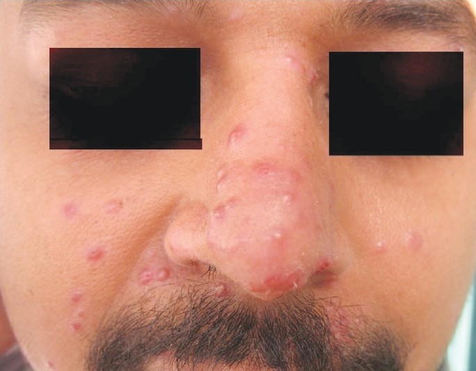
A 28-year-old male presented with the chief complaint of itchy, red, papulopustular eruptions on the central part of the face for the last 4 months. He had been treated with fluconazole, doxycycline, and azithromycin but without any response. There was no history of photosensitivity or flushing episodes. There was no history of pets in the house. Routine general physical examination was normal. On cutaneous examination, the central part of the face was mainly involved, with mild involvement of the external ear. The lesions were erythematous papulopustular eruptions with mild scaling [Figure 1]. There were no telangiectasia, and hair, nail, eye, and mucosal surfaces were normal. Routine hematological examination was within normal limits. Scale scrapings were examined under low-power microscopy after maceration with 40% KOH. No fungal elements were seen but Demodex mites were detected. Subsequently, more skin surface scrapings were taken from different affected sites and almost all samples showed the elongated bodies of the Demodex mite (five or more in a single low-power field). With these findings, the diagnosis of demodicidosis was confirmed and the patient was advised a single oral dose of ivermectin 12 mg and daily application of topical metronidazole gel. After a few days, the pustules started to subside. However, the response was slow and so the patient was put on daily oral metronidazole. Rapid recovery was achieved with oral metronidazole therapy. Subsequent follow-up evaluation showed excellent control of the disease and repeat skin scrapings were negative for Demodex mites.
Figure 1.

Papulopustular lesions over the central part of face
The causative organism of demodicidosis is a saprophytic mite that usually resides in the human pilosebaceous unit. This mite belongs to family Demodicidae of class Arachnida in the order Acarina.[1] The prevalence of infestation with Demodex mites is highest in the 20–30 years age-group, when the sebum secretion rate is at its highest.[2] The infestation may be clinically inapparent but under favorable circumstances these mites may multiply rapidly, leading to the development of different pathogenic conditions such as suppurative or granulomatous skin reactions resembling suppurative folliculitis, rosacea, or perioral dermatitis.[3]
Our patient was in good health, with no signs suggestive of any kind of immunosuppression; this is in contrast with earlier reports, which have suggested that there is an association between demodicidosis and some kind of immunosuppression, e.g., HIV, leukemia.[4]
Our patient responded rapidly after oral metronidazole for few weeks. It has been suggested that metronidazole acts on the mite through one or more of its active metabolites formed in vivo.[5]
To summarize, demodicidosis should be considered in the differential diagnosis of recurrent or recalcitrant facial eruptions. Correct diagnosis, which is imperative for proper treatment, can only be made by finding extrafollicular Demodex mites in the KOH examination, skin surface biopsy, skin biopsy specimens, or a combination of these.
References
- 1.Burns DA. Follicle mites and their role in disease. Clin Exp Dermatol. 1992;17:152–5. doi: 10.1111/j.1365-2230.1992.tb00192.x. [DOI] [PubMed] [Google Scholar]
- 2.Zomorodian K, Geramishoar M, Saadat F, Tarazoie B, Norouzi M, Rezaie S. Facial demodicosis. Eur J Dermatol. 2004;14:121–2. [PubMed] [Google Scholar]
- 3.Hsu CK, Hsu MM, Lee JY. Demodicosis: A clinicopathological study. J Am Acad Dermatol. 2009;60:453–62. doi: 10.1016/j.jaad.2008.10.058. [DOI] [PubMed] [Google Scholar]
- 4.Benessahraoui M, Paratte F, Plouvier E, Humbert P, Aubin F. Demodicidosis in a child with xantholeukaemia associated with type 1 neurofibromatosis. Eur J Dermatol. 2003;13:311–2. [PubMed] [Google Scholar]
- 5.Baima B, Sticherling M. Demodicidosis revisited. Acta Derm Venereol. 2002;82:3–6. doi: 10.1080/000155502753600795. [DOI] [PubMed] [Google Scholar]


