Summary
We describe a patient with a small ruptured azygos anterior cerebral artery aneurysm located at a non-bifurcation distal site on the artery treated successfully with simple coiling.
Key words: subarachnoid hemorrhage, anterior cerebral artery, aneurysm
Introduction
The vast majority of saccular aneurysms associated with azygos anterior cerebral artery (AACA) have been reported to be located at the distal bifurcation of the artery1,2. Rarely they may also be located in the proximal part of the AACA, the A1-A2 junction2. However no information exists regarding a saccular aneurysm located in the middle part of the artery or distally but not at the bifurcation of the artery. Herein we describe a patient who presented with a ruptured distal non-bifurcation AACA aneurysm treated successfully with endovascular approach using a single coil of the smallest size available on the market.
Case Report
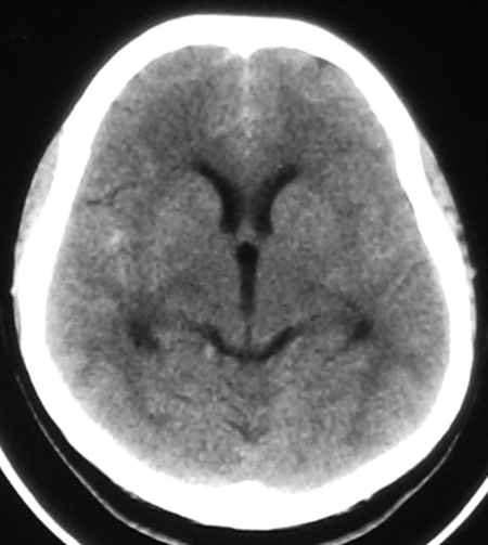
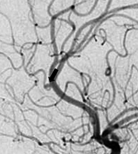
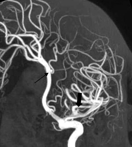
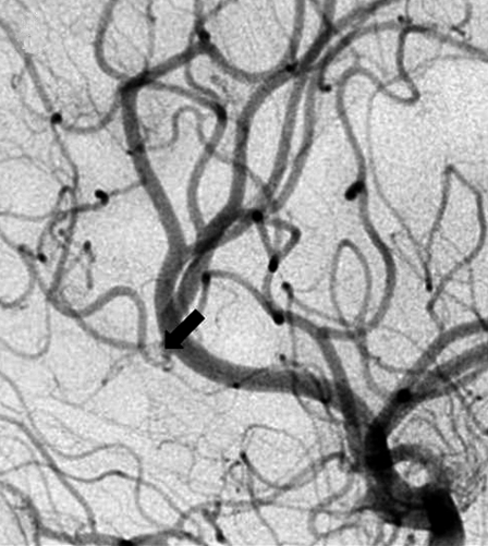
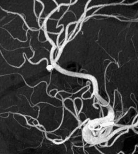
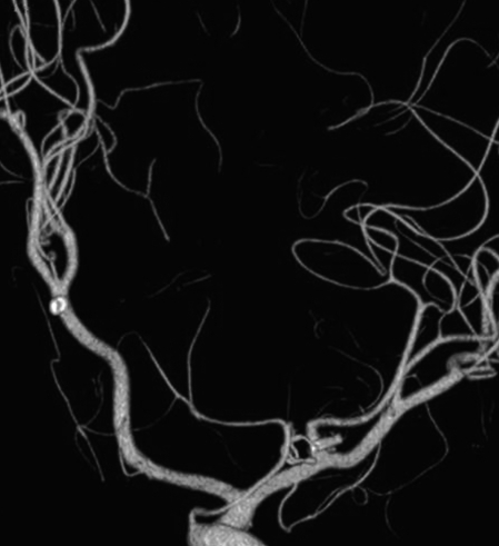
A 52-year-old woman presented with subarachnoid hemorrhage (SAH). She complained of a sudden worsening headache for several hours. Physical examination was unremarkable except some neck stiffness leading to Hunt-Hess grade 2. Subarachnoid hemorrhage involving both sylvian fissures and interhemispheric fissure consistent with a Fisher grade 2 hemorrhage was evident on the non-contrast head CT (Figure 1A). Cerebral DSA showed an azygos anterior cerebral artery with agenesis of the A1 segment of the right anterior cerebral artery. There was a saccular aneurysm measuring a mean diameter of 1.5 mm located on the distal part of the azygos trunk with a frontopolar artery arising close to its neck. A second aneurysm was located at the distal middle cerebral artery which showed a trifurcating pattern (Figure 1B,C). Since the first aneurysm was considered to be ruptured, an endovascular treatment option was recommended to the patient and next of kin in emergency settings and the one on the middle cerebral artery was thought to be treated electively. Under general anesthesia, via right femoral approach, a 6F guiding catheter (Guider Softip; Boston Scientific Target, Fremont, CA, USA) was placed in the right internal carotid artery at about the level of C2 and a microcatheter (Echelon-10; ev3, Irvine, CA, USA) was deployed into the aneurysm over a 0.011 inch with a 45° curved tip micro guidewire (Terumo, Tokyo, Japan). With the intention of preserving the branch arising from aneurysmal neck, only one helical coil with a diameter of 1.5 mm and 20 mm in length (Axium; ev3, Irvine, CA, USA), the smallest diameter available, was sufficient to secure the aneurysm. After surgical clipping of the left MCA aneurysm four days later, patient was eventually discharged from the hospital symptom-free. Control angiography findings three months after the discharge were excellent with no residual filling in either of the aneurysms (Figure 1D-F).
Figure 1.
A) Noncontrast head CT at admission. Note subarachnoid hemorrhage in the interhemispheric fissure and the left sylvian fissure. Left sylvian fissure is not visible at this section due to slight asymmetry of the head position. B,C) Angiography images. B) Close-up lateral view of the left internal carotid artery injection, small distal azygos anterior cerebral artery (AACA) aneurysm with a 1.5 mm diameter on the long axis is shown by the arrow. Note also the branch arising close to its neck. C) 3D angiography sagittal oblique MIP view, thin arrow indicating the distal AACA aneurysm, thick arrrow shows the MCA aneurysm. D-F) Control angiography three months after the discharge. D) Close-up lateral view, left ICA injection arrow shows the aneurysm site. E) Oblique sagittal MIP view of the 3D angiography, coils in the AACA and clip on the MCA aneurysms are seen. F) Volume rendered image, the position of the clip is seen with no residual filling in the MCA aneurysm.
A.
B.
C.
D.
E.
F.
Discussion
Azygos (occurring singly) anterior cerebral (or pericallosal) artery (AACA) is a rare anatomic variation with an incidence of approximately 1.1%, though a wide range of incidence has been reported in different series1-3. Baptista defined three distinct types of distal ACA variations after reviewing 381 brain specimens. Ayzgos ACA constitutes type I anomaly. Type II anomaly is bihemispheric distribution of an A2 segment. The most common anomaly was Type III with a third artery originating from the AcoA. This artery may vary in caliber from the median artery of the corpus callosum to a hyperplastic vessel, which may constitute a diagnostic pitfall and resemble azygos ACA especially if the two A2 segments are small in caliber with early termination4. AACA has also been known to be associated with several midline malformations such as holoprosencephaly and agenesis of the corpus callosum1.
The association of AACA with an aneurysm is less than 13%. The aneurysms found are almost always located at the distal end of the artery, with a few exceptional cases located at the proximal part of the artery1.2. The site of the AACA aneurysm in our case was unusual since, even though it was located distally, it was not at the bifurcation.
Distal ACA aneurysms not associated with an azygos anomaly are usually located at the so-called pericallosal-callosomarginal bifurcation. Distal ACA aneurysms are known to have a worse prognosis than aneurysms at other sites of the anterior circulation, being fragile due to lack of resistent arachnoid membranes at the pericallosal cisterns and the high rate of association with other aneurysms5. Distal ACA aneurysms also pose special difficulties for the surgeon some of which include narrow working space of the interhemispheric fissure, dense adhesions between the cingulate gyri, difficulty in identifying the parent artery and a wide neck1.However to compare the effectiveness of endovascular therapy for distal ACA aneurysms with that of surgery is beyond the scope of this report.
Nevertheless, special emphasis should be placed on the management of very small ruptured aneurysms which were defined as ruptured aneurysms below 3 mm size. Suzuki et Al. performed endovascular treatment in patients with very small ruptured aneurysms based on a criterion of fundus/neck ratio of greater than 1.5. They treated 21 patients with aneurysms located at different sites and had good outcomes in 15 patients, with severe disability in only one patient. They used one coil for each aneurysm and did not make use of balloon remodeling or stenting. Twenty of the 21 coils used were GDC and one was ultrasoft. The smallest coil size they used was 2x40 mm6. Chen et Al. described 11 patients with very small aneurysms, all ruptured except one. They treated six patients with primary coiling, one with balloon remodeling, and they used stent implantation in three cases, two of which needed further coiling. They used only one or two coils in the coiled cases. One patient was lost due to pneumonia but they had good outcome for the remainder7. In a recently published retrospective cohort study, Nguyen et Al found embolization of very small ruptured aneurysms is five times more likely to result in a procedure-related rupture compared with larger aneurysms and they advocated balloon inflation for hemostasis8.
We believe it is worthwhile presenting our findings in this patient due to rather unusual site, very small size and excellent result without complex endovascular methods. However intravascular stent deployment should always be kept in mind especially with an unfavorable fundus/neck ratio7.
References
- 1.Topsakal C, Ozveren MF, et al. Giant aneurysm of the azygos pericallosal artery: case report and review of the literature. Surg Neurol. 2003;60:524–533. doi: 10.1016/s0090-3019(03)00319-7. [DOI] [PubMed] [Google Scholar]
- 2.Fujimoto Y, Yamanaka K, et al. Ruptured aneurysm arising from the proximal end of an azygos anterior cerebral artery: case report. Neurol Med Chir (Tokyo) 2004;44:242–244. doi: 10.2176/nmc.44.242. [DOI] [PubMed] [Google Scholar]
- 3.Huh JS, Park SK, et al. Saccular Aneurysm of the Azygos Anterior Cerebral Artery: Three Case Reports. J Korean Neurosurg Soc. 2007;42:342–345. doi: 10.3340/jkns.2007.42.4.342. [DOI] [PMC free article] [PubMed] [Google Scholar]
- 4.Baptista AG. Studies on the arteries of the brain. II. The anterior cerebral artery: some anatomic features and their clinical implications. Neurology. 1963;13:825–835. doi: 10.1212/wnl.13.10.825. [DOI] [PubMed] [Google Scholar]
- 5.de Sousa AA, Dantas FL, et al. Distal anterior cerebral artery aneurysms. Surg Neurol. 1999;52:128–135. doi: 10.1016/s0090-3019(99)00066-x. [DOI] [PubMed] [Google Scholar]
- 6.Suzuki S, Kurata A, et al. Endovascular surgery for very small ruptured intracranial aneurysms. Technical note. J Neurosurg. 2006;105:777–780. doi: 10.3171/jns.2006.105.5.777. [DOI] [PubMed] [Google Scholar]
- 7.Chen Z, Feng H, et al. Endovascular treatment of very small intracranial aneurysms. Surg Neurol. 2008;70:30–35. doi: 10.1016/j.surneu.2007.05.059. [DOI] [PubMed] [Google Scholar]
- 8.Nguyen TN, Raymond J, et al. Association of endovascular therapy of very small ruptured aneurysms with higher rates of procedure-related rupture. J Neurosurg . 2008;108:1088–1092. doi: 10.3171/JNS/2008/108/6/1088. [DOI] [PubMed] [Google Scholar]


