
Figure 6.
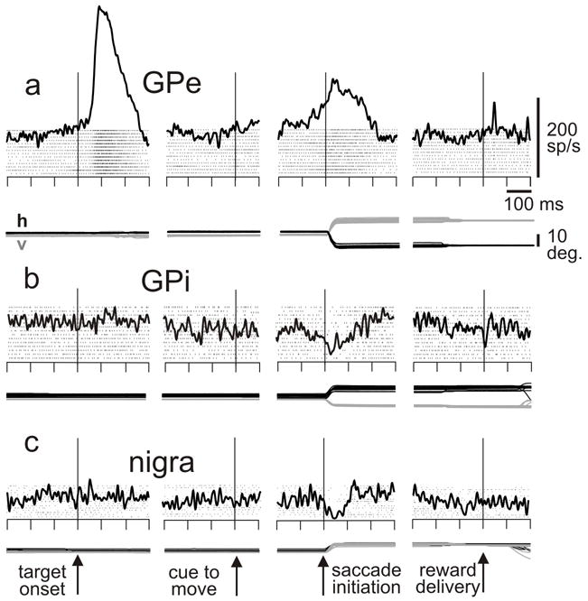
Saccade-related activity in example neurons from the a. external division of the globus pallidus, b. internal division of the globus pallidus, and c. nigra. The monkey was performing a memory-guided saccade task: monkey fixates a spot, a target is flashed in the response field for 50 ms, a delay of 500-1000 ms ensues, the fixation spot disappears (cue to move), the monkey makes a saccade to the remembered location of the target, and liquid reward is delivered for correct performance. Data are aligned as noted. h, v: horizontal and vertical components of eye position. Scales at top right. Modified from Shin and Sommer, 2010.