Summary
This paper reports the management of two clinical cases, in which the upper right central incisor was fused with a supernumerary tooth and the upper left central incisor was macrodontic. A radiographic examination revealed that the fused teeth had two separate roots. Hemisectioning of the fused teeth was performed, the supernumerary portion was extracted and the remaining part was reshaped to remove any sharp margins and to achieve a normal morphology. The macrodontic central incisors were not treated. At 12-months post-surgery there were no periodontal problems and no hypersensitivity. Orthodontic treatment was performed to appropriately align the maxillary teeth and to correct the malocclusion.
Keywords: fusion, fused tooth, macrodontia, hemisection, supernumerary tooth
Introduction
Developmental dental anomalies include abnormalities in tooth size (microdontia and macrodontia), shape (fusion, germination and concrescence), number (anodontia, oligodontia, hypodontia and hyperdontia) and structure (amelogenesis imperfect and dentinogenesis imperfecta), which are all the result of local and systemic factors that affect the initial morphological differentiation stage of dental germs (1,2).
Macrodontia is a rare abnormality in tooth size that manifests clinically as a tooth of normal morphology but of unusually larger size, with a single root and pulp chamber (4). This anomaly involves the entire dentition if it derives from a systemic disorder, and affects mainly the upper central incisors if it derives from a partial defect. The prevalence of macrodontia is calculated as 0.03% and there is no sex preference (3).
Several approaches to the treatment of this condition have been reported in the dental literature, such as restoring the crown shape or prosthetic treatment for esthetic reasons, or extracting the tooth for orthodontic or periodontal complications.
Fusion and germination are tooth shape abnormalities (1) that are often described using the terms “double teeth”, “double formations”, “joined teeth” and “dental twinning”. Fusion is the union of two or more dental germs, and can involve the permanent, primary or supernumerary dentition. Depending on the developmental stage at the time of union, fusion may be incomplete, involving only the tooth crowns, or complete, involving both the crowns and roots. Clinically the fused tooth usually has a wide crown and two independent root canals or, less often, a single root and one or two pulp chambers (4). The crown may exhibit an incised notch, a bifid aspect or an enamel groove between the medial and distal parts, which can extend to the root surface. The maxillary central incisors are the teeth that are most often affected. The prevalence of double teeth differs between studies, ranging from 0.05% to 5% in permanent dentition (2) and 0.7% in deciduous dentition (5). The discrepancy is probably due to racial and geographical differences in the population studied, variable sampling techniques and different diagnostic criteria (1,6,7).
Germination is considered to be incomplete division of a single tooth germ, starting at the incisal edge and aborting before cleavage is complete. Clinically, a germinated tooth is characterized by a bifid crown with a single root and a single or partly divided pulp space (3).
The aetiology and pathogenesis of fusion and germination remain unclear. It was assumed that trauma, heredity, genetic predisposition and environmental factors, such as foetal alcohol exposure, thalidomide embryopathy and hypervitaminosis A of the pregnant mother, may play a role in the formation of double teeth (3,6). Double teeth may also be part of certain syndromes, such as chondroectodermal dysplasia, achondrodysplasia, otodental dysplasia, focal dermal dysplasia and osteopetrosis (3,8,9,10). Double teeth usually occur in the anterior region of the jaws and cause aesthetic, periodontal, functional and orthodontic problems, such as diastema, crowding, protrusion, impaction or ectopic eruption of the adjacent teeth (11,12). Several treatment methods have been described in the literature according to the different types and morphologic variations of anomalous teeth. However the treatment of double teeth requires a multidisciplinary approach to achieve ideal functional and aesthetic results.
Herein we describe the treatment of two patients with dental fusion and macrodontia involving the central incisors.
Clinical cases
Case 1
An 8-year-old Italian boy was referred to the Paediatric Dentistry Unit of the Department of Oral and Maxillofacial Sciences, “Sapienza” University of Rome, with a complaint involving the right maxillary permanent central incisor. There was no history of dental anomalies, systemic diseases or dental trauma. A clinical examination (Fig. 1) revealed the following:
Figure 1.
Case 1. Intraoral photograph.
The right permanent central incisor had a bifid crown and had erupted buccal to the primary lateral incisor;
The right permanent lateral incisor had erupted in the oral position due to insufficient space;
The crown of the left permanent central incisor had a normal morphology but was abnormally large (macrodontia);
A diastema of 3 mm between the central incisors;
The normal number of teeth.
The response to thermal pulp testing and the periodontal probing of the permanent central incisors were normal. The patient was in the mixed dentition stage with a class I molar relationship on the basis of Angle’s classification. In-creased overjet, increased overbite and anterior lower dental crowding were also present.
A radiographic evaluation involving orthopantomography and computed tomography (Dentascan) indicated that the right central incisor had two distinct roots and two separate endodontic spaces, and that the left central incisor had a large pulp chamber and one root canal and macrodontia of the left central incisor (Figs. 2 and 3). The therapeutic options were discussed with the patient and his parents, and sectioning of the fused tooth was planned.
Figure 2.
Case 1. Orthopantomograph.
Figure 3.
Case 1. Computed tomography scan.
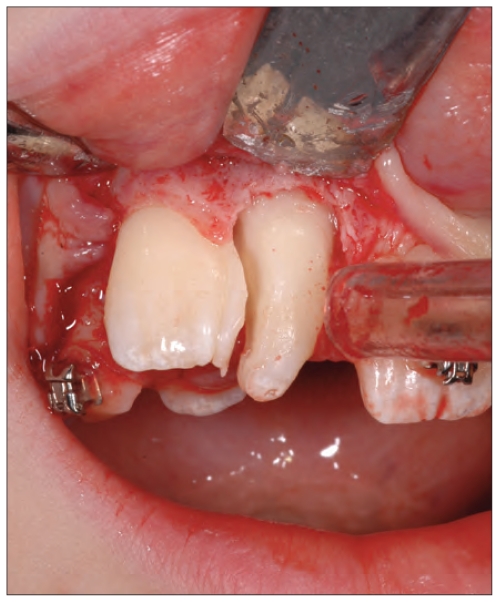
Under local anaesthesia (mepivacaine 2% with adrenalin 1:100,000), the right primary lateral incisor was extracted, and a mucoperiosteal flap, extended from the left permanent central incisor to the right primary canine, was raised until the whole crown of the anomalous tooth was exposed. The fused tooth was separated along the enamel groove, using a high-speed dental hand-drill and a diamond bur, and the mesial portion (supernumerary) was extracted (Figs. 4 and 5). The flap was sutured with 3-0 non-absorbable silk sutures (Ethicon) and the crown of the right maxillary central incisor was redefined with a flame-shaped finishing bur. Reshaping of the crown was necessary to remove the sharp margins and to establish an anatomy consistent with a normal central incisor.
Figure 4.
Case 1. Sectioning the tooth.
Figure 5.
Case 1. The extracted right primary lateral incisor and supernumerary tooth.
An orthodontic device (DAMON type MX3) was applied to allow realignment of the central and lateral incisors and resolution of the malocclusion (Fig. 6). At the 8-month post-surgery recall, the right maxillary central incisor exhibited healthy gingival tissues, no increase in probing depth and a positive response to pulp testing.
Figure 6.
Case 1. Intraoral view at 8-months post-surgery.
Case 2
An 8-year-old Italian boy was referred to the Paediatric Dentistry Unit of the Department of Oral and Maxillofacial Sciences, “Sapienza” University of Rome, with an anomaly of the right maxillary permanent central incisor. His medical history was negative for systemic diseases, dental trauma and familial history of dental anomalies. An intraoral examination revealed the following in the upper arch (Fig. 7):
Figure 7.
Case 2. Intraoral photograph.
A right central incisor had a bifid crown and was delineated by an enamel groove;
A wider than normal left central incisor;
A diastema of 4 mm between the central incisors;
A right lateral incisor that had erupted buccally due to insufficient space;
The normal number of teeth.
Both of the central incisors exhibited positive responses to thermal pulp testing, were asymptomatic to percussion and had no periodontal pockets on probing.
The patient was in the mixed dentition stage with a class I molar relationship and normal overjet. Upper dental crowding was present and the overbite was decreased, with a tendency to an open bite.
Periapical radiographs of the upper central incisors (Fig. 8) indicated that the right one had two separate roots, two pulp chambers and a single bifid crown and that the left one had a single root and one pulp chamber. The diagnosis was fusion of the right central incisor with a supernumerary tooth and a macrodontic left central incisor.
Figure 8.
Case 1. Periapical radiographs of the right (a) and left (b) side.
Surgical sectioning and extraction of the distal part of the fused tooth was scheduled to facilitate orthodontic repositioning of the lateral incisor. The left central incisor was left untreated. The treatment plan was explained to the patient and his family and informed consent was obtained. Before surgery, a fixed orthodontic device (Mc Laughin-Bennett-Trevisi) with 013 wire and elastic low friction ligatures (CuNiTi) was applied to facilitate repositioning of the teeth.
Under local anaesthesia (mepivacaine 2% with adrenalin 1:100,000), the tooth crown was sectioned was performed following the groove in the enamel, using a diamond cylindrical bur under saline irrigation, and the distal portion, aligned with the supernumerary, was extracted using a straight elevator. The remaining tooth was reshaped to remove the sharp edges and to improve the coronal morphology (Fig. 9). Compression was applied for a few minutes using wet gauze to control the bleeding.
Figure 9.
Case 2. Intraoral view of the tooth crown before (a) and after (b) extraction of the mesial portion (supernumerary).
After the surgical treatment, fixed orthodontic treatment was continued in the patient to align the teeth in the arch and to correct the malocclusion (Fig. 10). At the follow-up examinations performed at 3, 6 and 12-months post-surgery, the right central incisor had a positive response to thermal pulp testing and no periodontal pockets were observed on probing.
Figure 10.
Case 2. Intraoral view at 12-months post-surgery.
Discussion
Differentiating between fusion and germination can be difficult, especially when fusion occurs between permanent and supernumerary teeth. Levitas suggested that the number of teeth in the arch should be counted whilst considering the anomalous crown as a single tooth: fusion decreases this number and germination does not (13). This diagnostic criterion becomes untrustworthy either when the fusion involves a supernumerary tooth, because the number of teeth is not reduced, or when germination is associated with agenesis (3).
In the two cases presented here diagnosing fusion between the central incisor and a supernumerary tooth was not difficult, not only because of the normal number of elements in the arch, but also because of the coronal morphology, which was characterized by a groove separating the crown into two components, and the presence of two separate roots.
The diagnosis of the macrodontic tooth which had a single large-than-usual crown, one root and a single pulp chamber was more difficult because it was possible that this element could have represented either macrodontia or complete fusion with a supernumerary in the early stage of the morphological differentiation of the tooth germ. The association in the same dentition between fused teeth at one site and a macrodontic tooth at another site has been reported by several authors (3,4,6).
However the difficulty in differentiating between fusion, germination and macrodontia does not affect the choice of treatment plan, and is rather determined by the type of dentition, the crown morphology, the root and endodontic anatomy, and the orthodontic (diastema, crowding or protrusion), periodontal and aesthetic requirements of the patient.
Several treatments have been proposed in the literature to solve the problems related to an abnormally large element. In deciduous dentition, double or macrodontic teeth do not require therapy unless they interfere with the eruption of the homologous permanent dentition, in which case extraction is necessary (14). As for permanent dentition, there are four types of approaches for fusion and germination:
Extraction in cases where the roots are not separated and no endodontic treatment is possible;
Separation into two single units when the fusion is between two permanent teeth with two separate roots or if it is possible endodontic treatment if possible (9,15);
Hemisectioning and extraction of one tooth portion when the fusion is between a permanent and a supernumerary tooth with two distinct roots or endodontic treatment if possible (10,12,16);
Reshaping of the crown.
In the present cases, fusion of the right central incisors with a supernumerary and the macrodontic left central incisors required two different approaches. The right central incisors, with two roots and two independent endodontic spaces, were surgically separated without endodontic treatment, and the supernumerary tooth was extracted to allow correct alignment of the dental arch. The macrodontic left central incisors, characterized by a single root and single pulp, did not require any kind of therapy.
Hemisectioning of the abnormal teeth was performed under visual control: in case 1 it was necessary to raise a flap to expose the division of the roots, while in case 2 it was sufficient to section the crown using the enamel groove as a guide (14). During the hemisectioning procedure, every effort was made to remove the tooth structure only at the expense of the supernumerary part of the tooth.
The dislocation and extraction of the dental fragment were performed with delicate movements. The elevator was not inserted into the space between the two portions and force was applied only against the portion that was being extracted in order to preserve the remaining part.
More important for treatment outcome is periodic and long-term follow-up to check pulp vitality and periodontal health, since some authors have reported that the sectioning procedure may cause pulp disease as a result of exposing vascular canals in the conserved portion, that may have been shared between the two roots (10,11,17).
Conclusion
To achieve functional and aesthetic objectives double and macrodontic teeth usually require a multidisciplinary approach (endodontic, surgical, prosthetic and orthodontic). The choice of the appropriate treatment plan should be determined by the morphology and the endodontic anatomy of the anomalous tooth and the orthodontic, periodontal and aesthetic requirements of the patient.
References
- 1.Altug-Atac AT, Erdem D. Prevalence and distribution of dental anomalies in orthodontic patients. Am J Orthod Dentofacial Orthop. 2007;131(4):510–4. doi: 10.1016/j.ajodo.2005.06.027. [DOI] [PubMed] [Google Scholar]
- 2.Guttal KS, Naikmasur VG, Bhargava P, Bathi RJ. Frequency of developmental dental anomalies in the Indian population. Eur J Dent. 2010;4(3):263–9. [PMC free article] [PubMed] [Google Scholar]
- 3.Schuurs AH, van Loveren C. Double teeth: review of the literature. ASDC J Dent Child. 2000;67(5):313–25. [PubMed] [Google Scholar]
- 4.Karacay S, Guven G, Koymen R. Management of a fused central incisor in association with a macrodont lateral incisor: a case report. Pediatr Dent. 2006;28(4):336–40. [PubMed] [Google Scholar]
- 5.Wu CW, Lin YT, Lin YT. Double primary teeth in children under 17 years old and their correlation with permanent successors. Chang Gung Med J. 2010;33(2):188–93. [PubMed] [Google Scholar]
- 6.Mattos-Graner RO, Rontani RM, Gavião MB, de Souza Filho FJ, Granatto AP, de Almeida OP. Anomalies of tooth form and number in the permanent dentition: report of two cases. ASDC J Dent Child. 1997;64(4):298–302. [PubMed] [Google Scholar]
- 7.Guimarães Cabral LA, Firoozmand LM, Dias Almeida J. Double teeth in primary dentition: report of two clinical cases. Med Oral Patol Oral Cir Bucal. 2008;13(1):E77–80. [PubMed] [Google Scholar]
- 8.Karaçay S, Gurton U, Olmez H, Koymen G. Multidisciplinary treatment of “twinned” permanent teeth: two case reports. J Dent Child (Chic) 2004;71(1):80–6. [PubMed] [Google Scholar]
- 9.Olivan-Rosas G, López-Jiménez J, Giménez-Prats MJ, Piqueras-Hernández M. Considerations and differences in the treatment of a fused tooth. Med Oral. 2004;9(3):224–8. [PubMed] [Google Scholar]
- 10.Cetinbas T, Halil S, Akcam MO, Sari S, Cetiner S. Hemisection of a fused tooth. Oral Surg Oral Med Oral Pathol Oral Radiol Endod. 2007;104(4):e120–4. doi: 10.1016/j.tripleo.2007.03.029. [DOI] [PubMed] [Google Scholar]
- 11.Kim SY, Choi SC, Chung YJ. Management of the fused permanent upper lateral incisor: a case report. Oral Surg Oral Med Oral Pathol Oral Radiol Endod. 2011 May;111(5):649–52. doi: 10.1016/j.tripleo.2010.11.015. Epub 2011 Feb 18. [DOI] [PubMed] [Google Scholar]
- 12.Tsujino K, Shintani S. Management of a supernumerary tooth fused to a permanent maxillary central incisor. Pediatr Dent. 2010;32(3):185–8. [PubMed] [Google Scholar]
- 13.Levitas TC. Gemination, fusion, twinning and concrescence. ASDC J Dent Child. 1965;32:93–100. [PubMed] [Google Scholar]
- 14.Annibali S, Pippi R, Sfasciotti GL. Chirurgia orale a scopo ortodontico. Elvesier; 2007. [Google Scholar]
- 15.Braun A, Appel T, Frentzen M. Endodontic and surgical treatment of a geminated maxillary incisor. Int Endod J. 2003;36(5):380–6. doi: 10.1046/j.1365-2591.2003.00668.x. [DOI] [PubMed] [Google Scholar]
- 16.Gallo C, Borella L, Velussi C. Anomalie dentarie di numero e di sviluppo. Analisi della letteratura. Dent Cadmos. 2008;76(8):51–68. [Google Scholar]
- 17.Ozalp SO, Tuncer BB, Tulunoglu O, Akkaya S. Endodontic and orthodontic treatment of fused maxillary central incisors: a case report. Dent Traumatol. 2008;24(5):e34–7. doi: 10.1111/j.1600-9657.2008.00635.x. Epub 2008 Jun 28. [DOI] [PubMed] [Google Scholar]


