Type 1 diabetes is an autoimmune disease characterized by the destruction of pancreatic β-cells and an absolute deficiency of insulin. Patients with type 1 diabetes are insulin dependent for life and require multiple daily insulin injections or the use of an insulin pump. It has been considered that β-cell dysfunction and death in type 1 diabetes results from a combination of inflammation, autoimmunity, β-cell stress, and insulin resistance (1–5). Clinical and experimental evidence has indicated that defects in β-cell function precede the massive death of β-cells by severe infiltration of T cells into the islets and the clinical onset of type 1 diabetes (6–9). However, the mechanisms involved in β-cell dysfunction before the onset of clinical type 1 diabetes are unclear. In this issue of Diabetes, Tersey et al. (10) add a new dimension to the progression of type 1 diabetes by demonstrating that endoplasmic reticulum (ER) stress in β-cells precedes the clinical onset of type 1 diabetes.
ER stress as a trigger for β-cell dysfunction in type 1 diabetes
The ER performs a number of important cellular tasks, including protein folding, calcium regulation, redox regulation, and life or death decisions (11,12). Within the β-cell, insulin production and secretion depend on the processing capacity of the ER network. Thus, the ER of the β-cell must maintain homeostatic balance in order to efficiently produce insulin and maintain viability. Perturbations to the ER by genetic or environmental factors can disrupt ER homeostasis and lead to the development of ER stress. Unresolved ER stress has deleterious effects on pancreatic β-cell function and survival, and it can influence the progression of diabetes (11,13). Using NOD mice, a well-established model of type 1 diabetes, Tersey et al. (10) demonstrated activation of ER stress pathways in β-cells prior to the onset of type 1 diabetes. Isolated islets from prediabetic NOD mice displayed age-dependent increases in expression of ER stress markers, morphologic alteration in ER structure by electron microscopy, and activation of the nuclear factor (NF)-κB pathway, which is known to be linked to ER stress. Tersey et al. also showed that MIN6 β-cells treated with a mixture of proinflammatory cytokines, a condition that mimics the immunological microenvironment of type 1 diabetes, displayed evidence of polyribosomal runoff, a finding consistent with ER stress–mediated blockage of translational initiation. Recent clinical and genetic evidence indicates that acquired or inherited ER dysfunction can lead to β-cell death in Wolfram syndrome, a rare genetic form of diabetes, as well as β-cell death in both type 1 and type 2 diabetes. While the role of ER stress in the pathogenesis of type 2 diabetes is well established (14,15), the involvement of this pathway in type 1 diabetes remained elusive. The results of Tersey et al. clearly indicate that ER stress is an important pathogenic component of β-cell dysfunction before the onset of type 1 diabetes.
Targeting ER stress for preventing β-cell dysfunction and autoimmunity in type 1 diabetes
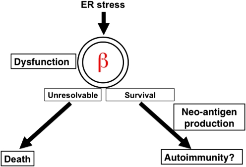
As always, new findings raise new questions. The most intriguing question might be, what are the consequences of ER stress–mediated β-cell dysfunction in type 1 diabetes? The authors (10) provide compelling evidence that the NF-κB pathway is activated in ER-stressed β-cells prior to the onset of type 1 diabetes. NF-κB has been shown to play an important role in β-cell death during progression of type 1 diabetes. Thus, ER-stressed β-cells in prediabetic NOD mice could eventually die through NF-κB signaling and lead to frank diabetes. Another interesting possibility is that ER stress in β-cells may trigger autoimmunity and the severe infiltration of T cells into the islets (Fig. 1). Type 1 diabetes is an autoimmune disease that evolves over years in genetically susceptible individuals who are exposed to unknown environmental triggers. Importantly, all major β-cell autoantigens, including insulin, GAD65, IA-2, ZnT-8, and chromogranin A, traffic through the ER (16). ER dysfunction may cause aberrant changes in the folding and posttranslational modifications of these proteins, leading to the production of neo-self antigens. Neo-self antigens produced from ER-stressed β-cells may trigger autoimmunity in individuals with a susceptible genetic background. In either case, now it is possible to determine subjects with high risk for type 1 diabetes with greater precision using biomarkers for ER-stressed β-cells. The fact that ER function is altered in type 1 diabetes suggests the possibility of using secreted molecules from ER-stressed β-cells as early biomarkers of type 1 diabetes progression. The data by Tersey et al. (10) also suggest that chemical or biological compounds that maintain ER homeostasis could be used for therapeutic purposes. To date, there are no effective therapies targeting the ER for preventing type 1 diabetes. A novel therapeutic strategy that aims to target the common molecular processes that are altered in ER-stressed β-cells may therefore offer promise. Because preventing type 1 diabetes before β-cell function decreases below critical levels may prove to be less challenging than curing established type 1 diabetes (17), further studies on this topic are particularly important for the development of novel prevention, diagnostic, and therapeutic strategies for type 1 diabetes.
FIG. 1.
ER stress as a trigger for β-cell dysfunction and autoimmunity in type 1 diabetes. ER-stressed β-cells in early type 1 diabetes could eventually die through NF-κB signaling and lead to frank diabetes. Another possibility is that ER-stressed β-cells may produce neo-autoantigens and trigger autoimmunity, leading to a severe infiltration of T cells into islets. (A high-quality color representation of this figure is available in the online issue.)
ACKNOWLEDGMENTS
Research in Dr. Urano’s laboratory is supported by grants from the National Institutes of Health, National Institute for Diabetes and Digestive and Kidney Diseases (R01 DK067493); the Diabetes and Endocrinology Research Center at the University of Massachusetts Medical School (5 P30 DK32520); and the Juvenile Diabetes Research Foundation International (40-2011-14).
No potential conflicts of interest relevant to this article were reported.
The authors are grateful to Dr. Aldo Rossini for critical reading of the manuscript and suggestions.
Footnotes
See accompanying original article, p. 818.
REFERENCES
- 1.Atkinson MA, Eisenbarth GS. Type 1 diabetes: new perspectives on disease pathogenesis and treatment. Lancet 2001;358:221–229 [DOI] [PubMed] [Google Scholar]
- 2.Bluestone JA, Herold K, Eisenbarth G. Genetics, pathogenesis and clinical interventions in type 1 diabetes. Nature 2010;464:1293–1300 [DOI] [PMC free article] [PubMed] [Google Scholar]
- 3.Atkinson MA, Bluestone JA, Eisenbarth GS, et al. How does type 1 diabetes develop? The notion of homicide or β-cell suicide revisited. Diabetes 2011;60:1370–1379 [DOI] [PMC free article] [PubMed] [Google Scholar]
- 4.Skyler JS, Ricordi C. Stopping type 1 diabetes: attempts to prevent or cure type 1 diabetes in man. Diabetes 2011;60:1–8 [DOI] [PMC free article] [PubMed] [Google Scholar]
- 5.Eizirik DL, Colli ML, Ortis F. The role of inflammation in insulitis and beta-cell loss in type 1 diabetes. Nat Rev Endocrinol 2009;5:219–226 [DOI] [PubMed] [Google Scholar]
- 6.Strandell E, Eizirik DL, Sandler S. Reversal of beta-cell suppression in vitro in pancreatic islets isolated from nonobese diabetic mice during the phase preceding insulin-dependent diabetes mellitus. J Clin Invest 1990;85:1944–1950 [DOI] [PMC free article] [PubMed] [Google Scholar]
- 7.Keskinen P, Korhonen S, Kupila A, et al. First-phase insulin response in young healthy children at genetic and immunological risk for type I diabetes. Diabetologia 2002;45:1639–1648 [DOI] [PubMed] [Google Scholar]
- 8.Ferrannini E, Mari A, Nofrate V, Sosenko JM, Skyler JS; DPT-1 Study Group Progression to diabetes in relatives of type 1 diabetic patients: mechanisms and mode of onset. Diabetes 2010;59:679–685 [DOI] [PMC free article] [PubMed] [Google Scholar]
- 9.Sreenan S, Pick AJ, Levisetti M, Baldwin AC, Pugh W, Polonsky KS. Increased beta-cell proliferation and reduced mass before diabetes onset in the nonobese diabetic mouse. Diabetes 1999;48:989–996 [DOI] [PubMed] [Google Scholar]
- 10.Tersey SA, Nishiki Y, Templin AT, et al. Islet β-cell endoplasmic reticulum stress precedes the onset of type 1 diabetes in the nonobese diabetic mouse model. Diabetes 2012;61:818–827 [DOI] [PMC free article] [PubMed] [Google Scholar]
- 11.Oslowski CM, Urano F. The binary switch that controls the life and death decisions of ER stressed β cells. Curr Opin Cell Biol 2011;23:207–215 [DOI] [PMC free article] [PubMed] [Google Scholar]
- 12.Walter P, Ron D. The unfolded protein response: from stress pathway to homeostatic regulation. Science 2011;334:1081–1086 [DOI] [PubMed] [Google Scholar]
- 13.Eizirik DL, Cnop M. ER stress in pancreatic beta cells: the thin red line between adaptation and failure. Sci Signal 2010;3:pe7. [DOI] [PubMed] [Google Scholar]
- 14.Ozcan U, Cao Q, Yilmaz E, et al. Endoplasmic reticulum stress links obesity, insulin action, and type 2 diabetes. Science 2004;306:457–461 [DOI] [PubMed] [Google Scholar]
- 15.Ozcan U, Yilmaz E, Ozcan L, et al. Chemical chaperones reduce ER stress and restore glucose homeostasis in a mouse model of type 2 diabetes. Science 2006;313:1137–1140 [DOI] [PMC free article] [PubMed] [Google Scholar]
- 16.Sherr J, Sosenko J, Skyler JS, Herold KC. Prevention of type 1 diabetes: the time has come. Nat Clin Pract Endocrinol Metab 2008;4:334–343 [DOI] [PubMed] [Google Scholar]
- 17.Insel RA, Deecher DC, Brewer J. Juvenile Diabetes Research Foundation: mission, strategy, and priorities. Diabetes 2012;61:30–35 [DOI] [PMC free article] [PubMed] [Google Scholar]


