Abstract
Background
Preliminary evidence suggests selective voluntary motor control (SVMC), defined as performance of isolated voluntary joint movement on request, may be an important factor affecting functional movement tasks. Individuals with poor SVMC are unable to dissociate hip and knee synergistic movement during the swing phase of gait and have difficulty extending their knee while the hip is flexing during terminal swing regardless of hamstring length. This pattern may limit their ability to take advantage of hamstring-lengthening surgery (HLS) and may explain a lack of improved stride length postoperatively.
Questions/purposes
Provide a preliminary clinical and conceptual framework for using SVMC to predict swing phase parameters of gait after HLS.
Patients and Methods
We contrasted two patients with spastic diplegia of similar age, gross motor function, and spasticity but with different SVMC scores using the Selective Control Assessment of the Lower Extremity (SCALE). The patients underwent bilateral HLS. Popliteal angles, joint kinematics, step length, stride length, and walking velocity were assessed pre- and postoperatively.
Result
Popliteal angles, terminal knee extension, and knee range of motion improved for both patients. However, only the patient with higher SCALE scores improved stride length postoperatively.
Conclusion
Although preliminary, the data suggest that SVMC, as measured by SCALE, may be a prognostic factor for improved stride length after HLS in patients with spastic diplegia.
Level of Evidence
Level IV, therapeutic study. See Guidelines for Authors for a complete description of levels of evidence.
Keywords: Medicine & Public Health; Conservative Orthopedics; Orthopedics; Sports Medicine; Surgery; Surgical Orthopedics; Medicine/Public Health, general
Introduction
Hamstring contractures, evidenced by increased popliteal angles, are a frequent finding in patients with spastic diplegic cerebral palsy (CP). The clinical picture may also include excessive knee flexion during stance (crouch), decreased knee ROM, and a shortened stride length [2, 3, 6, 19, 20]. For those cases recalcitrant to nonoperative measures, distal hamstring lengthening surgery has been the mainstay of treatment while addressing concomitant contractures at other levels simultaneously through single event multilevel surgery [10, 14]. Despite surgery, not all patients improve stride length or walking speed postoperatively [19, 20]. One study found that one-third of the patients who improved terminal swing knee extension after hamstring-lengthening surgery (HLS) did not walk with increased hamstring lengths or faster hamstring velocities [1]. Another group of investigators attempted to identify the preoperative characteristics of patients likely to improve after surgical hamstring release; however, they found no major relationships between preoperative and postoperative parameters, including velocity, cadence, and stride length [19].
Although spasticity and contractures are more obvious features of spastic CP, impaired selective voluntary motor control (SVMC) can also negatively affect function [15, 21]. Recently, there has been an effort to provide more precise and comprehensive definitions of pediatric movement disorders. A National Institutes of Health Task Force, created for this purpose, defined selective motor control as the “ability to isolate the activation of muscles in a selected pattern in response to demands of a voluntary movement or posture” [17]. Selective voluntary motor control is the deliberate performance of isolated movement on request as opposed to habitual selective muscle activation during a functional task such as walking [9]. Recently, a clinical assessment tool was developed for SVMC called the Selective Control Assessment of the Lower Extremity (SCALE) [9]. SCALE evaluates the ability of a patient to isolate joint movement without the use of flexor and extensor synergy patterns and without other undesired movements. Good validity and interrater reliability were reported. SVMC has been described as an important predictor of improvement after other CP interventions such as selective posterior rhizotomy [4, 18]. However, SVMC has not been examined as a criterion to improve the selection of patients undergoing hamstring lengthening.
Preliminary evidence indicates SVMC may be an important factor affecting functional movement tasks [8, 15, 21]. Fowler and Goldberg [8] found that individuals with poor SVMC were unable to dissociate hip and knee synergistic movement during the swing phase of gait. Variations in SVMC may explain why knee extension improves to a greater extent during stance than during swing after HLS [2, 3]. During stance, the hip and knee normally extend (synergistic movement), whereas during terminal swing, the hip normally flexes while the knee extends (nonsynergistic movement). Previous research suggests individuals with poor SVMC simultaneously extend the hip and knee during late swing [8]. If a patient undergoing bilateral hamstring lengthening walks in such a pattern, stride length may not improve postoperatively despite improved terminal knee extension. Therefore, hamstring lengthening may improve terminal swing knee extension and stride length only if the individual has the underlying ability to isolate hip and knee movement.
The purpose of this report is to investigate SVMC as a prognostic factor for improved post-HLS stride length in two patients with similar clinical characteristics.
Patients and Methods
We retrospectively analyzed two children with spastic diplegic CP who underwent bilateral HLS. These two patients were selected because they had a similar age and clinical picture with the exception of SVMC (Table 1). In addition to HLS, Participant 1 (P1) underwent bilateral hip flexor and adductor releases and bilateral gastrocnemius recessions. Participant 2 (P2) underwent bilateral hip flexor releases and bilateral calcaneal osteotomies. These simultaneous surgeries were included as part of a single-event multilevel surgery approach to care. This approach minimizes the number of hospitalizations during childhood [10, 14]. Both patients had spastic diplegic CP, were GMFCS Level III, had hamstring spasticity scores of 2, and were of similar age (Table 1). Informed consent and assent, approved by the Institutional Review Board Human Subject’s Protection Committee of our institution, was obtained for both participants.
Table 1.
Patient characteristics and time between analyses for participants who underwent hamstring lengthening surgery
| Participant number | Gender | Preoperative age (years) | Preoperative height (cm) | Preoperative weight (kg) | Time between analyses (years) | GMFCS | Hamstring spasticity score [16] | SCALE score | |
|---|---|---|---|---|---|---|---|---|---|
| Left | Right | ||||||||
| 1 | Male | 10.5 | 123.2 | 26.8 | 1.8 | III | 2 | 2 | 3 |
| 2 | Female | 9.6 | 144.2 | 43.9 | 2.3 | III | 2 | 6 | 5 |
GMFCS = Gross Motor Function Classification System; SCALE = Selective Control Assessment of the Lower Extremity.
The goal of surgical hamstring lengthening is to lengthen the musculotendinous unit by dividing the aponeurotic tissues overlying muscle. The general recommendation for HLS in CP includes sparing the lateral hamstrings. However, as a result of excessive residual tightness in both of these patients, the fascia of the biceps femoris was incised along with the fascia of the semitendinosus, semimembranosus, and gracilis. The surgical method was similar to that described by Horstmann and Bleck [13]. Lengthenings were performed 4 to 6 cm above the popliteal crease through a midline incision of 4 to 6 cm, a location where more muscle substance was likely to be encountered accompanying the tendons and enveloping aponeuroses. The supine position was used, flexing the hip to 90°, but taking care not to forcefully extend the knee beyond its initial excursion. The semitendinosus was lengthened by dividing the tendon and leaving the underlying muscle. The semimembranosus aponeurosis was then exposed by sweeping away the fat from the medial aspect of the muscle and dividing the sheet of overlying fascia. We thinned this muscle somewhat in both these cases because it was quite tight and voluminous. The gracilis was then exposed in the soft tissues medial to the semimembranosus and its tendon divided, again sparing the underlying muscle. Bilateral long leg casts with a removable crossbar were applied.
Postoperatively the patients were kept flat in bed until fully awake. The next day, they were allowed to gradually sit up and begin stretching the hamstrings. Typically, the patients were allowed to sit up to 30° on Postoperative Day 2 and 60° by Day 3. In this manner, we were careful to avoid sciatic or tibial nerve stretch injury while the patients were sedated in the immediate postoperative period. P2 had bilateral calcaneal lengthening surgery and was, therefore, nonweightbearing for 6 weeks. P1 was allowed to bear weight daily in the long leg casts, which were removed at the third week. The patients were instructed to lay prone 50% of the time to keep the hips in extension. Weightbearing was begun within 3 days for P1 and at 6 weeks for P2 (as a result of the calcaneal procedures). The wounds were checked at 2 weeks. At 6 weeks, the casts discontinued for P1 and for P2 were changed to short leg walking casts for an additional 6 weeks. Night splints were used for 6 months. Patients were monitored at 6-month intervals thereafter. Physical therapy was prescribed immediately postoperatively and continued for 6 months at two to three times weekly and then twice weekly for the subsequent 18 months. Outpatient therapy included muscle strengthening, hip and knee ROM, and gait training.
Popliteal angles (knee extension with the hip flexed to 90° while the patient is supine) and knee flexion contractures (maximum knee extension with the hip extended) were evaluated pre- and postoperatively. SVMC was assessed using SCALE [9]. Specific isolated movement patterns at the hip, knee, ankle, subtalar, and toe joints were observed bilaterally. Hip flexion and extension with the knee extended was tested in side lying and the following tests were performed in sitting: (1) knee extension and flexion; (2) ankle dorsiflexion and plantar flexion with the knee extended; (3) subtalar inversion and eversion; and (4) toe flexion and extension. Participants were instructed to move in a reciprocating pattern to a verbal cadence (eg, “flex, extend, flex”). SVMC was graded at each joint as “normal” (2 points), “impaired” (1 point), or “unable” (0 points). An overall SCALE score was calculated by summing the points assigned to each joint for a maximum of 10 points per limb.
Gait analysis was performed in the Kameron Gait and Motion Analysis Laboratory before and after surgery using an eight-camera motion analysis system (Motion Analysis Corporation, Santa Rosa, CA). P1 was studied at 1.8 years postoperatively and P2 at 2.3 years. Participants wore shorts and walked barefoot at a self-selected pace. Each walked back and forth on a 25-foot walkway until at least 10 gait cycles were recorded for each limb. Handheld assistance was provided for balance if required during walking. All data were normalized to 100% of the gait cycle for comparison across participants. The gait cycle was defined from foot strike to foot strike. Each trial was divided into gait cycles and an average obtained for each participant. Kinematics were checked for consistency, and aberrant trials were disregarded. Aberrant trials were those that did not reflect the normal and usual gait of the patient for a very short amount of time, as a result of patient distraction, momentary loss of balance, or other perturbations. No more than two trials were aberrant for each analysis session.
We compared terminal swing knee extension (TKE) and swing knee ROM (KROM) before and after hamstring lengthening. TKE was defined as the knee angle just before initial contact. KROM was calculated by subtracting TKE from peak knee flexion during swing. Step length, stride length, and walking speed were also compared before and after surgery, and were normalized to account for the change in body height (BH) of the patients using the following equations [12]:
Normalized step length = step length *

Normalized stride length = stride length *

Normalized speed = speed *

Step length, stride length, and body height are in meters. Speed is in m/s. Gravity (g) is in m/s2. Normalized parameters are dimensionless.
Medial hamstring lengths were estimated by combining kinematic data obtained from gait analysis with a three-dimensional computer model of the lower extremity using the approach described by Delp et al. [6] and software developed for this purpose (OpenSim 1.9.1 and OpenSMAC 0.9, Simbios; Stanford University, Stanford, CA). The semimembranosus was chosen to represent the medial hamstrings. One representative gait cycle per limb (from foot strike to foot strike) was chosen for analysis and a model was created for each participant [5]. For the scaling of models, muscle origin and insertion points for a generic musculoskeletal model based on cadaver studies were calculated using regression equations [5]. Muscle-tendon lengths of the semimembranosus muscle were calculated throughout the gait cycle pre- and postoperatively. Muscle-tendon lengths for each participant were created based on established relationships between muscle length and joint kinematics based on normative data. Lengths were normalized by dividing by averaged peak length during normal gait to ensure that changes were the result of the surgery and not the growth of the participant.
Results
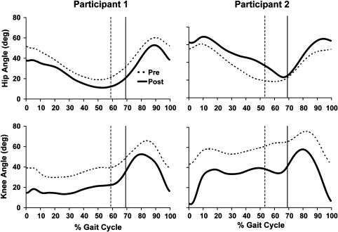
P1 had SCALE scores of 2 and 3 on the left and right, respectively, and P2 had SCALE scores of 6 and 5 (Table 1). Popliteal angles and knee kinematics all showed improvement (Table 2; Fig. 1); P1, with lower SCALE scores, walked slower with a shorter stride length postoperatively. In contrast, P2 with the higher SCALE scores walked at a faster speed and a longer stride length (Table 3).
Table 2.
Knee kinematics for participants before and after surgery
| Participant number | Popliteal angle (degrees) | Terminal knee extension (degrees) | Knee swing ROM (degrees) | |||||||||
|---|---|---|---|---|---|---|---|---|---|---|---|---|
| Left | Right | Left | Right | Left | Right | |||||||
| Pre | Post | Pre | Post | Pre | Post | Pre | Post | Pre | Post | Pre | Post | |
| 1 | 65 | 55 | 65 | 50 | 39 | 16 | 43 | 15 | 26 | 36 | 32 | 38 |
| 2 | 80 | 35 | 60 | 30 | 44 | 7 | 41 | 0 | 32 | 51 | 21 | 49 |
Angle convention: 0 = full knee extension.
Fig. 1.
Hip and knee angles on the left before and after surgery for both participants is shown. The vertical lines indicate toe-off. Positive indicates flexion.
Table 3.
Walking speed, stride length, and step length before and after surgery
| Participant number | Normalized speed | Normalized stride length | Normalized step length | |||||
|---|---|---|---|---|---|---|---|---|
| Pre | Post | Pre | Post | Left | Right | |||
| Pre | Post | Pre | Post | |||||
| 1 | 0.12 | 0.10 | 0.62 | 0.57 | 0.33 | 0.27 | 0.28 | 0.29 |
| 2 | 0.22 | 0.23 | 0.72 | 0.79 | 0.36 | 0.41 | 0.36 | 0.37 |
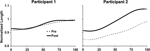
Preoperatively, both participants walked with peak hamstring lengths that were close to the peak length of an individual without disability. P1 walked with essentially the same hamstring lengths during swing both pre- and postsurgery, whereas P2 increased the peak hamstring lengths by 10% and 13% on the left and right, respectively (Table 4; Fig. 2). No knee flexion contractures were present before or after surgery for P1. A 20° knee flexion contracture that was present before the surgery was eliminated postoperatively for P2. Although TKE and KROM improvements occurred for both participants, improved TKE was accompanied by increased hip extension during late swing in P1 (Fig. 1). Although the hip extended slightly at the end of swing for P2, the magnitude of hip extension was much less than that in P1 and occurred later during the swing phase (Fig. 1).
Table 4.
Peak normalized hamstring lengths during swing before and after surgery
| Participant number | Peak hamstring length | |||
|---|---|---|---|---|
| Left | Right | |||
| Pre | Post | Pre | Post | |
| 1 | 1.00 | 0.99 | 0.99 | 1.00 |
| 2 | 0.98 | 1.08 | 0.94 | 1.06 |
Fig. 2.
Normalized hamstring lengths for the left limb for Participants 1 and 2 are shown.
Discussion
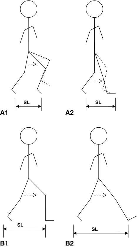
The purpose of these case studies was to investigate SVMC as a potential prognostic factor for improved stride length after HLS. The rationale was that a lack of selective motor control indicates an inability to break out of abnormal synergy (simultaneous flexor and extensor movement patterns of the hip and knee), which has particular importance during terminal swing when the knee normally extends while the hip is flexing. This critical combination of movements results in an increased stride length and occurs independently of total knee excursion or terminal knee extension (Fig. 3). These illustrative cases demonstrate a potential explanation for the variations in response to HLS [1, 19, 20] that has not been previously studied.
Fig. 3A–B.
Stick figures demonstrating how hip-knee coordination can influence step length. Representations of subjects with poor (A) and good SVMC (B) before (1) and after (2) hamstring-lengthening surgery. Only one limb is represented, but it is shown moving from toe-off to initial contact. The limb in the dotted line indicates the position of the limb at maximum hip flexion. This is not present for the figure representing good SVMC because maximum hip flexion occurs at the end of the swing phase. Step length (SL) is labeled in each figure. SVMC = selective voluntary motor control.
This report is limited in a number of ways. First, we studied only two patients; however, they were similar in age, functional classification, spasticity scores, and time to final followup.
Although both patients received additional surgical procedures according to a single-event multilevel surgical protocol, we believe the increased stride length found for P2 was primarily influenced by greater lower limb SVMC. This participant received bilateral hip flexion releases, which can potentially increase step length through improved stance hip extension; however, peak hip extension did not improve postoperatively for either limb. P1 with poor SVMC underwent adductor releases and gastrocnemius recessions in addition to hip flexor releases. These additional procedures could potentially improve stride length by decreasing “scissoring” and providing an improved foot position at initial contact. Postoperatively, stance hip extension increased and equinus decreased in this patient. Despite these improvements, stride length decreased postoperatively.
This study was limited to the swing phase of gait. A second goal of HLS is to diminish crouch during the stance phase. During stance, simultaneous extension of both the hip and knee normally occurs. Regardless of differences in SVMC, both patients benefited from HLS by increasing midstance knee extension.
Another limitation of this study was musculoskeletal modeling is not a direct measure muscle-tendon lengths during gait. Although this technology has been used for prior analysis of hamstring lengthening in children with CP [1], the limitations and accuracy are still being defined under an open-source software policy [5]. More precise techniques such as MRI are currently being developed to determine in vivo muscle lengths [11], but estimates of lengths based on kinematic data are sufficient to gain insight into the mechanisms used during gait after HLS.
These cases illustrate a potential relationship between SCALE scores and postoperative improvements in gait parameters in two children with spastic CP. Although SVMC has been suggested as a prognostic factor for surgical selection in patients with cerebral palsy [4, 7, 8, 18], this is the first study that examines its role using a valid and reliable tool. Both participants improved KROM and TKE during the swing phase of walking, supporting previous studies that showed improved knee kinematics after HLS [2, 3, 19, 20]. Carney et al. [3] and Baumann et al. [2] reported a large improvement in TKE after hamstring lengthening in individuals with CP but did not report hip kinematics, temporal-spatial parameters, nor estimate the degree of SVMC. Simply improving the ROM during gait or the static examination of a contracted joint by HLS may not affect gait dynamics because neural constraints are unchanged. Despite improved knee kinematics in both patients, only the patient with higher SCALE scores improved stride length and walking speed. Examination of the peak hamstring lengths in P1 during walking revealed they were within normal limits before surgery and did not change substantially postoperatively. Although the hamstrings were mechanically lengthened by the surgery, the increased length was not used during terminal swing knee extension. In contrast, P2 used the increased hamstring lengths dynamically. Peak hamstring lengths were longer than that reported for individuals without disability [1] in this participant, which is most likely the result of excessive anterior pelvic tilt throughout the gait cycle.
These case studies illustrate the potential importance of SVMC in predicting outcomes and setting realistic goals for HLS. A larger retrospective study of hamstring lengthening is underway and a prospective study with greater surgical controls and pre-/post-SCALE assessment is currently being planned.
Acknowledgments
We gratefully acknowledge Marcia Greenberg and Loretta Staudt for their assistance with data collection.
Footnotes
One or more of the authors (WLO, EGF) received funding from a grant of the Pediatric Orthopaedic Society of North America.
Each author certifies that his or her institution approved the human protocol for this investigation, that all investigations were conducted in conformity with ethical principles of research, and that informed consent for participation in the study was obtained.
References
- 1.Arnold AS, Liu MQ, Schwartz MH, Ounpuu S, Dias LS, Delp SL. Do the hamstrings operate at increased muscle-tendon lengths and velocities after surgical lengthening? J Biomech. 2006;39:1498–1506. doi: 10.1016/j.jbiomech.2005.03.026. [DOI] [PubMed] [Google Scholar]
- 2.Baumann JU, Ruetsch H, Schurmann K. Distal hamstring lengthening in cerebral palsy. An evaluation by gait analysis. Int Orthop. 1980;3:305–309. doi: 10.1007/BF00266026. [DOI] [PubMed] [Google Scholar]
- 3.Carney BT, Oeffinger D, Meo AM. Sagittal knee kinematics after hamstring lengthening. J Pediatr Orthop B. 2006;15:348–350. doi: 10.1097/01202412-200609000-00008. [DOI] [PubMed] [Google Scholar]
- 4.Chicoine MR, Park TS, Vogler GP, Kaufman BA. Predictors of ability to walk after selective dorsal rhizotomy in children with cerebral palsy. Neurosurgery. 1996;38:711–714. doi: 10.1227/00006123-199604000-00016. [DOI] [PubMed] [Google Scholar]
- 5.Delp SL, Anderson FC, Arnold AS, Loan P, Habib A, John CT, Guendelman E, Thelen DG. OpenSim: open-source software to create and analyze dynamic simulations of movement. IEEE Transactions on Biomedical Engineering. 2007;54:1940–1950. doi: 10.1109/TBME.2007.901024. [DOI] [PubMed] [Google Scholar]
- 6.Delp SL, Arnold AS, Speers RA, Moore CA. Hamstrings and psoas lengths during normal and crouch gait: implications for muscle-tendon surgery. J Orthop Res. 1996;14:144–151. doi: 10.1002/jor.1100140123. [DOI] [PubMed] [Google Scholar]
- 7.Engsberg JR, Ross SA, Collins DR, Park TS. Predicting functional change from preintervention measures in selective dorsal rhizotomy. J Neurosurg. 2007;106:282–287. doi: 10.3171/ped.2007.106.4.282. [DOI] [PMC free article] [PubMed] [Google Scholar]
- 8.Fowler EG, Goldberg EJ. The effect of lower extremity selective voluntary motor control on interjoint coordination during gait in children with spastic diplegic cerebral palsy. Gait Posture. 2009;29:102–107. doi: 10.1016/j.gaitpost.2008.07.007. [DOI] [PubMed] [Google Scholar]
- 9.Fowler EG, Staudt LA, Greenberg MB, Oppenheim WL. Selective Control Assessment of the Lower Extremity (SCALE): development, validation, and interrater reliability of a clinical tool for patients with cerebral palsy. Dev Med Child Neurol. 2009;51:607–614. doi: 10.1111/j.1469-8749.2008.03186.x. [DOI] [PubMed] [Google Scholar]
- 10.Gage JR. Surgical treatment of knee dysfunction in cerebral palsy. Clin Orthop Relat Res. 1990;253:45–54. [PubMed] [Google Scholar]
- 11.Gold GE, Asakawa DS, Blemker SS, Delp SL. Magnetic resonance imaging findings after rectus femoris transfer surgery. Skeletal Radiol. 2004;33:34–40. doi: 10.1007/s00256-003-0702-5. [DOI] [PubMed] [Google Scholar]
- 12.Hof AL. Scaling gait data to body size. Gait Posture. 1996;4:222–223. doi: 10.1016/0966-6362(95)01057-2. [DOI] [Google Scholar]
- 13.Horstmann HM, Bleck EE. Orthopaedic Management in Cerebral Palsy. 2. London: Blackwell Publishing; 2007. [Google Scholar]
- 14.Nene AV, Evans GA, Patrick JH. Simultaneous multiple operations for spastic diplegia. Outcome and functional assessment of walking in 18 patients. J Bone Joint Surg Br. 1993;75:488–494. doi: 10.1302/0301-620X.75B3.8496229. [DOI] [PubMed] [Google Scholar]
- 15.Ostensjo S, Carlberg EB, Vollestad NK. Motor impairments in young children with cerebral palsy: relationship to gross motor function and everyday activities. Dev Med Child Neurol. 2004;46:580–589. doi: 10.1111/j.1469-8749.2004.tb01021.x. [DOI] [PubMed] [Google Scholar]
- 16.Peacock WJ, Staudt LA. Functional outcomes following selective posterior rhizotomy in children with cerebral palsy. J Neurosurg. 1991;74:380–385. doi: 10.3171/jns.1991.74.3.0380. [DOI] [PubMed] [Google Scholar]
- 17.Sanger TD, Chen D, Delgado MR, Gaebler-Spira D, Hallett M. Mink JW and the Taskforce on Childhood Motor Disorders. Definition and classification of negative motor signs in childhood. Pediatrics. 2006;118:2159–2167. doi: 10.1542/peds.2005-3016. [DOI] [PubMed] [Google Scholar]
- 18.Staudt LA, Peacock W. Selective posterior rhizotomy for the treatment of spastic cerebral palsy. Pediatric Physical Therapy. 1989;1:3–9. doi: 10.1097/00001577-198901010-00002. [DOI] [Google Scholar]
- 19.Thometz J, Simon S, Rosenthal R. The effect on gait of lengthening of the medial hamstrings in cerebral palsy. J Bone Joint Surg Am. 1989;71:345–353. [PubMed] [Google Scholar]
- 20.Linden ML, Aitchison AM, Hazlewood ME, Hillman SJ, Robb JE. Effects of surgical lengthening of the hamstrings without a concomitant distal rectus femoris transfer in ambulant patients with cerebral palsy. J Pediatr Orthop. 2003;23:308–313. doi: 10.1097/00004694-200305000-00007. [DOI] [PubMed] [Google Scholar]
- 21.Voorman JM, Dallmeijer AJ, Knol DL, Lankhorst GJ, Becher JG. Prospective longitudinal study of gross motor function in children with cerebral palsy. Arch Phys Med Rehabil. 2007;88:871–876. doi: 10.1016/j.apmr.2007.04.002. [DOI] [PubMed] [Google Scholar]


