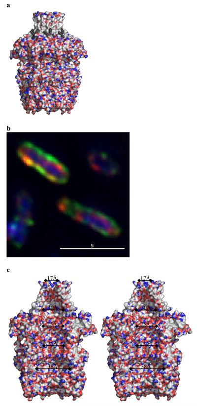
Figure 3. The central cavity of Wza.
a, Wza is shown as a surface with polar atoms colored (colors are Fig. 2c.). Wza is oriented as the Fig. 2b. The lipid molecules are shown as black spheres and are located near the top of the structure. There are no gaps in the through the walls of the structure. The helical barrel is clearly non-polar and a band of tryptophan residues is exposed at the base of helical barrel (Fig S4b). b Green fluorescent anti-PK antibody binds to the surface of the cells which express Wza with a PK tag added to the C-terminus (Wza-PK). The periplasm is located by a red anti- alkaline phosphatase antibody and the nucleus is stained blue by DAPI. This confirms the C-terminus of Wza-PK is exposed on the surface of cells and thus the orientation of Wza is as shown in Fig. 2b. c, A stereo diagram of the internal cavity. The surface is colored according to polarity, (oxygen red, nitrogen blue and carbon, selenium, sulfur white) and reveals the interior of Wza is polar. The cavity is open at top through the helical barrel.

