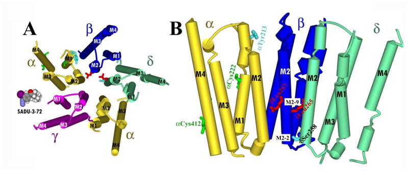
Figure 9. Molecular model of sites of [125I]-SADU-3-72 labeling in the Torpedo nAChR structure (PDB # 2BG9).
Residues photolabeled by [125I]-SADU-3-72 within the transmembrane domain of the Torpedo nAChR. Views of the membrane-spanning helices (shown as cylinders) of the Torpedo nAChR structure (PDB # 2BG9): A, looking down the channel from the base of the extracellular domain; and B, looking parallel to the membrane with 2 subunits removed for clarity, rotated 90° from (A). Subunits are color-coded: α, yellow; β blue; and δ, green. Residues photolabeled by [125I]-SADU-3-72 are included in stick format, color-coded by domain and conformation: ion channel, resting state (red); ion channel, desensitized state (cyan); lipid-protein interface (green). A Connolly surface model of [125I]-SADU-3-72 is included in A for scale.

