Abstract
Bardet-Biedl Syndrome (BBS, MIM#209900) is a genetically heterogeneous disorder with pleiotropic phenotypes that include retinopathy, mental retardation, obesity and renal abnormalities. Of the 15 genes identified so far, seven encode core proteins that form a stable complex called BBSome, which is implicated in trafficking of proteins to cilia. Though BBS9 (also known as PTHB1) is reportedly a component of BBSome, its direct function has not yet been elucidated. Using zebrafish as a model, we show that knockdown of bbs9 with specific antisense morpholinos leads to developmental abnormalities in retina and brain including hydrocephaly that are consistent with the core phenotypes observed in syndromic ciliopathies. Knockdown of bbs9 also causes reduced number and length of cilia in Kupffer's vesicle. We also demonstrate that an orthologous human BBS9 mRNA, but not one carrying a missense mutation identified in BBS patients, can rescue the bbs9 morphant phenotype. Consistent with these findings, knockdown of Bbs9 in mouse IMCD3 cells results in the absence of cilia. Our studies suggest a key conserved role of BBS9 in biogenesis and/or function of cilia in zebrafish and mammals.
Introduction
The cilium is a specialized organelle projecting from plasma membrane of most polarized cell types in vertebrates [1], [2]. The cilium develops from the basal body that is in turn derived from the mother centriole and participates in many fundamental signaling pathways, including those associated with sonic hedgehog [3], Wnt [4] and planar cell polarity (PCP) [5]. The vertebrate cilium is estimated to contain over 1000 proteins for its structural and/or functional integrity [6] (http://www.ciliaproteome.org). Defects in cilia biogenesis are associated with pleiotropic syndromic phenotypes, collectively referred as ciliopathies; these include Bardet-Biedl Syndrome (BBS), Meckel-Gruber Syndrome (MKS), Joubert Syndrome (JBTS), and Nephronophthesis (NPHP) [7].
Bardet-Biedl syndrome (MIM#209900) is typically an autosomal recessive disorder that exhibits variable expressivity and phenotypes including retinopathy, mental retardation, obesity, polydactyly, and renal abnormalities [8], [9]. Mutations in fifteen genes are reported to account for 80% of the BBS cases [9], [10]; a few of these are also associated with the pathogenesis of related ciliopathies. Despite tremendous genetic heterogeneity, all BBS proteins are localized to centrosome, basal body or the ciliary transition zone [9], [11], [12], [13], [14], [15]. Investigations using mouse and zebrafish models have demonstrated the cilia-associated functions of BBS8, BBS4 and other BBS proteins [5], [13], [16], [17]. Similarities in clinical phenotypes and cellular localization have suggested interaction(s) among different BBS proteins and their participation in cilia biogenesis, signaling or transport. Identification of two multiprotein complexes that include BBS proteins has provided key biochemical and functional insights into cilia biology and disease. BBSome, a stable complex of seven core BBS proteins (BBS1, BBS2, BBS4, BBS5, BBS7, BBS8, BBS9) is implicated in cilia trafficking and biogenesis [18], whereas the chaperonin complex (comprising of BBS6, BBS10, and BBS12) seems to mediate the assembly of BBSome [19].
BBS9 (also called PTHB1) was originally identified by differential display analysis as a gene (B1) down regulated by parathyroid hormone (PTH) in an osteoblastic cell line [20]. Multiple variant isoforms of PTHB1 are expressed in different tissues, and the gene is interrupted in a translocation associated with Wilms' Tumor 5 [21]. More recently, haplotypes in the region of PTHB1 have been associated with the pathogenesis of premature ovarian failure, a complex multifactorial disease that causes female infertility [22]. Independent genetic studies, involving comparative mapping and gene expression analysis, led to the identification of PTHB1 as a novel BBS gene – BBS9 [23]. Mutations in BBS9 account for 6% of BBS mutations [9].
Though BBS9 protein is shown to be a part of BBSome core [18], its precise physiological function is not delineated, and the mechanism of disease pathogenesis caused by BBS9 mutations is poorly understood [23]. The zebrafish (Danio rerio) has been used as an excellent system to model human diseases, especially those involving ciliary protein functions, by knockdown using morpholino (MO) technology [16]. Knockdown of many cilia genes in zebrafish are reported to cause developmental abnormalities in the eye, brain and somites [24], [25], [26]. Here we report that bbs9 knockdown results in ciliogenesis defects in zebrafish and in mouse IMCD3 cells.
Results
A BBS9 ortholog is expressed in the zebrafish during development
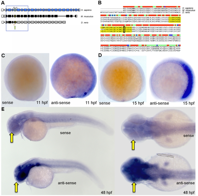
In order to test the in vivo function of bbs9 using zebrafish, we first identified the zebrafish ortholog of human BBS9 gene (NM_198428) using available resources from GenBank (XM_002664792.1), Zfin (AL845419) and GENESCAN program (http://genes.mit.edu/GENESCAN.html). The predicted transcript codes for a protein of 904 amino acids that shows 63% identity and 79% similarity with human BBS9 protein (Fig. 1A, B). A multi-species comparison of BBS9 protein sequences revealed high level of conservation from exons 2 to 8 among human, mouse and zebrafish (Fig. 1B). Furthermore, the region of zebrafish bbs9 on chromosome 16 is syntenic with the human BBS9 locus on chromosome 7. Currently, there is no evidence for additional copies of bbs9 in zebrafish genome.
Figure 1. Zebrafish bbs9 gene: structural comparison and expression pattern.
(A) A comparison of BBS9 exon:intron structure between human (H. sapiens, top blue), mouse (M. musculus, middle black) and zebrafish (D. rerio, bottom gray/black). The filled and open boxes indicate coding exons and UTRs, respectively. The blue and black boxes represent validated exons. The gray boxes represent exons present in provisional sequence XM_002664792.1. Exons 2 to 8 are highly conserved across species (boxed area within hatched square). The yellow arrow points to yellow mark on exon 5, which represents the missense mutation G→A (p.G141R) in human BBS9 protein. Under the zebrafish bbs9 transcript, the red line represents bbs9-spMO targeting site at intron4:exon5 boundary. (B) The protein sequence alignment (clustalW) between human (NP_940820.1), mouse (NP_848502.1) and predicted zebrafish BBS9 (904 amino acids). Exon 5 is highlighted (yellow), and the position of missense mutation (p.G141R) in human is highlighted by a black rectangle. The bar coding on top of the sequences represents degree of conservation (red and blue represent maximum and minimum conservation, respectively). (C, D) In situ hybridization analysis at 11 hpf and 15 hpf. Left and right panels represent the sense and anti-sense probes generated from bbs9 cDNA. (E) In situ hybridization analysis at 48 hpf. Expression of bbs9 in the eye, brain and somites gives a strong signal with the anti-sense probe compared to the background signal from the sense probe. Compare the strong signal in the head regions (arrows). Left and right panels represent lateral and dorsal views, respectively.
To analyze the expression of bbs9 during development, we generated a partial cDNA spanning exons 2–5 by RT-PCR from 72 hour post-fertilization (hpf) embryos. In situ hybridization with antisense mRNA probe generated from this cDNA to 11 hpf zebrafish embryos showed almost ubiquitous expression (Fig. 1C); however, by 15 hpf, bbs9 expression became restricted to the anterior portion of the embryo (Fig. 1D). At 48 hpf, bbs9 transcripts were expressed in high levels in eyes and brain, while the somites displayed low level expression (Fig. 1E).
Validation of bbs9 morpholinos
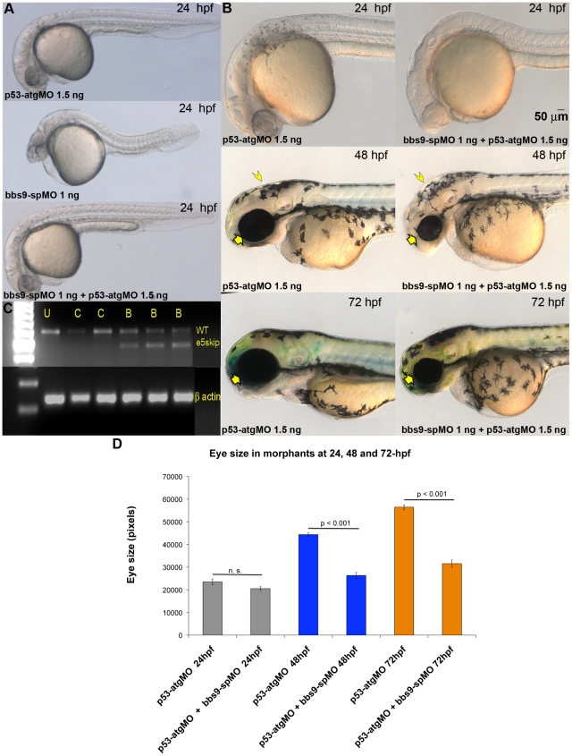
As the only known missense mutation in BBS9 patients is in exon 5 and another frame-shift mutation in intron 4 affected exon 5 [23], we designed an exon-skipping morpholino (bbs9-spMO) targeting the intron 4:exon 5 boundary of zebrafish bbs9 gene (Fig. 1A, red underline). To examine whether bbs9-spMO indeed blocked splicing, we performed RT-PCR analysis using RNA from control-MO and bbs9-spMO injected morphants (Fig. 2C). A single 575 bp wild type RT-PCR product was observed in control-MO injected morphants, whereas in bbs9-spMO injected morphants a shorter product (461 bp, presumably generated by exon 5 skipping, termed e5skip) was detected in addition to the 575 bp band (Fig. 2C). The presence of two bands in the latter indicated an incomplete effect of bbs9-spMO in the morphants.
Figure 2. Exon 5-targeted bbs9 splice morpholino affects eye development independent of p53 pathway.
(A) At 24 hpf, the p53-atgMO (1.5 ng) alone injection did not elicit a phenotype. The bbs9-spMO (1 ng) injection alone caused developmental defects in the eye, brain and tail of morphants. However, co-injection of p53-atgMO reduced the defects seen by the bbs9-spMO injection alone, though mild eye defect remained the tail becomes normal (bottom panel). (B) Higher magnification of morphants' head region. Top, middle and bottom rows are 24-, 48- and 72-hpf, respectively. Left and right column of panels are p53-atgMO without and with bbs9-spMO, respectively. At 48 hpf the effect of bbs9-spMO injection on eye size visible (compare the arrows). The bbs9-spMO injection also resulted in hydrocephalous (compare the arrow heads). The defects seen at 48 hpf are weaker at 72 hpf. (C) The gel photograph of RT-PCR showing exon-skipping by bbs9-spMO. mRNA isolated from individual embryos was used for RT-PCR. U, C (4 and 6 ng) and B (1, 4, 6 ng) represent un-injected, control, and bbs9-spMO, respectively. Splice blocking gave an additional smaller (marked e5skip) band along with the original WT band. The bottom panel shows β-actin control for respective samples. (D) Quantification of the effect of morpholino(s) injection on eye size. X-axis shows the morpholinos used and time (hpf) of scoring. Y-axis shows eye size in pixels (mean ± SEM).
bbs9 knockdown causes developmental defects in zebrafish
Microinjection of bbs9-spMO into wild type embryos resulted in severe morphogenesis defects (Fig. 2). At 48 hpf, embryos injected with 1 ng bbs9-spMO showed a striking defect in the eye and a conspicuous hydrocephaly in most cases (Fig. 2B, middle panels). In initial studies with the bbs9-spMO extensive cell death was seen at 24 hpf and there was dose dependent malformation of the trunk and tail (Fig. 2A). As some morpholinos are known to produce p53 dependent cell death [27], we co-injected p53-atgMO to determine if this cell death is responsible for a subset of the observed phenotypes. In embryos co-injected with 1 ng bbs9-spMO and 1.5 ng p53-atgMO, trunk and tail malformations was suppressed (Fig. 2A, bottom panel) compared to embryos injected with bbs9-spMO only (Fig. 2A, middle panel). However, reduction in the size of the eye and hydrocephaly was consistently observed at 48 hpf in embryos co-injected with p53-atgMO (Fig. 2B, middle row, right panel), and a statistically significant reduction in eye size was seen at 48 and 72 hpf (Fig. 2D), confirming that these changes are bona fide effects of reduced bbs9 function. The bbs9-spMO morphant phenotype observed with p53-atgMO co-injection is reminiscent of what has been reported for BBS patients with clinical manifestations in multiple organs - including eye and brain (see MIM ID #209900).
To confirm the results obtained using the splice blocking bbs9 morpholino, we designed a bbs9-atgMO to target the first translational initiation site, aug, in the predicted zebrafish bbs9 open reading frame (Fig. S1). After 48 hpf, bbs9-atgMO injected morphants, co-injected with p53-atgMO, displayed a similar reduction in eye size (data not shown) though, overall, bbs9-atgMO injected morphants displayed a slightly milder phenotype, with no hydrocephaly.
Human BBS9 mRNA rescues bbs9 knockdown phenotype in the zebrafish
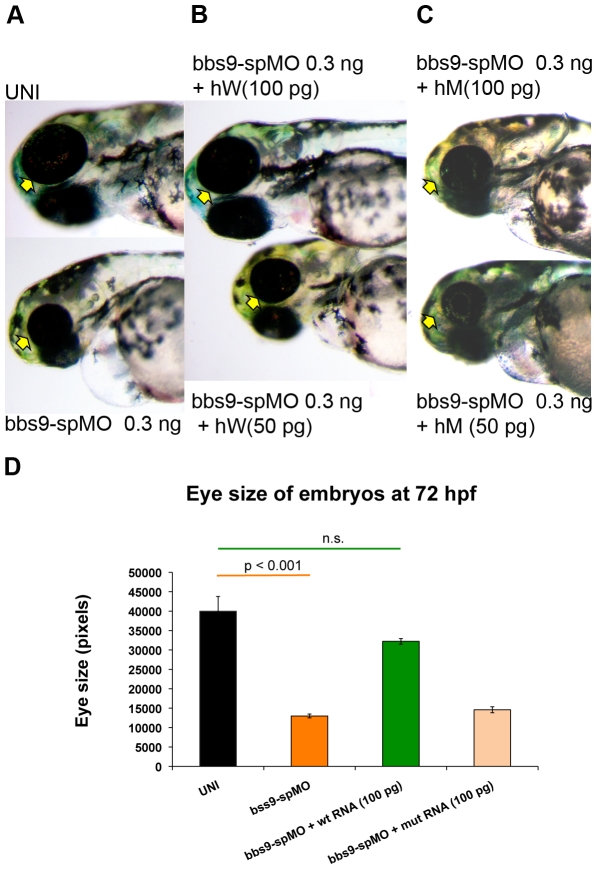
To confirm the specificity of the bbs9-spMO morphant phenotype, we asked whether wild type human BBS9 mRNA could rescue the bbs9-spMO injected morphants. Co-injection of 0.3 ng bbs9-spMO along with wild type human BBS9 mRNA rescued the morphant phenotype in a dose-dependent manner (Fig. 3). The bbs9-spMO injection alone resulted in morphants with reduced eye size. Co-injection of bbs9-spMO with 100 pg of wild-type human mRNA significantly improved the eye size (Fig. 3B, D). Analysis of RNA from the rescued zebrafish revealed an effective splice blocking of zebrafish bbs9 transcript (data not shown), suggesting that the phenotypic rescue was indeed by the human mRNA.
Figure 3. Human mRNA rescues zebrafish bbs9-spMO phenotype.
hW and hM represent wild type and mutant human mRNA, respectively. The arrows indicate eye phenotype. (A) The uninjected control (top) and bbs9-spMO alone injected (bottom) zebrafish at 72 hpf. (B) Rescue of bbs9-spMO eye phenotype by hW 100 pg (top), but not by lower dose of 50 pg (bottom). (C) The bbs9-spMO phenotype is not rescued by hM as the eye defect remains in the morphants. (D) The quantification of embryos' eye size at 72 hpf in rescue experiment using human mRNAs co-injected with bbs9-spMO. X-axis shows category of embryos scored. Y-axis shows the eye size in pixels. Data are presented as mean ± SEM. Statistically significant and non-significant observations are indicated with p value and n.s., respectively.
A comparable phenotype produced by exon 5 mutation in a BBS9 patient and by the bbs9-spMO in zebrafish prompted us to evaluate whether BBS9 mRNA carrying the exon 5 missense mutation (amino acid change, G141R) [23] could complement the abnormal morphant phenotypes. As predicted, the co-injection of 100 pg of missense mutant mRNA with bbs9-spMO failed to rescue the defects in morphants (Fig. 3C, D). Our data further suggest that human and zebrafish BBS9 proteins are highly conserved at the functional level.
bbs9 is required for photoreceptor and brain development
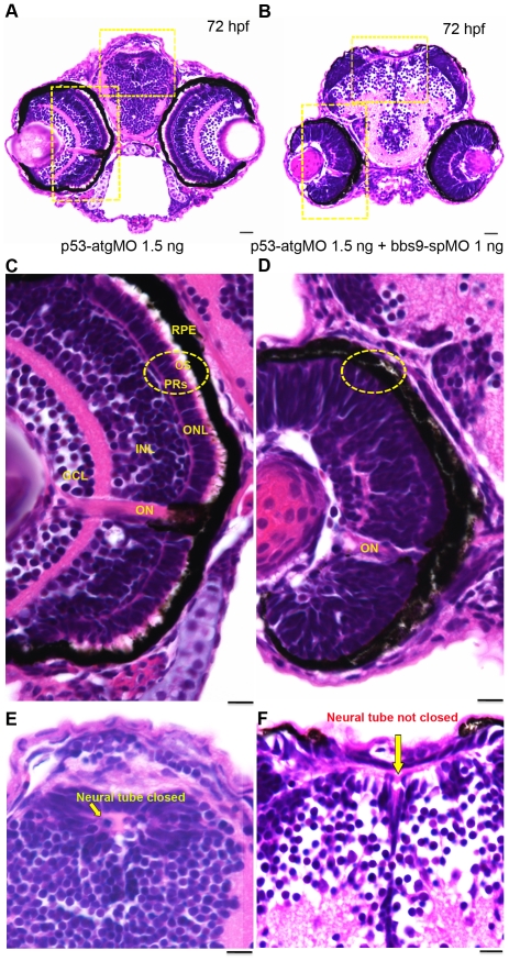
bbs9-spMO morphants displayed developmental defects in eye and brain, and often with hydrocephaly. These are reminiscent of the clinical features reported in BBS patients [23], [28], [29]. Histological analysis of the morphants' eyes revealed a dose dependent effect of bbs9-spMO on photoreceptors compared to uninjected or the control-MO injected embryos (data not shown). To examine whether the eye and brain defects are caused by widespread cell death, we co-injected p53-atgMO (1.5 ng) with bbs9-spMO (1 ng). At 72 hpf, p53-atgMO morphant showed a normal eye and brain development (Fig. 4A), whereas morphants co-injected with bbs9-spMO and p53-atgMO showed severely malformed eye and brain (Fig. 4B). Our results demonstrate that the eye and brain defects are indeed bona fide effects of bbs9 knockdown.
Figure 4. bbs9-spMO morphant shows defects in retina lamination and neural tube closure.
Zebrafish head sections (72 hfp) stained with H& E. (A) The morphant injected with p53-atgMO (1.5 ng) shows normal retinal lamination and neural tube (areas highlighted with hatched yellow rectangles). (B) The morphant co-injected with bbs9-spMO (1 ng) and p53-atgMO (1.5 ng) shows lack of retinal lamination and incomplete closure of neural tube (areas highlighted with hatched yellow rectangles). (C) The morphant retina in higher magnification - area boxed in ‘A’. The retina shows all the 5 layers: Retinal pigment epithelium (RPE) abutting close to photoreceptors (PRs). The PRs (hatched circle) have visible outer segments (OS). Next to PRs are the outer nuclear layer (ONL) followed by an intact inner nuclear layer (INL) and the ganglion cell layer (GCL). The optical nerve (ON) is used as a reference point. (D) The bbs9-spMO injected morphant retina in higher magnification - area boxed in ‘B’. The retina shows no clear lamination and it lacks photoreceptor outer segments (hatched circle). (E) The morphant neural tube in higher magnification from ‘A’ shows normal closure (arrow). (F) The bbs9-spMO injected morphant neural tube in higher magnification from ‘B’ shows incomplete closure of neural tube (arrow). The scale bars indicate 100 µm.
In p53-atgMO, the retina displayed proper lamination with all five layers (Fig. 4, C). The photoreceptors' outer segments were clearly visible (Fig. 4C, circle) abutting the retinal pigment epithelium (RPE). In contrast, co-injection of bbs9-spMO caused altered retinal layer stratification apparently forming an amalgam (Fig. 4D). The photoreceptor layer was indistinguishable from RPE, which often displayed denudation from the inner retinal layer due to lack of photoreceptor outer segments (Fig. 4D, circle). The morphants did not develop a full size eye as in the controls (Fig. 4A, B). Absence of properly formed photoreceptors, especially without defined outer segments, argues for compromised cilia function in the bbs9-spMO morphants.
We then examined the morphant brain anatomy because of the hydrocephaly, a known sign of ciliary abnormality in the ventricles [26], [28]. Histological analysis of the morphants revealed an effect of bbs9-spMO on brain structure. At 72 hpf, the p53-atgMO morphants showed normal closed neural tube (Fig. 4A, E), whereas the addition of bbs9-spMO resulted in failure of complete neural tube closure (Fig. 4B, F, arrow). Ciliary dysfunction is one of the reasons for incomplete neural tube closure as seen in some of the ciliary mutants [30].
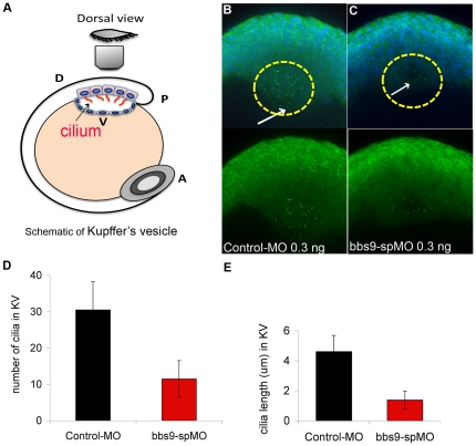
The preceding data prompted us to evaluate whether bbs9 knockdown affected the cilia in Kupffer's vesicle (KV; Fig. 5A), a structure often afflicted by ciliary dysfunction. We stained morphant (injected with either control-MO or bbs9-spMO 0.3 ng) KV cilia with antibodies against acetylated alphaα-tubulin and gammaγ-tubulin. The number of cilia was reduced in bbs9-spMO injected morphants compared to control-MO injected morphants (Fig. 5B, C). In bbs9-spMO injected morphants, the cilia were less in number and of shorter length compared to the cilia in control-MO injected morphants (Fig. 5D, E). These data further show that cilia biogenesis is compromised in bbs9-spMO morphants.
Figure 5. Knockdown of bbs9 affects cilia in Kupffer's vesicle.
(A) Schematic view of Kupffer's vesicle (KV) in zebrafish embryo at 12 hpf. A, P, D and V indicate anterior, posterior, dorsal and ventral sides, respectively. (B) Morphant injected with control-MO (0.3 ng). (C) Morphant injected with bbs9-spMO (0.3 ng). In the morphants, KV cilia were visualized by staining with both anti-α-tubulin and anti-γ-tubulin (green), between 10–13 hpf. In ‘B’, cilia are more in number and are longer (cf. white arrows) than in ‘C’. In B and C, upper panels show the nuclei visualized with DAPI. (D) and (E) show quantification of KV cilia number and length, respectively. The Y-axis represents the mean ± SEM. The X-axis represents the indicated category of morphants analyzed.
BBS9 participates in cilia biogenesis in IMCD3 cells
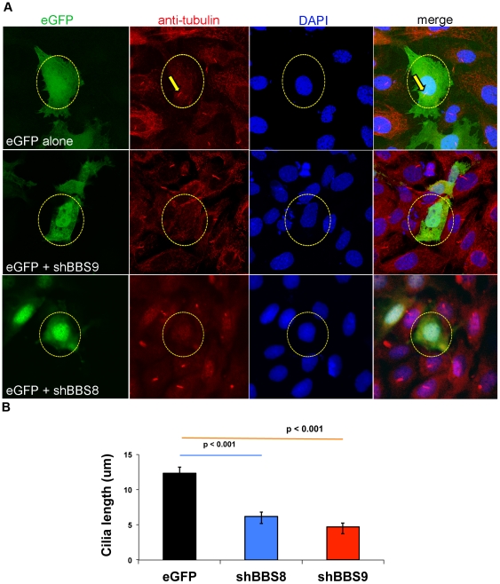
To further validate the contribution of BBS9 to cilia function, we took advantage of an in vitro ciliogenesis assay using IMCD3 cells, which normally grow cilia. Knockdown of Bbs9 using mouse specific shRNA constructs negatively affected ciliogenesis in IMCD3 cells, resulting in more cells with no cilia compared to the control transfection (Fig. 6A, B; Fig. S2). Some of the transfected cells retained their cilia but these were relatively shorter than the controls (Fig. 6B). As BBS9 interacts with BBS8 in biochemical assays [18], knockdown of Bbs8 also resulted in similar defects in IMCD3 cells (Fig. 6A, B; Fig. S2). The mouse Bbs9 knockdown data is in concordance with bbs9-spMO morphant KV cilia results (see Fig. 5D, E).
Figure 6. Knockdown of Bbs9 affects ciliogenesis in IMCD3 cells.
(A) Bbs8 and Bbs9 shRNA transfection in IMCD3 cells. The top row shows eGFP control transfection, whereas middle and bottom rows represent eGFP co-transfected with shRNA against Bbs9 or Bbs8, respectively. The nuclei are visualized with DAPI (blue). Transfection is visualized with eGFP (green). Cilia are visualized with both anti-alpha-tubulin and gamma-tubulin (red). shRNA transfected cells (green) have no cilia (red) - highlighted with yellow circle (broken). In the top control panel, eGFP alone-transfected cell shows a cilium (highlighted with yellow arrows). Images are taken at 60× magnification. (B) The quantification of cilia length after Bbs8 and Bbs9 shRNA transfection in IMCD3 cells (obtained from A). The X and Y axes respectively show transfection category and length (micrometer) of cilia in eGFP transfected cells per seven fields. Data are presented as mean ± SEM, and statistical significance is indicated with p values.
Discussion
Pioneering studies during the last decade have begun to delineate the molecular pathways leading to BBS and other ciliopathies. As BBS patients share similar clinical features, it is believed that BBS proteins function through common molecular pathways. The existence and interdependence of multimeric BBS protein complexes and their influence on ciliogenesis further supports this view. BBS9 is a component of the BBSome complex and reportedly interacts with several BBS proteins [18]. Our studies provide strong evidence in support of the role of BBS9 in cilia development as its knockdown results in BBS-like syndromic phenotype in zebrafish. An orthologous human BBS9 mRNA rescued the morphant phenotype, but a mutant mRNA (carrying a missense change observed in a BBS9 patient) failed to provide functional complementation, suggesting an evolutionary conservation of BBS9 function.
In humans, a total of seven mutations have been reported in the BBS9 gene [23]; all are homozygous except one, a compound heterozygote. Our data show the functional conservation of BBS9 protein domain that includes the missense mutation during evolution. However, additional investigations will be necessary to identify the consequence of other known human mutations within the conserved region of zebrafish bbs9.
One important question is how the mutations in BBS9 lead to a syndromic phenotype. BBS9 is required for the assembly of the BBSome [18], which in turn is needed for targeting membrane proteins to the cilium [31]. Hence, loss of (or severely reduced) function of BBS9 could affect the integrity of the BBSome complex and compromise cilia function. Our KV cilia data demonstrate that like many other BBS genes, Bbs9 is required for cilia development [32]. The defective cilia in KV can affect the left-right laterality [32]. Since we did not analyze heart looping or laterality markers it remains to be tested whether bbs9 morphants have any laterality defects. The retinal degeneration in zebrafish, exhibited by bbs9-spMO morphants, is a further indication of cilia dysgenesis similar to the phenotype produced by Rpgr knockdown, which causes abnormal ciliary transport [33].
BBS patients suffer varying degrees of cognitive dysfunctions [34], possibly due to dyskinesia [35] and subsequent development of hydrocephalus, observed in BBS3 patients [29] and its rodent models [36]. Similar phenotypes are reported in rodents [28], [37] and in zebrafish [26]. Thus, the hydrocephaly in bbs9-spMO morphant could be attributed to ciliary dyskinesia. Hydrocephaly in bbs9-spMO morphants suggested a possible ciliary abnormality in the ventricles and ependymal canal. However, we did not analyze the cilia in these structures. bbs9-spMO morphants show defects in neural tube closure, which could be due to defective non-canonical Wnt (PCP) pathway mediated via Vangl2 [38]. Mice having mutations in BBS1, BBS4 or BBS6 reportedly display a phenotype resembling a mutation in Vangl2, which includes neural tube defects [30]. Interestingly, VANGL2 and BBS proteins co-localize in the basal body and ciliary axoneme [5]. We therefore propose that bbs9 knockdown results in ciliary dysfunction in the morphants, resulting in open neural tubes.
Several components of the BBSome are critical for ciliogenesis. The roles of BBS1, BBS5, and BBS8 in ciliogenesis have been demonstrated in RPE cells [18], [39]. BBS9 has been shown to interact with BBS1 and BBS8, with variable strength [18]. Though an earlier ciliogenesis assay using RPE cells showed a weak effect of BBS9 siRNA [39], our assay using IMCD3 cells and BBS9 shRNA conclusively demonstrated that BBS9 is required for the development of cilia. Defects in cilia function can account for abnormalities in eye and brain of bbs9-spMO morphants. Notably, an association between an amino acid change in PTHB1 and premature ovarian failure in human has been reported [22]. The exact pathogenic mechanism is unclear; however, ciliary dysfunction has been associated with ovarian function [40].
In summary, we provide in vivo evidence of bbs9 function in cilia biogenesis and/or transport. Loss of BBS9 leads to defects in organogenesis, presumably because of its crucial role in BBSome assembly and cilia formation. Further investigations are necessary to elucidate the precise biochemical role of BBS9 within the BBSome complex and in cilia biogenesis and/or function.
Materials and Methods
Morpholino injections in zebrafish
Fluorescein-tagged morpholinos (MOs) were procured from Gene Tools Inc. (OR, USA). A standard negative control (control-MO), p53-atgMO (5′- GCGCCATTGCTTTGCAAGAATTG - 3′) and custom-designed translation blocking (bbs9-atgMO - 5′-CGCTGAAGCCAGAACTGTGGAACAT - 3′) and splice blocking (bbs9–spMO - 5′-CGGTGCCTGAGAAAACCATACATAT - 3′) MOs against zebrafish bbs9 were obtained in lyophilized form, re-suspended in distilled water, and quantified spectrophotometrically (NanoDrop Tech Inc, DE, USA).
Zebrafish (Danio rerio) were maintained under an approved National Institutes of Health animal use protocol. Staged wild type embryos of EK strain between 2–8 cells were microinjected 0.4–1.2 nL of morpholinos into the yolk sac using pneumatic pico pump (WPI, FL, USA). Before injection, a fresh glass capillary needle was pulled with Kopf needle/pipette puller (Model 750, Tujunga, CA, USA), and calibrated against a micrometer to determine the volume delivered per pulse. Microinjected embryos were incubated at 28°C overnight and scored for survival the following day. Live embryos were ascertained of successful injection by fluorescein signal and followed until 48–72 hpf to observe any overt phenotype under Leica Microscopes MZ16F and ICA (Leica Microsystems, IL, USA). Phenotypes were captured with Leica DC500 camera attached to Leica microscope MZ16F (total magnification employed: 0.63××4× or 8×). Eye size was determined by imaging embryos on a Zeiss Axioskop upright microscope using a 10× objective. Total eye area in pixels was quantified in imageJ using the known scaling factor for this objective. p values were obtained using students t-test (two tailed, unpaired).
mRNA isolation, RT-PCR and verification of bbs9 sequence
Total RNA was extracted from individual embryos at 72 hpf with Trizol Reagent (Invitrogen, CA, USA). RNA sample (200 ng) was reverse transcribed with random primers using Superscript III (Invitrogen, CA, USA.). The cDNA was then amplified according to standard protocol using Taq polymerase (New England Biolabs, MA, USA). The following PCR primers were used for splice verification:
5′-TTTGTTTAAGGCCCGTGATT-3′ and 5′-TGAAGGAGTCTGTGCGAATG-3′. Exon 1 to 5 were generated by PCR using primers: 5′-ATGTTCCACAGTTCTGGCTTCAG-3′ and 5′-CTGTAACACCACCGAATGGGCCATA-3′, and the PCR product was TA cloned in pGEMTEasy (Promega Corp, WI, USA). The presence of exons 2 to 5 in pGEMTEasy was verified (Fig. S1) by sequencing using T7 primer.
In vitro RNA preparation for rescue and mutation synthesis
For rescue experiment, a human BBS9 full-length cDNA (Clone ID 5519851) was obtained from Open Biosystems (AL, USA) and sequence-verified. To recreate the missense mutation (G141R) in human BBS9 protein, a corresponding wild type zebrafish nucleotide was subjected to site-directed mutagenesis using QuikChange II kit (Stratagene; Agilent Tech, CA, USA). The wild type and mutant cDNA from SPORT6 were subcloned into pcDNA3.1(+) vector at EcoRV-XhoI sites. Subcloned cDNAs were linearized with XmaI and used for in vitro synthesis of capped mRNA using mMESSAGE mMACHINE T7 Ultra Kit (Ambion, Applied Biosystems, CA, USA). The quality of in vitro synthesized mRNA was checked on Bioanalyser (Agilent Tech, CA, USA) before co-injection with bbs9-spMO.
In situ hybridization using zebrafish embryos or larvae
RT-PCR product corresponding to zebrafish bbs9 was cloned into pGEMT-easy (Promega Corp, WI, USA) and sequence-verified. The vector was linearized with Sal I or Nco I and used to generate the sense (T7 RNA polymerase) or antisense (SP6 RNA polymerase) probes, respectively, with DIG RNA labeling Mix (Roche Applied Science, IN, USA). In situ hybridization was performed as described [41].
H&E staining of eye and brain
At 72 hpf, zebrafish larvae were fixed with 4% glutaraldehyde for 30 min at RT, then fixed with 4% paraformaldehyde (PFA) overnight at 4°C. Subsequently, they were washed with PBS and embedded in OCT compound Tissue-Tek (SakuraFinetek USA, Inc, CA, USA) and 10 µm sections were cut. The sections were stained with standard H&E staining protocol.
Staining of Kupffer's vesicle cilia
The embryos aged 10–13 hpf were fixed with 4% PFA and stained with antibodies against acetylated-alpha- tubulin and gamma-tubulin to visualize the cilia. The embryos were then embedded in 2% low melting agarose and positioned for confocal microscopy. The images were taken with Leica TCS SP2 using a water immersion lens (40×) and processed for maximum projection and quantification of cilia using LCM software.
Ciliogenesis assay
Adult mouse kidney Inner Medullary Collecting Duct cells -3 (IMCD3; ATCC Number: CRL-2123; ATCC, VA, USA.) were grown near confluence overnight on 6-well plate by seeding 200×10∧3 cells and transfected with 750 ng of each plasmid DNA (eGFP and shRNA) in serum free medium using fugene6 (Roche Applied Science, IN, USA). After 8 hr, the serum free medium was replaced with complete medium. Twelve hr later, the cells were washed twice for 5 min each with 0.5 mL PBS and fixed for 15 min at RT with 0.5 mL of 4% PFA in PBS. After fixation, PFA was removed and the cells were washed twice with 0.5 mL PBS. Subsequently, the cells were incubated at RT in 0.5 mL of 5% normal goat serum in PBT (0.1%) for 30 min for blocking. The cells were then incubated with 0.2 mL of a primary antibody (anti-acetylated alpha-tubulin, Sigma -T7451 and anti-gamma-tubulin, Sigma-T6557 (Sigma-Aldrich Corp., MO, USA), both 1∶1000 diluted in blocking solution) for 1 hr at RT. Subsequently, the cells were washed 3× with 0.1% PBT for 5 min each, and incubated with 0.2 mL secondary antibody (anti-mouse Alexa 568 (Invitrogen, CA, USA), 1∶500 diluted in blocking solution) for 1 hr at RT. Finally, nuclear staining was performed with 0.2 mL DAPI (diluted 1∶1000) for 5 min at RT. The cells were washed with 0.5 mL PBS twice for 5 min. The slides were mounted with flouromount and imaged on Olympus FluoView FV1000 (Tokyo, Japan) confocal microscope.
shRNA construct sets against mouse Bbs8 and Bbs9 were obtained from Open Biosystems (AL, USA): (Bbs8: TRCN0000113210 - 14), (Bbs9: TRCN0000178683; TRCN0000181485; TRCN0000182069; TRCN0000182387; TRCN0000182647). The following shRNA constructs gave the best knockdown results - Bbs8.3: TRCN0000113213, and Bbs9.5 (TRCN0000182387) (shown in Fig. 6). The presence or absence of cilia in a transfected (eGFP alone or co-transfected with shRNA construct) cell was manually scored under an epifluorescence microscope (Olympus, BX50F4; 40×; Olympus, Japan). The raw data for all shRNA constructs are presented in Fig. S2. The cilia length in transfected cells (eGFP, BBS8.3 and BBS9.5) was quantified using ImageJ software.
Supporting Information
Validated zebrafish bbs9 sequence. The bbs9 cDNA sequences are aligned to see the degree of matching (top - t. and the bottom - b. sequences were obtained from sequencing data and the provisional version, respectively. The bbs9 specific product was amplified by PCR using cDNA generated from zebrafish total mRNA. RT-PCR and sequencing data show that exons 2 to 5 are expressed in zebrafish. Exons 2 and 5 are highlighted in yellow; the sequences are perfectly matched until exon 5 (indicated by red bar on top).
(TIF)
Bbs8 and Bbs9 knockdown compromised ciliogenesis in IMCD3 cells. Knockdown of Bbs8 and Bbs9 in IMCD3 cells with 4 different shRNA constructs (#1, 3, 4, 5). Green cells represent the cells transfected with shRNA construct. X-axis displays the analysis categories. Y-axis displays the number of green cells. BBS8 or BBS9 shRNA construct (as indicated) was used along with eGFP. Control transfection was performed with eGFP (shown on the right) without any shRNA construct. Only green cells were counted for obtaining the raw data.
(TIF)
Acknowledgments
The authors thank Chun Y. Gao and Robert N. Farris, NEI imaging core, for help on KV cilia imaging, and Joby Joseph for assistance with the zebrafish imaging.
Footnotes
Competing Interests: The authors have declared that no competing interests exist.
Funding: The study was supported by intramural funds of the National Eye Institute, National Human Genome Research Institute and National Institute of Child Health and Development, National Institutes of Health. The funders had no role in study design, data collection and analysis, decision to publish, or preparation of the manuscript.
References
- 1.Fliegauf M, Benzing T, Omran H. When cilia go bad: cilia defects and ciliopathies. Nat Rev Mol Cell Biol. 2007;8:880–893. doi: 10.1038/nrm2278. [DOI] [PubMed] [Google Scholar]
- 2.Gerdes JM, Davis EE, Katsanis N. The vertebrate primary cilium in development, homeostasis, and disease. Cell. 2009;137:32–45. doi: 10.1016/j.cell.2009.03.023. [DOI] [PMC free article] [PubMed] [Google Scholar]
- 3.Huangfu D, Liu A, Rakeman AS, Murcia NS, Niswander L, et al. Hedgehog signalling in the mouse requires intraflagellar transport proteins. Nature. 2003;426:83–87. doi: 10.1038/nature02061. [DOI] [PubMed] [Google Scholar]
- 4.Simons M, Gloy J, Ganner A, Bullerkotte A, Bashkurov M, et al. Inversin, the gene product mutated in nephronophthisis type II, functions as a molecular switch between Wnt signaling pathways. Nat Genet. 2005;37:537–543. doi: 10.1038/ng1552. [DOI] [PMC free article] [PubMed] [Google Scholar]
- 5.Ross AJ, May-Simera H, Eichers ER, Kai M, Hill J, et al. Disruption of Bardet-Biedl syndrome ciliary proteins perturbs planar cell polarity in vertebrates. Nat Genet. 2005;37:1135–1140. doi: 10.1038/ng1644. [DOI] [PubMed] [Google Scholar]
- 6.Gherman A, Davis EE, Katsanis N. The ciliary proteome database: an integrated community resource for the genetic and functional dissection of cilia. Nat Genet. 2006;38:961–962. doi: 10.1038/ng0906-961. [DOI] [PubMed] [Google Scholar]
- 7.Hurd TW, Hildebrandt F. Mechanisms of nephronophthisis and related ciliopathies. Nephron Exp Nephrol. 118:e9–14. doi: 10.1159/000320888. [DOI] [PMC free article] [PubMed] [Google Scholar]
- 8.Beales PL, Elcioglu N, Woolf AS, Parker D, Flinter FA. New criteria for improved diagnosis of Bardet-Biedl syndrome: results of a population survey. J Med Genet. 1999;36:437–446. [PMC free article] [PubMed] [Google Scholar]
- 9.Zaghloul NA, Katsanis N. Mechanistic insights into Bardet-Biedl syndrome, a model ciliopathy. J Clin Invest. 2009;119:428–437. doi: 10.1172/JCI37041. [DOI] [PMC free article] [PubMed] [Google Scholar]
- 10.Kim SK, Shindo A, Park TJ, Oh EC, Ghosh S, et al. Planar cell polarity acts through septins to control collective cell movement and ciliogenesis. Science. 329:1337–1340. doi: 10.1126/science.1191184. [DOI] [PMC free article] [PubMed] [Google Scholar]
- 11.Ansley SJ, Badano JL, Blacque OE, Hill J, Hoskins BE, et al. Basal body dysfunction is a likely cause of pleiotropic Bardet-Biedl syndrome. Nature. 2003;425:628–633. doi: 10.1038/nature02030. [DOI] [PubMed] [Google Scholar]
- 12.Craige B, Tsao CC, Diener DR, Hou Y, Lechtreck KF, et al. CEP290 tethers flagellar transition zone microtubules to the membrane and regulates flagellar protein content. J Cell Biol. 190:927–940. doi: 10.1083/jcb.201006105. [DOI] [PMC free article] [PubMed] [Google Scholar]
- 13.Kim JC, Badano JL, Sibold S, Esmail MA, Hill J, et al. The Bardet-Biedl protein BBS4 targets cargo to the pericentriolar region and is required for microtubule anchoring and cell cycle progression. Nat Genet. 2004;36:462–470. doi: 10.1038/ng1352. [DOI] [PubMed] [Google Scholar]
- 14.Kim JC, Ou YY, Badano JL, Esmail MA, Leitch CC, et al. MKKS/BBS6, a divergent chaperonin-like protein linked to the obesity disorder Bardet-Biedl syndrome, is a novel centrosomal component required for cytokinesis. J Cell Sci. 2005;118:1007–1020. doi: 10.1242/jcs.01676. [DOI] [PubMed] [Google Scholar]
- 15.Li JB, Gerdes JM, Haycraft CJ, Fan Y, Teslovich TM, et al. Comparative genomics identifies a flagellar and basal body proteome that includes the BBS5 human disease gene. Cell. 2004;117:541–552. doi: 10.1016/s0092-8674(04)00450-7. [DOI] [PubMed] [Google Scholar]
- 16.Badano JL, Leitch CC, Ansley SJ, May-Simera H, Lawson S, et al. Dissection of epistasis in oligogenic Bardet-Biedl syndrome. Nature. 2006;439:326–330. doi: 10.1038/nature04370. [DOI] [PubMed] [Google Scholar]
- 17.May-Simera HL, Kai M, Hernandez V, Osborn DP, Tada M, et al. Bbs8, together with the planar cell polarity protein Vangl2, is required to establish left-right asymmetry in zebrafish. Dev Biol. 345:215–225. doi: 10.1016/j.ydbio.2010.07.013. [DOI] [PubMed] [Google Scholar]
- 18.Nachury MV, Loktev AV, Zhang Q, Westlake CJ, Peranen J, et al. A core complex of BBS proteins cooperates with the GTPase Rab8 to promote ciliary membrane biogenesis. Cell. 2007;129:1201–1213. doi: 10.1016/j.cell.2007.03.053. [DOI] [PubMed] [Google Scholar]
- 19.Seo S, Baye LM, Schulz NP, Beck JS, Zhang Q, et al. BBS6, BBS10, and BBS12 form a complex with CCT/TRiC family chaperonins and mediate BBSome assembly. Proc Natl Acad Sci U S A. 2010;107:1488–1493. doi: 10.1073/pnas.0910268107. [DOI] [PMC free article] [PubMed] [Google Scholar]
- 20.Adams AE, Rosenblatt M, Suva LJ. Identification of a novel parathyroid hormone-responsive gene in human osteoblastic cells. Bone. 1999;24:305–313. doi: 10.1016/s8756-3282(98)00188-4. [DOI] [PubMed] [Google Scholar]
- 21.Vernon EG, Malik K, Reynolds P, Powlesland R, Dallosso AR, et al. The parathyroid hormone-responsive B1 gene is interrupted by a t(1;7)(q42;p15) breakpoint associated with Wilms' tumour. Oncogene. 2003;22:1371–1380. doi: 10.1038/sj.onc.1206332. [DOI] [PubMed] [Google Scholar]
- 22.Kang H, Lee SK, Kim MH, Song J, Bae SJ, et al. Parathyroid hormone-responsive B1 gene is associated with premature ovarian failure. Hum Reprod. 2008;23:1457–1465. doi: 10.1093/humrep/den086. [DOI] [PubMed] [Google Scholar]
- 23.Nishimura DY, Swiderski RE, Searby CC, Berg EM, Ferguson AL, et al. Comparative genomics and gene expression analysis identifies BBS9, a new Bardet-Biedl syndrome gene. Am J Hum Genet. 2005;77:1021–1033. doi: 10.1086/498323. [DOI] [PMC free article] [PubMed] [Google Scholar]
- 24.Ghosh AK, Murga-Zamalloa CA, Chan L, Hitchcock PF, Swaroop A, et al. Human retinopathy-associated ciliary protein retinitis pigmentosa GTPase regulator mediates cilia-dependent vertebrate development. Hum Mol Genet. 19:90–98. doi: 10.1093/hmg/ddp469. [DOI] [PMC free article] [PubMed] [Google Scholar]
- 25.Wolff C, Roy S, Ingham PW. Multiple muscle cell identities induced by distinct levels and timing of hedgehog activity in the zebrafish embryo. Curr Biol. 2003;13:1169–1181. doi: 10.1016/s0960-9822(03)00461-5. [DOI] [PubMed] [Google Scholar]
- 26.Zhou W, Dai J, Attanasio M, Hildebrandt F. Nephrocystin-3 is required for ciliary function in zebrafish embryos. Am J Physiol Renal Physiol. doi: 10.1152/ajprenal.00043.2010. [DOI] [PMC free article] [PubMed] [Google Scholar]
- 27.Robu ME, Larson JD, Nasevicius A, Beiraghi S, Brenner C, et al. p53 activation by knockdown technologies. PLoS Genet. 2007;3:e78. doi: 10.1371/journal.pgen.0030078. [DOI] [PMC free article] [PubMed] [Google Scholar]
- 28.Banizs B, Pike MM, Millican CL, Ferguson WB, Komlosi P, et al. Dysfunctional cilia lead to altered ependyma and choroid plexus function, and result in the formation of hydrocephalus. Development. 2005;132:5329–5339. doi: 10.1242/dev.02153. [DOI] [PubMed] [Google Scholar]
- 29.Smith UM, Consugar M, Tee LJ, McKee BM, Maina EN, et al. The transmembrane protein meckelin (MKS3) is mutated in Meckel-Gruber syndrome and the wpk rat. Nat Genet. 2006;38:191–196. doi: 10.1038/ng1713. [DOI] [PubMed] [Google Scholar]
- 30.Torban E, Wang HJ, Groulx N, Gros P. Independent mutations in mouse Vangl2 that cause neural tube defects in looptail mice impair interaction with members of the Dishevelled family. J Biol Chem. 2004;279:52703–52713. doi: 10.1074/jbc.M408675200. [DOI] [PubMed] [Google Scholar]
- 31.Jin H, White SR, Shida T, Schulz S, Aguiar M, et al. The conserved Bardet-Biedl syndrome proteins assemble a coat that traffics membrane proteins to cilia. Cell. 141:1208–1219. doi: 10.1016/j.cell.2010.05.015. [DOI] [PMC free article] [PubMed] [Google Scholar]
- 32.Yen HJ, Tayeh MK, Mullins RF, Stone EM, Sheffield VC, et al. Bardet-Biedl syndrome genes are important in retrograde intracellular trafficking and Kupffer's vesicle cilia function. Hum Mol Genet. 2006;15:667–677. doi: 10.1093/hmg/ddi468. [DOI] [PubMed] [Google Scholar]
- 33.Shu X, Zeng Z, Gautier P, Lennon A, Gakovic M, et al. Zebrafish Rpgr is required for normal retinal development and plays a role in dynein-based retrograde transport processes. Hum Mol Genet. 19:657–670. doi: 10.1093/hmg/ddp533. [DOI] [PubMed] [Google Scholar]
- 34.Green JS, Parfrey PS, Harnett JD, Farid NR, Cramer BC, et al. The cardinal manifestations of Bardet-Biedl syndrome, a form of Laurence-Moon-Biedl syndrome. N Engl J Med. 1989;321:1002–1009. doi: 10.1056/NEJM198910123211503. [DOI] [PubMed] [Google Scholar]
- 35.Vieira JP, Lopes P, Silva R. Primary Ciliary Dyskinesia and Hydrocephalus With Acqueductal Stenosis. Journal of child neurology. 2012 doi: 10.1177/0883073811429856. [DOI] [PubMed] [Google Scholar]
- 36.Gattone VH, 2nd, Tourkow BA, Trambaugh CM, Yu AC, Whelan S, et al. Development of multiorgan pathology in the wpk rat model of polycystic kidney disease. Anat Rec A Discov Mol Cell Evol Biol. 2004;277:384–395. doi: 10.1002/ar.a.20022. [DOI] [PubMed] [Google Scholar]
- 37.Tissir F, Qu Y, Montcouquiol M, Zhou L, Komatsu K, et al. Lack of cadherins Celsr2 and Celsr3 impairs ependymal ciliogenesis, leading to fatal hydrocephalus. Nat Neurosci. 13:700–707. doi: 10.1038/nn.2555. [DOI] [PubMed] [Google Scholar]
- 38.Ciruna B, Jenny A, Lee D, Mlodzik M, Schier AF. Planar cell polarity signalling couples cell division and morphogenesis during neurulation. Nature. 2006;439:220–224. doi: 10.1038/nature04375. [DOI] [PMC free article] [PubMed] [Google Scholar]
- 39.Loktev AV, Zhang Q, Beck JS, Searby CC, Scheetz TE, et al. A BBSome subunit links ciliogenesis, microtubule stability, and acetylation. Dev Cell. 2008;15:854–865. doi: 10.1016/j.devcel.2008.11.001. [DOI] [PubMed] [Google Scholar]
- 40.Johnson ET, Nicola T, Roarty K, Yoder BK, Haycraft CJ, et al. Role for primary cilia in the regulation of mouse ovarian function. Dev Dyn. 2008;237:2053–2060. doi: 10.1002/dvdy.21612. [DOI] [PubMed] [Google Scholar]
- 41.Lyons SE, Shue BC, Oates AC, Zon LI, Liu PP. A novel myeloid-restricted zebrafish CCAAT/enhancer-binding protein with a potent transcriptional activation domain. Blood. 2001;97:2611–2617. doi: 10.1182/blood.v97.9.2611. [DOI] [PubMed] [Google Scholar]
Associated Data
This section collects any data citations, data availability statements, or supplementary materials included in this article.
Supplementary Materials
Validated zebrafish bbs9 sequence. The bbs9 cDNA sequences are aligned to see the degree of matching (top - t. and the bottom - b. sequences were obtained from sequencing data and the provisional version, respectively. The bbs9 specific product was amplified by PCR using cDNA generated from zebrafish total mRNA. RT-PCR and sequencing data show that exons 2 to 5 are expressed in zebrafish. Exons 2 and 5 are highlighted in yellow; the sequences are perfectly matched until exon 5 (indicated by red bar on top).
(TIF)
Bbs8 and Bbs9 knockdown compromised ciliogenesis in IMCD3 cells. Knockdown of Bbs8 and Bbs9 in IMCD3 cells with 4 different shRNA constructs (#1, 3, 4, 5). Green cells represent the cells transfected with shRNA construct. X-axis displays the analysis categories. Y-axis displays the number of green cells. BBS8 or BBS9 shRNA construct (as indicated) was used along with eGFP. Control transfection was performed with eGFP (shown on the right) without any shRNA construct. Only green cells were counted for obtaining the raw data.
(TIF)


