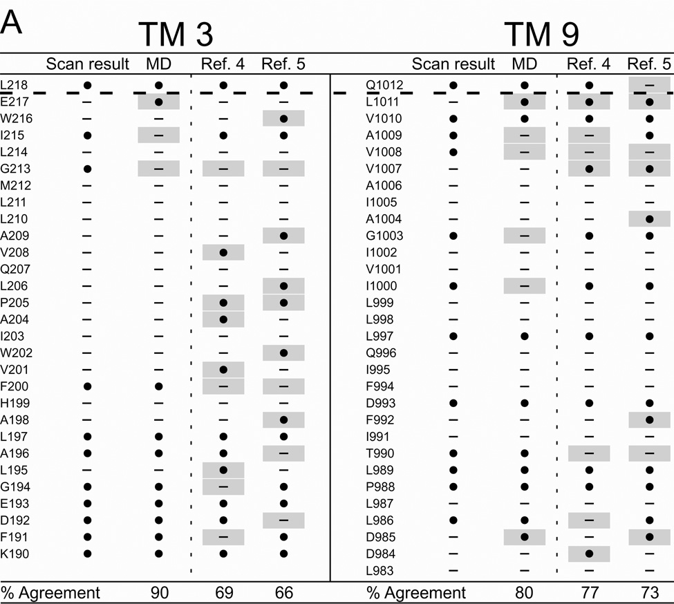
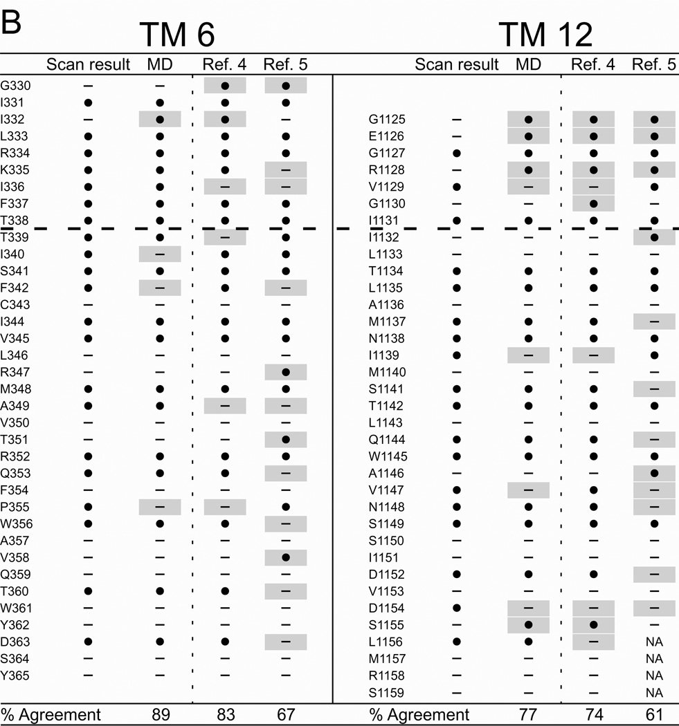
Figure 6. Comparing results of cysteine-scanning using channel-permeant and channel-impermeant probes with predictions of three molecular models of the pore domain of CFTR.
(A) TMs 3 and 9. (B) TMs 6 and 12. Cysteine-scan outcome labeled “Scan result”. “MD” column contains the consensus predictions of 27 MD frames for pore-lining amino acids (see Methods for details). Columns labeled “Ref. 4” and “Ref. 5” contain predictions from Sav-based homology models developed by Mornon et al. (4) and Serohijos et al. (5) (see Methods for details). (●): Residues where engineered cysteines were reactive by scanning or predicted to be pore-lining. (—): Residues where cysteines were not reactive or predicted to face away from the pore. Sites where scanning data reveals a model prediction to be inaccurate are indicated by a gray box. Amino acids that are not included in a particular model are indicated as “NA.” Dashed lines indicate the boundaries demarcated by reactivity toward channel-permeant and channel-impermeant reagents as described in the text.


