Abstract
This case highlights the need for cautious management and serial regular examination of trauma patients. A 22-year-old Caucasian male presented to the emergency department 4 h following an injury sustained during football training. He complained of the immediate onset of severe left upper quadrant and left flank pain. He subsequently developed frank haematuria. On initial review, he was haemodynamically stable. CT of the abdomen and pelvis showed a grade 4 renal trauma. Over the following 36 h, he remained haemodynamically stable. On serial abdominal examinations however, he developed a rigid abdomen and was noted to have a haemoglobin drop. Interval CT scan showed a progression of his injury and the presence of a haemoperitoneum. An emergency laparotomy was performed resulting in a left nephrectomy. He made an uneventful recovery.
Background
Renal trauma accounts for approximately 3% of all trauma admissions and as many of 10% of cases of traumatic abdominal injury. In the UK, 90% of renal traumas are secondary to blunt trauma. Approximately 40% of cases have a coexistent abdominal injury. Isolated renal trauma as illustrated in this case, is rare. The direct trauma causes the kidney to be crushed against the ribs, while indirect trauma can result in vascular or pelvis-ureteric disruption.
This case highlights how life-threatening injuries can be sustained by relatively unremarkable trauma, how serial clinical examination is critical for the evaluation of trauma patients and how there should be no hesitation in intervening operatively when a patient shows signs of clinical deterioration.
The radiological images, which accompany this case, clearly illustrate for medical students and junior doctors the pertinent urological anatomy, acting as a teaching exercise.
Case presentation
A 22-year-old Caucasian male presented to the emergency department 4 h following an injury sustained during football training. He had been referred to the hospital following review by his general practitioner. During football training, he sustained a knock to the left upper quadrant. He complained of the immediate onset of severe left upper quadrant and left flank pain. There was associated weakness but no syncope. He subsequently developed frank haematuria.
There was no medical or surgical history. He did not take any regular medications. He had no known drug allergies. There was no significant family history. He was a non-smoker, consumed approximately 10 units of alcohol per week and worked in a warehouse. He lived with his mother and siblings.
On initial review, he was haemodynamically stable. His heart rate (HR) was 73 beats per min (BPM), blood pressure (BP) 147/71, temperature (T) 36.4°C, respiratory rate (RR) 14 and oxygen saturations (O2 sats) 100% on room air. Abdominal examination showed tenderness in the left upper quadrant. There was no guarding or rigidity.
Investigations
Blood sampling showed a haemoglobin (Hb) of 13.3 g/dl, white cell count of 14.9 and a creatinine (Cr) of 112. Mid-stream urine showed evidence of frank haematuria. He was treated with intravenous analgesia and fluids. Intravenous antibiotics were commenced. Blood was cross-matched and he was kept fasting. A 3-way urethral catheter was placed.
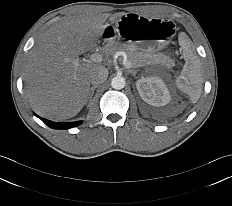
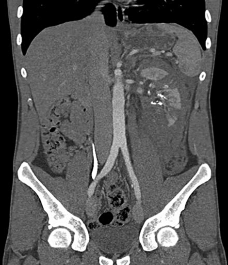
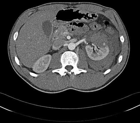
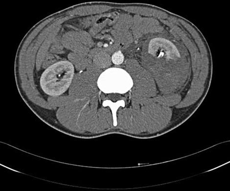
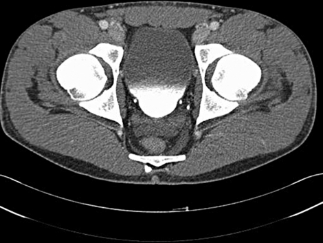
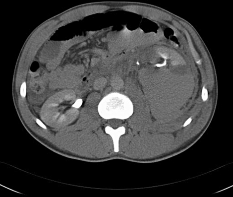
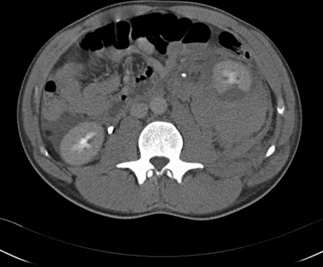
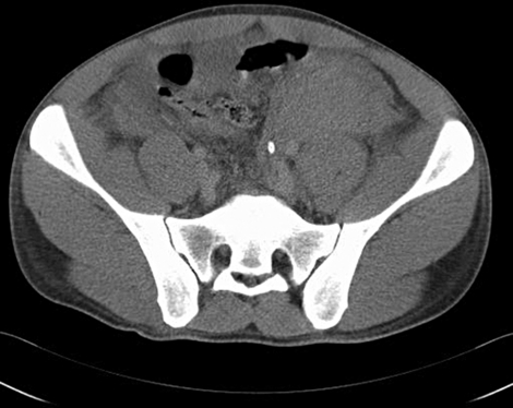
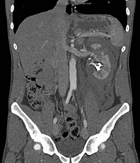
A contrast CT scan of the abdomen and pelvis was obtained (figures 1–6). A left perirenal haematoma with multiple deep cortical lacerations greater than 1 cm in depth was seen. Multiple areas of subsegmental infarction were seen particularly in the mid and lower poles. The haematoma tracked inferiorly along the psaos muscle. No arterial injury was seen. Extravasation of urine to the renal capsule consistent with a ruptured mid-polar calyx was seen, but it appeared to be confined by the renal capsule. The ureter was intact from the renal pelvis to the bladder, with no extravasation of contrast. The right kidney, liver, spleen, pancreas and bones were all normal. There was no free air or fluid seen in the peritoneum. These radiological findings were consistent with a grade 4 left renal injury.
Figure 1.
Transverse CT abdomen/pelvis image showing a perirenal haematoma at the upper pole of the left kidney.
Figure 6.
Sagittal CT abdomen/pelvis image showing the large left perirenal haematoma with cortical lacerations greater than 1 cm in depth, extending inferiorly along the left psoas muscle. Contrast is seen in the left renal pelvis and in right ureter.
Figure 2.
Transverse CT abdomen/pelvis image showing a perirenal haematoma at the hilar region of the left kidney. Contrast is seen in the left renal artery.
Figure 3.
Transverse CT abdomen/pelvis image showing a perirenal haematoma at the lower pole of the left kidney. Contrast is seen in the proximal ureters bilaterally, with the left ureter seen to be pushed anteriorly.
Figure 4.
Transverse CT abdomen/pelvis image showing contrast in the distal ureters bilaterally and also within the bladder.
Figure 5.
Sagittal CT abdomen/pelvis image showing a left perirenal haematoma. Contrast is seen in the left renal pelvis, at the pelvi-ureteric junction and in the middle third of the ureter.
Twelve hours post admission he remained haemodynamically stable with a HR 72 BPM, BP 130/62, T 37.2°C, RR 18 and O2 sats of 97% on room air. His urine output remained haematuric and was on average 60 mls per hour. His HB dropped to 12.6 g/dl and he was transfused 1 unit of blood.
Twenty-four hours post admission measurement of his vital signs showed HR 82 BPM, BP 143/83, T 37.8 degrees C, RR 18 and O2 sats of 98% on room air. His urine remained haematuric. Hb was 11.4 g/dl.
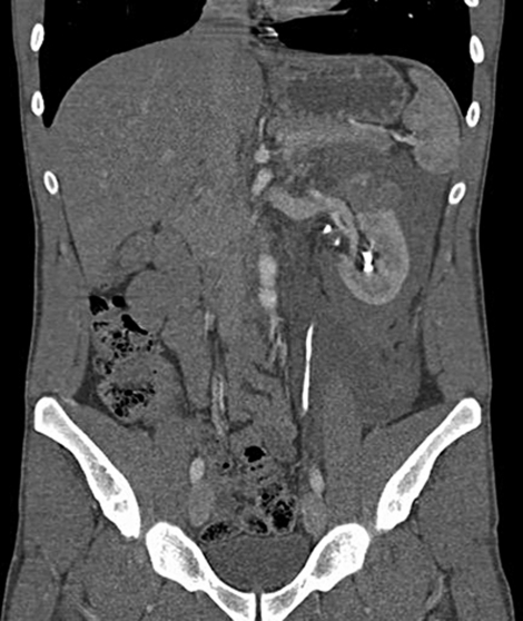
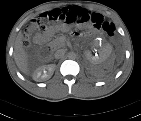
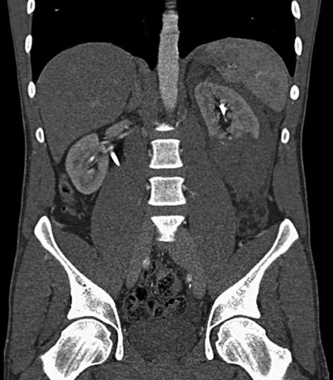
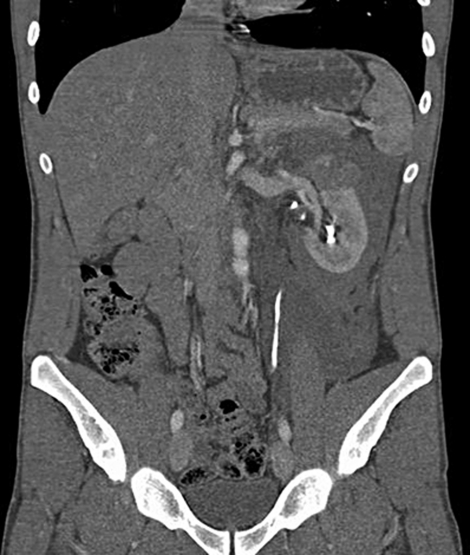
Thirty-six hours post admission measurement of his vital signs showed HR 95 BPM, BP 139/81, T 36.7°C, RR 16 and O2 sats of 98% on room air. Hb dropped to 7.7 g/dl and Cr increased to 179. Three units of blood were transfused. Serial clinical examinations showed the development of a tender, rigid abdomen with evidence of guarding. We proceeded to repeat CT scan (figures 7–13). Interval progression of the large left perirenal haematoma with interval development of free fluid within the peritoneum was seen, consistent with a haemoperitoneum. The left ureter was displaced anteriorly by the large haematoma but appeared intact.
Figure 7.
Transverse CT abdomen/pelvis image showing a perirenal haematoma at the mid-polar region of the left kidney. Contrast is seen in the renal pelvis and is also seen to extravasate anteriorly through a ruptured calyx. Free fluid is evident in the peritoneum.
Figure 13.
Sagittal CT abdomen/ pelvis image showing the left perirenal haematoma distorting the lower renal pole. Contrast is seen in the renal pelvis bilaterally and in the proximal right ureter.
Figure 8.
Transverse CT abdomen/pelvis image showing a largely posteriorly placed perirenal haematoma at the lower pole of the left kidney, pushing the kidney anteriorly. Contrast is seen in the proximal left ureter and within the right pelvi-ureteric junction. Free fluid is evident in the peritoneum.
Figure 9.
Transverse CT abdomen/pelvis image showing a largely posteriorly placed perirenal haematoma at the lower pole of the left kidney, pushing the kidney and left ureter anteriorly. Contrast is seen in the proximal ureters bilaterally. Free fluid is evident in the peritoneum.
Figure 10.
Transverse CT abdomen/pelvis image showing a large haematoma extending inferiorly along the left psoas muscle. Contrast is seen in the distal left ureter. Free fluid is evident in the peritoneum.
Figure 11.
Sagittal CT abdomen/pelvis image showing the large left perirenal haematoma with cortical lacerations greater than 1 cm in depth, extending inferiorly along the left psoas muscle. Contrast is seen in the left renal pelvis. The left renal artery and splenic vein are clearly seen.
Figure 12.
Sagittal CT abdomen/pelvis image showing the large left perirenal haematoma with cortical lacerations greater than 1 cm in depth, extending inferomedially along the left psoas muscle. The left upper pole is distorted. Contrast is seen in the renal pelvis and in the middle third of the left ureter.
Treatment
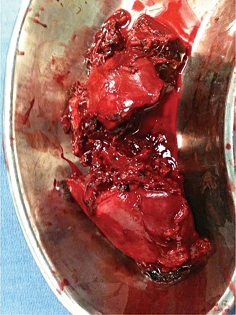
We proceeded to immediate surgical exploration. A midline incision from the xiphisternum to the pubic symphysis was made. Free blood was seen within the peritoneal cavity. A very large left expanding perirenal haematoma was also seen. No associated abdominal organ was seen to be damaged. We gained vascular control of the left renal vessels at their junction with the aorta and inferior vena cava, via a transperitoneal approach. The retroperitoneum was opened and the left kidney was deemed non-salvageable. It was lacerated at the junction of the upper and midpolar regions. A left nephrectomy was performed (figure 14). Cumulative blood loss and clot evacuation was 2650 mls during the procedure. Intraoperatively a further two units of blood were transfused.
Figure 14.
Posterior view of the left nephrectomy specimen. Deep cortical lacerations at the junction of the upper and mid-polar regions are seen.
Outcome and follow-up
He made a good recovery and was discharged home on the seventh postoperative day.
Discussion
Renal trauma accounts for approximately 3% of all trauma admissions and as many of 10% of cases of traumatic abdominal injury. 80–90% of renal traumas are secondary to blunt trauma.1 Approximately 40% of cases have a coexistent abdominal injury, with multi-organ involvement reported in up to 75% of patients with major renal injuries secondary to blunt trauma.2 Isolated renal trauma as illustrated in this case, is rare. The large majority of isolated renal traumas are minor.3 The direct trauma causes the kidney to be crushed against the ribs, while indirect trauma can result in vascular or pelvi-ureteric junction disruption.
Haematuria is present in over 95% of patients who sustain renal trauma.1 Notably however, the absence of haematuria does not rule out significant renal injury. The absence of haematuria has been reported in up to a quarter of patients with renal artery thrombosis and in one-third of cases of pelvi-ureteric junction injury.4 5
Renal trauma classification as advocated by the American Association of Surgery for Trauma is from grades 1 to 5. The classification system is as follows:
-
▶
Grade 1 Contusion – Microscopic.
-
▶
Haematoma – Subcapsular non-expanding without parenchymal laceration.
-
▶
-
▶
Grade 2 Haematoma – Non-expanding perirenal haematoma confirmed to the retroperitoneum.
-
▶
Laceration – <1 cm parenchymal depth of renal cortex without urinary extravasation.
-
▶
-
▶
Grade 3 Laceration – <1 cm parenchymal depth of renal cortex without collecting system rupture or urinary extravasation.
-
▶
Grade 4 Laceration – Parenchymal laceration extending through renal cortex, medulla and collecting system.
-
▶
Vascular – Main renal artery or vein injury with contained haemorrhage.
-
▶
-
▶
Grade 5 Laceration – Completely shattered kidney.
-
▶
Vascular – Avulsion of the renal hilum which devascularises the kidney.
-
▶
Advance one grade for bilateral injuries up to grade 3.
Management of renal trauma varies from emergent laparotomy in the setting of a haemodynamically unstable patient to careful surveillance for more minor injuries. Close clinical observation is critical in the surveillance of haemodynamically stable patients with more severe injuries.
Learning points.
-
▶
Isolated renal trauma is rare. However even trival trauma can lead to severe injury especially in kidneys with prior abnormality such as a pelvi-ureteric obstruction.
-
▶
Clinical features suggestive of renal trauma include flank pain and tenderness, a flank mass, flank bruising or skin laceration, loss of the flank contour and haematuria.
-
▶
Approximately 80% of renal injuries are grade 1 or 2, and can be managed conservatively.
-
▶
Serial clinical examination is absolutely critical for the evaluation of trauma patients.
-
▶
CT is the primary imaging modality for suspected renal injuries.
Footnotes
Competing interests None.
Patient consent Obtained.
References
- 1.McAninch JW. Renal injuries. In: Gillenwater JY, Grayhack JT, Howards SS, Duckett JW,eds. Adult and Pediatric Urology. Third Edition St Louis, Missouri: Mosby; 1996:539–53 [Google Scholar]
- 2.Dunnick NR, Sandler CM, Amis ES, Jr, et al. Urinary tract trauma. Textbook of Uroradiology. Second Edition Baltimore, Maryland: Williams & Wilkins; 1997:297–324 [Google Scholar]
- 3.Cass AS, Luxenberg M, Gleich P, et al. Clinical indications for radiographic evaluation of blunt renal trauma. J Urol 1986;136:370–1 [DOI] [PubMed] [Google Scholar]
- 4.Stables DP, Fouche RF, de Villiers van Niekerk JP, et al. Traumatic renal artery occlusion: 21 cases. J Urol 1976;115:229–33 [DOI] [PubMed] [Google Scholar]
- 5.Boone TB, Gilling PJ, Husmann DA. Ureteropelvic junction disruption following blunt abdominal trauma. J Urol 1993;150:33–6 [DOI] [PubMed] [Google Scholar]


