Abstract
Primary cardiac tumours are rare in the paediatric age group. Bronchogenic cysts, although relatively rare, represent the most common cystic lesion of the mediastinum. Intracardiac bronchogenic cysts however, are extremely rare. The authors are unaware of any case previously reported in a Nigerian child and hence report the case of a 2-year-old boy for its rarity and interest. The boy was referred for evaluation of a cardiac murmur. Clinical, radiological and electrographic findings were suggestive of mild pulmonary stenosis or an atrial septal defect (ASD). 2-dimensional echocardiography however, revealed in addition to a small ASD, an intracardiac mass attached to the tricuspid valve. The mass was surgically removed and found on histology to be a bronchogenic cyst. Our experience highlights the importance of echocardiography in the evaluation of asymptomatic patients with cardiac murmurs, in whom a rare lesion might have otherwise been missed.
Background
Primary cardiac tumours are rare in the paediatric age group, with a prevalence of 0.0017 to 0.28 in autopsy series.1 Bronchogenic cysts, which result from anomalous development of the ventral foregut, although relatively rare, represent the most common cystic lesion of the mediastinum.2 In children they make up 6–15% of primary mediastinal tumours.3 These thin-walled, fluid or mucus-filled cysts are mostly encountered along the tracheo-oesophageal axis or within the lung parenchyma, with a predilection for the carinal region. They may also occur in remote sites such as the interatrial septum, neck, abdomen and retroperitoneal space.4 They are often connected to the tracheobronchial tree, although they do not communicate with the lumen.
Bronchogenic cysts involving the heart are very rare, the intracardiac variety being extremely rare. The authors are aware of a few reports of intracardiac bronchogenic cysts occurring in adults,5–13 involving the right atrium,5 left atrium,6 7 interatrial septum,8–10 right ventricle11 12 and left ventricle,13 but could find only one previous report of an intracardiac bronchogenic cyst occurring in a child.14 The only other reports of bronchogenic cysts related to the heart in children we found15–18 were mostly of extracardiac origin and at best, intrapericardial. One of these was reportedly in the left ventricle in a 2-year-old boy,18 but was subepicardial, not within the cardiac cavity. The authors are unaware of any case previously reported in a Nigerian child. We therefore report the case of a 2-year-old Nigerian boy with a right ventricular bronchogenic cyst for its rarity and interest.
Case presentation
A 2-year-old boy was referred to the Paediatric Cardiology Unit of the Lagos State University Teaching Hospital for evaluation of a cardiac murmur detected during a febrile illness. Cardiac-wise, he was completely asymptomatic. His medical history was unremarkable.
Physical examination revealed a healthy-looking, well nourished, afebrile boy weighing 14 kg. Chest examination was normal. Auscultation of the heart yielded a single, soft S2 with a grade 3/6 ejection systolic murmur loudest at the left upper sternal edge and another Grade 3/6 pan systolic murmur loudest at the left lower sternal edge, leading to a clinical diagnosis of ventricular septal defect (VSD) with right ventricular outflow tract obstruction (RVOTO).
Investigations
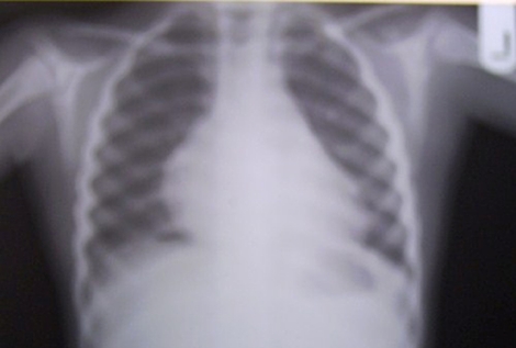
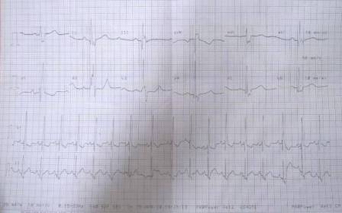
Chest x-ray (figure 1) showed situs solitus, laevocardia and a bulky cardiac silhouette with normal pulmonary vascular markings. ECG (figure 2) showed right atrial enlargement, right ventricular hypertrophy and right bundle branch block, raising the possibility of the presence of an atrial septal defect (ASD).
Figure 1.
Chest radiograph, showing a normal shaped, though bulky cardiac silhouette.
Figure 2.
Electrocardiograph, showing right atrial enlargement, right bundle branch block and right ventricular hypertrophy.
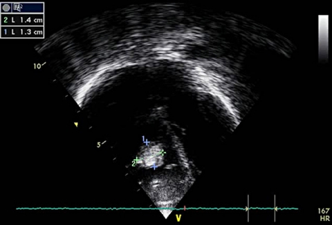
2-dimensional (2-D) echocardiography (figure 3) revealed normal intracardiac connections and a small secundum ASD. In addition, there was a 13×14 mm mass moving with the tricuspid valve, flipping between the right atrium and right ventricle (figure 3). This was thought to be either a myxoma or a vegetation. There was also moderate tricuspid regurgitation.
Figure 3.
2-dimensional echocardiograph showing the tumour in the right ventricle.
Differential diagnosis
-
▶
Myxoma
-
▶
Vegetation.
Treatment
As the options for surgical intervention in Nigeria were limited, the patient was referred to India for further evaluation and treatment. He subsequently underwent open-heart surgery at the Fortis Escorts Heart Institute, India, at which a ‘large, broad-based mass attached to the interventricular septum, adherent to the septal leaflet of the tricuspid valve’ was found. The mass ‘caused distortion of the tricuspid valve apparatus and obstruction to the right ventricle inflow and outflow tracts’. The tumour was removed and the secundum ASD closed.
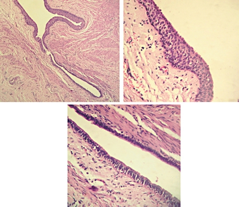
Histology of the tumour (figure 4) showed ‘cystic areas lined by pseudo-stratified ciliated mucous secreting columnar lining along the surrounding muscular wall. Also seen were focal areas of cartilage, small glandular luminae, squamous metaplasia of lining. There were also areas of ulceration with surrounding connective tissue fragments with interspersed vessels. There were no ectodermal components or 3rd germ layer components seen. There were no immature or malignant components’. These findings led to a diagnosis of bronchogenic cyst.
Figure 4.
Histological section of the tumour.
Outcome and follow-up
He made an uneventful postoperative recovery and returned to Nigeria 8 days after the surgery. He has remained well and symptom free 1 year following the surgery, with normal parameters of cardiac function, though still with significant tricuspid regurgitation.
Discussion
Bronchogenic cysts are often asymptomatic, being discovered incidentally, as was the case in our patient. They usually draw attention to themselves on account of compression on adjacent structures such as the tracheobronchial tree,19 or the heart,15 16 a situation which, particularly in childhood could be life-threatening. They may also be a cause of recurrent chest infections or chest pain.
In our patient, attention was only drawn to the heart by an incidental murmur discovered during routine evaluation of a febrile illness. In the only other report of an intracardiac bronchogenic cyst that we are aware of, the cyst was discovered incidentally in a 5-year-old girl during surgery for an aneurysm of the pars membranacea septi,14 which itself was thought to be the cause of premature ventricular contractions with which the patient presented. In the case reported by Somwaru et al,17 in a 3-year-old girl, the intrapericardial cyst, arising from pulmonary artery, was also discovered incidentally during surgery for a sinus venosus ASD.
In other reports we found of bronchogenic cysts related to the heart in children, the patients were mostly symptomatic. In Mosquera et al’s15 case, a 10-month-old girl presented with intractable heart failure. The cases of Göksel et al16 and Wei et al18 both presented with chest pain.
In retrospect, the auscultatory findings suggestive of RVOTO in the patient are likely to have been produced by the mass as it obstructed the outflow during systole, while the murmur suggestive of VSD was most probably due to tricuspid regurgitation consequent on the mass being adherent to it. In the similar case of a 48-year-old woman with a right ventricle bronchogenic cyst, reported by Prates et al,11 the mass similarly caused RVOTO, but was not adherent to the tricuspid valve, and there was therefore no regurgitation.
The importance of echocardiography in the evaluation of children suspected to have cardiac lesions cannot be gainsaid. The discovery of the lesion in this child was unexpected, particularly as he was asymptomatic. A similar experience of unexpected discovery on echocardiography, of an intracardiac tumour in a Nigerian neonate was reported in 1992,20 which turned out to be a rhabdomyoma, the most common cardiac tumour encountered in children.2 In Nigeria, particularly when patients are asymptomatic, the pressure to perform echocardiography is often downplayed because of lack of the equipment or the expertise to perform the procedure. Diagnosis of most common lesions is often therefore based on clinical features, supported by chest radiography and electrocardiography alone. Thus, a number of asymptomatic patients with a murmur, who might have significant pathology, might be misdiagnosed, as would have been the case in our patient.
From a review of the previously reported adult cases of intracardiac bronchogenic cyst, it would appear that these cysts occur most commonly in the interatrial septum, (three cases),8–10 followed by the left atrium,6 7 (two cases), right ventricle,,11 12 (two cases), with one case each in the right atrium,5 and left ventricle.13 Associated lesions reported in these cases include ASD,10 as is the case with our patient, and persistent left superior vena cava (SVC) (two cases).6 9
Even when asymptomatic, surgery is the preferred option of treatment for all cases of intracardiac bronchogenic cyst, the main reason being that there is no other way of ascertaining the histology of any such tumour. Even though it is a benign tumour, it may pose dangers by reason of its site or size, and there is always a possibility, though remote, of future malignant change.
In conclusion, we wish to submit that bronchogenic cyst should be considered as a rare differential of intracardiac masses.
Learning points.
-
▶
This is the first report of intracardiac bronchogenic cyst in an African child.
-
▶
There is a need for echocardiography in the evaluation of all asymptomatic children with cardiac murmurs, even in resource-poor settings. This would facilitate the identification of seemingly rare, asymptomatic cardiac lesions.
-
▶
Bronchogenic cyst should be considered as a differential diagnosis of a mass on the cardiac valve seen on echocardiography in the absence of fever.
-
▶
Intracardiac bronchogenic cyst, though rare, is a potential cause of RVOTO in childhood.
Acknowledgments
Surgery and histological diagnosis were carried out at the Fortis Escorts Heart Institute, India. The authors therefore acknowledge the contributions of Dr S Radhakrishnan (Paediatric Cardiology), Dr KS Iyer (Cardiothoracic Surgery) and Histopatology Division, SRL Reference lab, Gurgaon, India.
Footnotes
Competing interests None.
Patient consent Obtained.
References
- 1.Uzun O, Wilson DG, Vujanic GM, et al. Cardiac tumours in children. Orphanet J Rare Dis 2007;2:11. [DOI] [PMC free article] [PubMed] [Google Scholar]
- 2.Beghetti M, Gow RM, Haney I, et al. Pediatric primary benign cardiac tumors: a 15-year review. Am Heart J 1997;134:1107–14 [DOI] [PubMed] [Google Scholar]
- 3.Ribet ME, Copin MC, Gosselin B. Bronchogenic cysts of the mediastinum. J Thorac Cardiovasc Surg 1995;109:1003–10 [DOI] [PubMed] [Google Scholar]
- 4.Cataletto ME. Bronchogenic Cyst. http://emedicine.medscape.com/article/1005440-overview (accessed 14 November 2011).
- 5.Martínez-Mateo V, Arias MA, Juárez-Tosina R, et al. Permanent third-degree atrioventricular block as clinical presentation of an intracardiac bronchogenic cyst. Europace 2008;10:638–40 [DOI] [PubMed] [Google Scholar]
- 6.Lee T, Tsai IC, Tsai WL, et al. Bronchogenic cyst in the left atrium combined with persistent left superior vena cava: the first case in the literature. AJR Am J Roentgenol 2005;185:116–9 [DOI] [PubMed] [Google Scholar]
- 7.Soeda T, Matsuda M, Fujioka Y, et al. [A case report of huge bronchogenic cyst originated in the atrial septum]. Nihon Kyobu Geka Gakkai Zasshi 1996;44:1781–6 [PubMed] [Google Scholar]
- 8.Kawase Y, Takahashi M, Takemura H, et al. Surgical treatment of a bronchogenic cyst in the interatrial septum. Ann Thorac Surg 2002;74:1695–7 [DOI] [PubMed] [Google Scholar]
- 9.Chen CC. Bronchogenic cyst in the interatrial septum with a single persistent left superior vena cava. J Chin Med Assoc 2006;69:89–91 [DOI] [PubMed] [Google Scholar]
- 10.Borges AC, Knebel F, Lembcke A, et al. Bronchogenic cyst of the interatrial septum presenting as atrioventricular block. Ann Thorac Surg 2009;87:1920–3 [DOI] [PubMed] [Google Scholar]
- 11.Prates PR, Lovato L, Homsi-Neto A, et al. Right ventricular bronchogenic cyst. Tex Heart Inst J 2003;30:71–3 [PMC free article] [PubMed] [Google Scholar]
- 12.Weinrich M, Lausberg HF, Pahl S, et al. A bronchogenic cyst of the right ventricular endocardium. Ann Thorac Surg 2005;79:e13–4 [DOI] [PubMed] [Google Scholar]
- 13.Klass O, Hoffmann MH, Ludwig B, et al. Images in cardiovascular medicine. Left ventricular bronchogenic cyst. Circulation 2007;116:e385–7 [DOI] [PubMed] [Google Scholar]
- 14.Inzani F, Recusani F, Agozzino M, et al. Bronchogenic cyst: unexpected finding in a large aneurysm of the pars membranacea septi. J Thorac Cardiovasc Surg 2006;132:972–4 [DOI] [PubMed] [Google Scholar]
- 15.Mosquera VX, Rijlaarsdam M, Filippini L, et al. Early atrial septal defect surgery due to a bronchogenic cyst causing congestive heart failure by left atrium compression. Interact Cardiovasc Thorac Surg 2008;7:517–8 [DOI] [PubMed] [Google Scholar]
- 16.Göksel OS, Sayin OA, Cinar T, et al. Bronchogenic cyst invading right atrium in a 5-year-old. Thorac Cardiovasc Surg 2008;56:435–6 [DOI] [PubMed] [Google Scholar]
- 17.Somwaru LL, Midgley FM, Di Russo GB. Intrapericardial bronchogenic cyst overriding the pulmonary artery. Pediatr Cardiol 2005;26:713–4 [DOI] [PubMed] [Google Scholar]
- 18.Wei X, Omo A, Pan T, et al. Left ventricular bronchogenic cyst. Ann Thorac Surg 2006;81:e13–5 [DOI] [PubMed] [Google Scholar]
- 19.Ahrens B, Wit J, Schmitt M, et al. Symptomatic bronchogenic cyst in a six-month-old infant: case report and review of the literature. J Thorac Cardiovasc Surg 2001;122:1021–3 [DOI] [PubMed] [Google Scholar]
- 20.Ogunkunle OO, Omokhodion SI, Olasode BJ, et al. Rhabdomyoma of the heart associated with endocardial fibroelastosis in a Nigerian neonate. Trop Cardiol 1992;18:67–71 [Google Scholar]


