Fig. 6.
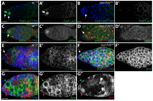
mei-P26 mutants display defects in BMP signal transduction. (A-B′) w1118 control (A,A′) and mei-P26mfs1 homozygotes (B,B′) stained for Dad-lacZ (green), Hts (red) and DNA (blue). (A′,B′) Dad-lacZ staining alone. Arrows indicate germ cells adjacent to cap cells. (C-D′) w1118 control (C,C′) and mei-P26mfs1 homozygous (D,D′) germaria stained for pMad (green), Hts (red) and DNA (blue). (C′,D′) pMad staining alone. Lack of Dad-lacZ and pMad expression in germline cells immediately adjacent to cap cells in mutant samples suggests that BMP signaling is compromised in the absence of mei-P26. Arrows indicate germ cells adjacent to cap cells. (E-F′) w1118 control (E) and mei-P26mfs1 (F) germaria stained for Brat (green), Hts (red) and DNA (blue). (E′,F′) Brat expression shown alone. Germline cells throughout mei-P26 mutant germaria express higher levels of Brat than the control samples. Arrows indicate germ cells adjacent to cap cells. (G-G″) mei-P26mfs1 clonal germarium stained for Brat (green), GFP (red) and DNA (blue) (G). The homozygous mei-P26 mutant GSC clone (green arrow) expresses more Brat protein than the heterozygous control clone (red arrow). (G′,G″) Brat and GFP staining alone. Scale bars: 10 μm.

