Abstract
Influenza A virus glycoprotein hemagglutinin (HA) binds to host cell surface sialic acid (SA)-terminated sugars in glycoproteins to initiate viral entry. It is thought that avian influenza viruses preferentially bind to N-acetylneuraminic acid α3 (NeuAcα3) sugars, while human influenza viruses exhibit a preference for NeuAcα6-containing sugars. Thus, species-specific SA(s) is one of the determinants in viral host tropism. The SA binding pocket of the HA1 subunit has been extensively studied, and a number of residues important for receptor binding have been identified. In this study, we examined the potential roles of seven highly conserved HA surface-located amino acid residues in receptor binding and viral entry using an H5 subtype. Among them, mutant Y161A showed cell-type-dependent viral entry without obvious defects in HA protein expression or viral incorporation. This mutant also displayed dramatically different ability in agglutinating different animal erythrocytes. Oligosaccharide binding analysis showed that substituting alanine at Y161 of HA changed the SA binding preference from NeuAc to N-glycolylneuraminic acid (NeuGc). Rescued mutant Y161A viruses demonstrated a 5- to 10-fold growth defect, but they were robust in viral replication and plaque forming ability. Our results demonstrate that Y161 is a critical residue involved in recognition of different SA species. This residue may play a role in determining influenza virus host tropism.
INTRODUCTION
Influenza A viruses, which have been isolated from a wide range of animals, including humans, swine, poultry, horses, canines, and wild waterfowl and other migrating birds, cause seasonal flu and occasionally pandemics. It is thought that wild waterfowl are the natural reservoir of these viruses. Nevertheless, these viruses can occasionally break the host barrier, i.e., they are transmitted from one host to another, sometimes leading to severe infection and illness in humans and birds (23, 45, 46). It is believed that the recent 2009 H1N1 influenza pandemic was the result of a reassortment of bird, swine, and human influenza viruses (3, 11, 16, 27).
The hemagglutinin (HA) of influenza A virus is one of the viral surface glycoproteins and is responsible for binding the virus to host cells and subsequent membrane fusion within the late endosome (2, 24, 35, 36, 49). HA also plays an important role in host immune responses by harboring the major antigenic sites responsible for the generation of neutralizing antibodies. The mature HA is a spike-like homotrimer, and each monomer is synthesized as a precursor HA0, which is then cleaved into HA1 and HA2 subunits by host enzymes and modified by multiple glycosylations. Most of the HA1 subunit forms the head region of HA, while the HA2 subunit is the primary feature of the stem region (38, 39, 48). The receptor binding site (RBS), which has been well characterized, is located on the head region of each HA monomer (15, 35).
One of the determinants of the influenza A virus host range is receptor recognition. The HA binds to glycans terminated with sialic acids (SAs), which play a crucial role as a receptor in influenza A virus entry. Amino acids involved in the interaction of SA are highly conserved among the 16 subtypes of HA, but substitutions of these residues have been detected and linked to receptor specificity during the adaptation of influenza virus to new hosts (6, 17, 22, 33, 47, 48). In particular, two HA residues at positions 226 and 228 are critical in receptor recognition shift or host switch for several influenza virus subtypes (28, 30). Human influenza virus strains preferentially bind to N-5-acetylneuraminic acid α2,6-glactose (Neu5Acα2,6Gal)-terminated glycans, while avian influenza viruses prefer N-5-acetylneuraminic acid α2,3-glactose (Neu5Acα2,3Gal) (4, 13, 26, 31, 32). Although Neu5Ac is recognized as an essential determinant of the cell surface receptor, influenza A viruses also differentially bind to receptors which are modified by N-glycolylneuraminic acid (NeuGc), another common SA found in mammals, which potentially serves as an entry and host determinant (19, 22, 40).
In this study, we have targeted and characterized the function in viral entry of 7 highly conserved, surface-located HA amino acids which have not been investigated previously. We have identified two interesting mutants. The double mutation K307A/K310A at amino acids located in the HA stem abolished virus erythrocyte binding, but the mutant retained reasonably good viral entry and replication, suggesting that the stem region is also important for the influenza virus receptor. Of particular interest, we have discovered that a critical residue, Y161, is essential in viral receptor recognition of NeuAc or NeuGc SAs, which is important in understanding the structure determinants of HA in limiting influenza virus host range.
MATERIALS AND METHODS
Cell lines, antibodies and plasmids.
Human embryonic kidney 293T cells and human lung epithelial A549 cells were maintained in Dulbecco's modified Eagle's medium (DMEM) supplemented with 10% fetal bovine serum, 100 μg/ml streptomycin, and 100 units of penicillin.
Goat polyclonal anti-influenza virus H5 HA (A/HongKong/213/03 [H5N1]; NR-163) was obtained from BEI Resources. The mouse anti-β-actin monoclonal antibody was purchased from Sigma (St. Louis, MO). The mouse anti-HIV p24 monoclonal antibody was obtained from the National Institutes of Health AIDS Research and Reference Reagent Program.
Mutant HAs were generated by the QuikChange site-directed mutagenesis kit from Stratagene by following the supplier's protocol and using custom-designed primers. All HA mutants were confirmed by DNA sequencing of the full-length HA gene.
Generation of pseudovirions.
Pseudotyped influenza viruses were produced by a polyethylenimine (PEI)-based transfection protocol (41, 44). Plasmids encoding hemagglutinin (HA), neuraminidase (NA), and vesicular stomatitis virus glycoprotein (VSV-G) and a replication-defective HIV vector (pNL4.3.Luc-R−E− or pNL4.3.GFP-R−E−) were used for transient cotransfection on 293T producer cells. Codon-optimized H5N1 A/Vietnam/1203/04 HA (CO-HA) was used as a parental HA. Seven hours after transfection, cells were washed with phosphate-buffered saline (PBS) and 6 ml of fresh medium was added to each plate. Forty-eight hours posttransfection, the supernatants were collected and filtered through a 0.45-μm-pore-size filter (Nalgene) and were used directly for infection. The remaining pseudovirions were stored at −80°C.
Infection assay.
The infection assay was performed as described previously (24, 28). Pseudovirions prepared as described above were incubated (500 μl/well) with 293T and A549 target cells, which were seeded 24 h prior to infection. The target cells were lysed in 150 μl lysis reagent (Promega) 48 h after infection. The luciferase activity was measured with a luciferase assay kit (Promega) and an FB12 luminometer (Berthold Detection Systems). Each experiment was repeated at least three times.
Western blot analysis.
To examine the expression of HA, Western blotting was carried out as described previously (24, 28). The 293T producer cells were lysed in 500 μl Triton X-100 lysis buffer and a protease inhibitor cocktail 48 h after transfection. To examine the incorporation of HA protein into pseudovirions, 4 ml of collected supernatants was layered onto a 1-ml cushion of 20% (wt/vol) sucrose in PBS and centrifuged at 55,000 rpm for 1 h in a Beckman SW55 rotor at 4°C. The pseudovirus pellets were lysed in 50 μl of Triton X-100 lysis buffer. The samples were then subjected to sodium dodecyl sulfate-polyacrylamide gel electrophoresis (SDS-PAGE) and transferred to a polyvinylidene difluoride membrane. The membrane was first incubated with NR163 antibody (1:5,000 dilution) for 2 h and then probed with peroxidase-conjugated rabbit anti-goat antiserum (Pierce) for 1 h. The bands were visualized by the chemiluminescence method according to the protocol of the supplier (Pierce). In these experiments, mouse anti-β-actin (1:10,000 dilution) and anti-HIV p24 (1:10,000 dilution) monoclonal antibodies were used as indicators for the cell lysate loading control and the relative amounts of the pseudovirions, respectively.
Hemagglutination assay.
The hemagglutination assay was performed as described previously (41). Supernatants from producer 293T cells were harvested 48 h posttransfection. Four milliliters of filter-sterilized pseudoparticles was concentrated over a 30% sucrose-NTE [sodium tris(hydroxymethyl)-aminomethane buffer containing EDTA] cushion. The samples were spun at 55,000 rpm for 1 h in a Beckman SW55 rotor at 4°C. Pseudovirion pellets were resuspended in 200 μl of Tris buffer. Twofold serial dilutions were mixed with an equal volume of 0.5% animal erythrocyte suspension (CRBCs; Lampire Biological Laboratories) in a U-bottom 96-well plate. HA titers were recorded after 2 h of incubation at 4°C. HA assay experiments were repeated at least three times.
Oligosaccharide binding assay.
Streptavidin-coated high-binding-capacity 384-well plates (Pierce) were incubated overnight at 4°C with 50 μl of 3 μM biotinylated saccharides. Saccharides were provided by the Consortium of Functional Glycomics (http://www.functionalglycomics.org). Pseudotyped viruses were concentrated as described above. Fifty microliters of viruses diluted in PBS with 1% (wt/vol) bovine serum albumin (PBS-BSA) was added to saccharide-coated wells and was incubated with oligosaccharides overnight at 4°C. Wells were then washed 3 times with PBST (PBS, 0.1% Tween 20) and 3 times with PBS and then blocked with PBS-BSA for 2 h at 4°C, before incubation with anti-HA antibody NR-163 diluted in PBS-BSA for 4 h at 4°C. After a washing as described above, wells were incubated with horseradish peroxidase (HRP)-conjugated rabbit anti-goat IgG secondary antibody in PBS-BSA. After a washing as described above, 50 μl of 1-step Ultra TMB enzyme-linked immunosorbent assay (ELISA) substrate (Thermo) was added to each well and the plate was incubated at room temperature for 20 min. Binding of the secondary antibody was detected by measuring the absorbance of each well at 450 nm after adding 50 ml of stop solution of 2 M sulfuric acid to each well. Appropriate negative controls were included. Assays were repeated at least three times.
Generation of recombinant viruses and viral growth.
Y161A and K307A/K310A mutations in the HA protein of influenza HAlo virus generated from influenza virus A/Vietnam/1203/04 (H5N1) were produced by PCR mutagenesis, and the resulting DNAs were cloned into pPolI transcription plasmids. The HAlo virus HA is identical to wild-type HA except for the removal of the multibasic cleavage site, responsible for its high pathogenicity (37). Generation of recombinant virus by reverse genetics was as described before (29). Briefly, a coculture of MDCK and 293T cells was transfected with four expression plasmids coding for the PB1, PB2, and PA proteins and nucleoprotein (NP) and with eight pPolI plasmids, each containing one of the HAlo virus viral RNA (vRNA) segments, including the wild-type HAlo segment, or their corresponding mutant HAlo genes. A total of 0.5 to 1 μg of each plasmid was transfected by using Lipofectamine 2000 (Invitrogen, Carlsbad, CA). At 12 h posttransfection the medium was replaced by DMEM containing 0.3% bovine serum albumin, 10 mM HEPES, and 1 μg of tosylsulfonyl phenylalanyl chloromethyl ketone (TPCK)-treated trypsin/ml. At 3 days posttransfection, virus within the cell supernatants was plaque purified by titration on MDCK cells. All the segments of the recovered mutant viruses were analyzed by reverse transcription-PCR (RT-PCR) and confirmed by sequencing.
MDCK cells were infected at a multiplicity of infection (MOI) of 0.01 with recombinant influenza A viruses expressing either HA Y161A, HA K307A/K310A, or parental HAlo. At 0, 12, 24, 36, 48, and 60 h postinfection, cell supernatants were harvested and titrated by plaque assay on fresh MDCK cells.
RESULTS
Design of the amino acid substitutions within influenza virus HA1.
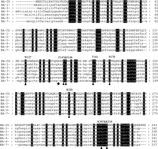
HA1 mediates the attachment of the viral particle to the target cell surface, which is the first step in viral entry, while the HA2 subunit mediates the fusion process after virus binding to the cell membrane (35). To further explore the potential roles of other critical amino acids in addition to those which are already known to be involved in the interaction with SA, we compared the HA1 sequences from all 16 HA subtypes (Fig. 1). Overall, 89 highly conserved residues were identified, discounting residues that were previously implicated in HA-SA interactions. Seven residues, which are located on the surface of the HA crystal structure, were chosen for further analysis. In addition, we also selected residues K144 and K307, which are located on the HA surface and on the interior surface of the stem region of HA, respectively, for this study. Together, 9 single-alanine-substitution mutants plus two more with double mutations at nearby positions of HA were generated by site-directed mutagenesis, and their phenotypes were characterized.
Fig 1.
Sequence alignment of HA1 from 16 different HA subtypes. For space restriction, only the alignment of H1 to H7 is shown. CO-HA is a representative of H5 HA. Residues shaded in black are the highly conserved ones in all 16 subtypes. Residues targeted for mutation are indicated by triangles. The black dot represents control residue K144. Each residue is numbered according to the H3 HA number.
Effects of alanine substitutions on pseudotyped virus entry.
To study the entry mechanism of the highly pathogenic influenza A virus while alleviating safety concerns, we have developed an HIV-based pseudotyping system to produce pseudotyped viruses (18). HIV particles pseudotyped with CO-HA or mutant HAs were used to infect the target cells (293T and A549). The luciferase activities of the transduced cells were used as a measure for viral entry (Fig. 2). In these experiments, virions pseudotyped with VSV-G were used as a positive control, as VSV-G can be incorporated into pseudotyped particles with a very high efficiency and the pseudovirions have a broad host range (1, 50). The effect of alanine substitution mutants of HA on viral entry can be classified into two groups: (i) mutants that behaved like the parental HA (W127A, K144A, F147A, and F148A) and (ii) mutants with impaired viral entry (F147A/F148A, Y161A, N170A, R220A, K307A, K310A, and K307A/K310A). Among them, Y161A and K307A/K310A appeared to display cell-type-dependent viral entry since there was roughly 1 log difference in luciferase activities between 293T and A549 target cells.
Fig 2.
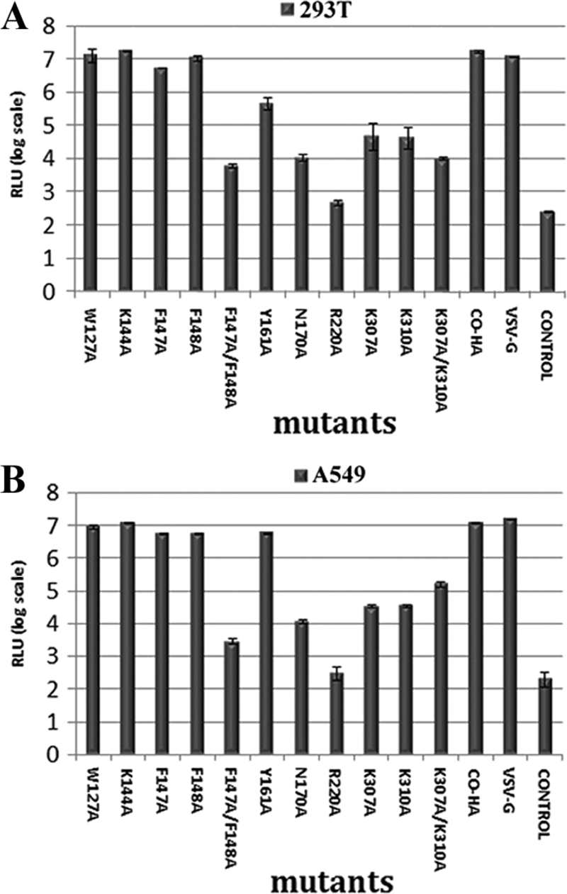
Mutational effects on viral entry as determined by measuring luciferase activity in two different cell lines. (A) Relative luciferase units (RLU) for 293T target cells. (B) RLU for A549 target cells. HIV NA pseudotyped with only the NA glycoprotein was used as a negative control. VSV-G pseudotyped viruses served as a positive control. The error bars are the standard deviations (SD) of three independent experiments from the transfection step.
Effects of alanine substitutions on envelope expression and incorporation.
We next examined the possible effect of alanine substitutions on HA expression, processing, and incorporation. First, cell lysates derived from the cells transiently transfected with the HIV-luc vector and different mutant HA plasmids were subjected to SDS-PAGE and Western blot analysis, as shown in Fig. 3A. Three bands, which corresponded to HA0, HA1, and HA2, with sizes of about 75 kDa, 50 kDa, and 25 kDa, respectively, were detected for CO-HA, the parental HA (lane 12), indicating that CO-HA was expressed and processed in 293T cells as predicted. Similarly, 8 of the 11 HA substitution mutants had the same pattern as CO-HA (lanes 1, 2, 3, 4, 6, 9, 10, and 11), suggesting that these substitutions did not greatly alter the expression and processing of HA in producer 293T cells. In contrast, F147A/F148A, N170A, and R220A (lanes 5, 7, and 8) displayed no or reduced cleavage products compared to CO-HA, suggesting that these alanine substitution mutations adversely affected the expression and/or proper folding of HA.
Fig 3.
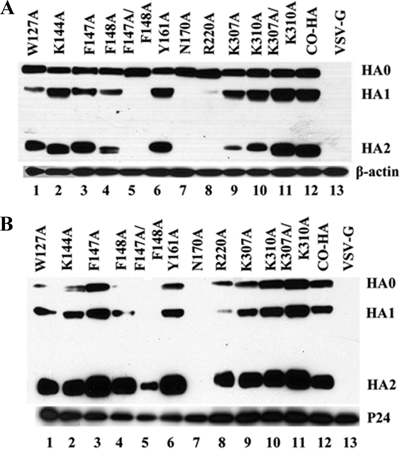
Western blot analysis of HA protein expression (A) and viral incorporation (B). The HA precursor (HA0) and proteolytic products HA1 and HA2 are shown. VSV-G was used to show the antibody specificity.
To test if alanine substitutions affected HA viral incorporation, the viral particles were pelleted by ultracentrifugation in a 20% sucrose cushion and subjected to SDS-PAGE and Western blot analysis (Fig. 3B). The pseudovirions carrying CO-HA showed the presence of correctly sized HA0, HA1, and HA2 (lane 12), indicating proper incorporation of HA into pseudoparticles. Mutants W127A, K144A, F147A, and F148A were detected on the viral particles (lanes 1, 2, 3, and 4). In contrast, there were no detectable HA0 and cleaved HA products from F147A/F148A or N170A on the pseudotyped viruses (lanes 5 and 7). Intriguingly, however, a band corresponding to HA2 was detectable from mutant F147A/F148A. Similarly, for mutant R220A, even though no HA2 was detected from the cell lysate (Fig. 3A, lane 8), there were still detectable but small amounts of HA1 and more HA2 in the viral particles (Fig. 3B, lane 8). Mutants Y161A, K307A, K310A, K307A/K310A were also detected on the viral particles (lanes 6, 9, 10, and 11).
In addition, immunostaining was used to examine mutational effects on HA surface expression. There was no significant difference in HA surface expression between each mutant and parental HA, except for F147A/F148A and N170A, which exhibited slightly weaker surface immunofluorescence (data not shown).
Together, our results suggest that alanine substitutions at positions 147/148, 170, and 220 affected HA protein processing and proper incorporation into viral particles; therefore, HA-mediated viral entry was extremely reduced. Alanine substitutions at positions 127, 144, 147, 148, 161, 307, and 310 did not dramatically influence HA protein processing and incorporation into viral particles. The reason for reduced viral entry for mutants Y161A, K307A, K310A, and K307/K310A was further studied.
Effect of alanine substitutions on sialic acid recognition.
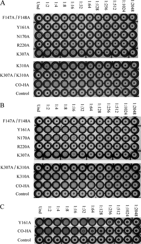
The binding of influenza virus to erythrocytes is mediated by the interaction of HA with cell surface receptors containing SA. As the influenza viruses attach to multiple erythrocytes, a lattice structure forms (21). We used the HA assay to further determine whether alanine substitutions changed SA binding preference and thus altered viral entry. Chicken, horse, and swine erythrocytes were chosen based on previous studies showing that erythrocytes from these three animals have different surface SA structures and that influenza A virus receptor specificity correlates with the agglutinating patterns from different animal erythrocytes (20). Briefly, 2-fold serial dilutions of purified pseudotyped viral particles were mixed with chicken or horse erythrocytes. Viral particle loading is standardized by HIV-P24. The hemagglutination results for parental HA (CO-HA) and the selected HA mutants (F147A/F148A, Y161A, R220A, K307A, K310A, and K307A/K310A) which displayed reduced entry (Fig. 2A) are shown in Fig. 4. The highest dilution of parental virus agglutination is 1:64 for both erythrocytes. HA assay of mutants from the first group of mutants (W127A, K144A, F147A, and F148A) showed a pattern similar to that for parental HA (data not shown). For mutants F147A/F148A, N170A, R220A, and K307A/K310A, alanine substitutions totally abolished SA binding as their HA titers are zero on both erythrocytes (Fig. 4A and B). K307A and K310A displayed lower HA titers than CO-HA on both animal erythrocytes (Fig. 4A and B). Interestingly, Y161A pseudotyped viruses showed dramatically different patterns of agglutination on chicken and horse erythrocytes: no agglutination was observed when virus was mixed with chicken erythrocytes, while a 1-unit-higher (1:128) titer than that for CO-HA pseudoparticles was revealed in the HA assay when horse erythrocytes were agglutinated.
Fig 4.
Hemagglutination assay of titrated pseudovirions mixed with chicken erythrocytes (A), horse erythrocytes (B), and pig erythrocytes (C). Mutants that didn't show reduced viral infectivity are not shown. HIV-NA virus pseudotyped with only the NA glycoprotein served as the control.
Considering that the surfaces of chicken and horse erythrocytes differ in SA structure and species, i.e., chicken erythrocytes contain 95 to 100% N-acetylneuraminic acid (NeuAc) SA of both α2,3-Gal and α2,6-Gal linkages, with slightly more abundant of α2,6-Gal SAs, and horse erythrocytes contain 95 to 100% N-glycolylneuraminic acid (NeuGc) SAs, with more abundant α2,3-Gal SAs than α2,6-Gal SAs (10, 20, 34), we hypothesized that alanine substitution at Y161 changed the viral receptor preference from NeuAc to NeuGc SA. We thus further tested the ability of Y161A mutant virus to agglutinate swine erythrocytes, which had been shown to contain about 66% NeuGc and 34% NeuAc with both α2,3-Gal and α2,6-Gal SAs but with more of the α2,6-Gal SAs (10). The results from the swine agglutination assay (Fig. 4C) showed a pattern similar to that for horse agglutination, supporting our hypothesis.
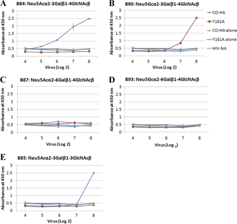
To further confirm our hypothesis, we used an enzyme-linked immunosorbent assay (ELISA) to investigate the direct binding of mutant Y161A and parental CO-HA pseudoparticles to five biotinylated saccharides bearing varied species of terminal SA and sialyl linkages (Fig. 5). Controls, including CO-HA virus alone (without incubation with any biotinylated oligosaccharides), Y161A virus alone, HIV-NA virus pseudotyped with only NA glycoprotein, oligosaccharides alone, and HRP-conjugated rabbit anti-goat IgG secondary antibody alone, were performed, and only the first 3 controls are shown in Fig. 5 since they all displayed similar levels of absorbance at 450 nm. CO-HA pseudotyped viruses bound to Neu5Acα2,3-linked SA (3′SLN) in a dose-dependent manner (Fig. 5A), but not to Neu5Acα2,6-linked SA (6′SLN) (Fig. 5C), which is consistent with the current notion that avian influenza viruses preferentially bind to NeuAcα2,3-linked SAs (4). The Y161A mutant showed only a background level of binding to 3′SLN (Fig. 5A). Interestingly, when Neu5Gcα2,3-linked SA [3′S(Gc)LN] was incubated with these two viruses, the binding pattern was totally divergent. CO-HA viruses showed background binding levels, while the Y161A mutant viruses displayed a dose-dependent binding to 3′S(Gc)LN (Fig. 5B), which strongly supports our hypothesis that alanine substitution at Y161 switched virus SA receptor preference from NeuAc to NeuGc. Neither of the two viruses showed binding to Neu5Gcα2,6-linked saccharide [6′S(Gc)LN] (Fig. 5D), which further confirmed that 2,6 linkage is not preferred by avian influenza virus.
Fig 5.
The switched HA binding preference of the Y161A virus. The biotinylated oligosaccharides indicated were used to measure saccharide binding of parental CO-HA and mutant Y161A pseudotyped viruses. The binding of graded amounts of virus (from 16 HA units to 256 HA units) to immobilized oligosaccharides was determined by measuring the binding of an anti-HA antibody as described in Materials and Methods. Wells with Y161A or CO-HA viruses alone without added oligosaccharides served as negative controls. HIV-NA pseudotyped virus binding to different saccharides was used as another negative control. All other controls, including oligosaccharides alone and the secondary antibody alone, were performed and displayed levels similar to that for the negative controls (data not shown).
It has been reported that influenza virus adaptation was determined not only by the receptor terminal head, which has the linkage of the α2,3 and α2,6 motifs, but also by the inner fragments of the carbohydrate chain (12, 14). Another oligosaccharide, 3′SLec, which differs from 3′SLN by a penultimate saccharide linkage, was used in the binding ELISA (Fig. 5E). Not surprisingly, the Y161A mutant did not show binding to 3′SLec since this oligosaccharide ends with Neu5Ac. CO-HA parental viruses also exhibited weaker binding to 3′SLec than 3′SLN at lower concentrations, suggesting that chicken H5 has higher relative affinity for binding to saccharide with β1-4 than to saccharide with β1-3 between the 2nd and 3rd sugars.
Growth properties of Y161A and K307A/K310A mutant influenza viruses.
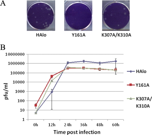
The ex vivo characteristics of the mutants Y161A and K307A/K310A were studied by generating recombinant viruses through plasmid transfection. We used HAlo virus HA as the parental HA. HAlo HA is a modified HA from the influenza virus H5 avian strain A/Vietnam/1203/04(H5N1) in which the HA polybasic cleavage peptide has been removed (29). The relative plaque size of the K307A/K310A mutant was morphologically similar to those produced by parental HAlo. In contrast, Y161A showed a 50% reduction in the plaque size (Fig. 6A), suggesting impaired cell-to-cell spread of Y161. The growth curve was assayed on MDCK cells, and the results are shown in Fig. 6B. Both Y161A and K307A/K310A mutant viruses replicated with a mild defect after 24 h (less than 10-fold).
Fig 6.
Growth proprieties of recombinant influenza HAlo viruses expressing parental HA and mutant Y161A or K307A/K310A proteins. MDCK cells were infected at an MOI of 0.01 with the different recombinant viruses. Viruses released to the supernatant were titrated at 0, 12, 24, 48, and 60 h postinfection by plaque assay on fresh MDCK cells.
DISCUSSION
The RBS of HA has been extensively studied; it consists of amino acids 98, 134 to 138, 153, 155, 183, 190, 194, and 224 to 228 (15, 35). Most of these residues are highly conserved among all 16 subtypes of HA. However, little is known regarding the function of other conserved amino acids in HA1 in viral entry. In this report, we have targeted 7 highly conserved residues of HA1. Alanine scanning mutagenesis was performed for 11 targets, including 7 selected residues, double alanine substitutions, and controls. Targets were analyzed for viral entry, HA protein expression and processing, and viral incorporation. We have identified several key residues (F147/F148, Y161, N170, R220, K307, K310, and K307/K310) that are important for HA cellular processing and receptor recognition. Of particular interest, we have identified one residue, Y161, that is critical for different SA species recognition. These findings have important implications for understanding the function of the HA structure determinants in influenza virus entry, pathogenesis, and host tropism.
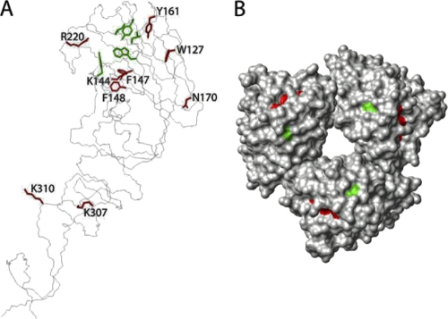
To better understand how the structural determinants of receptor properties and functional information of the influenza virus glycoprotein HA1 subunit affect viral entry, we have highlighted 7 selected residues and the RBS in the H5N1 HA crystal structure (Fig. 7). F147, F148, and R220 are in the vicinity of the RBS, and alanine substitutions at F147/F148 and R220 affected HA cleavage (Fig. 3, lanes 5 and 8), with most of the HA proteins retained in their precursor state in the producer cells. Note that the RBS is distant from the cleavage site, so it is somewhat surprising that these residues affected HA cleavage. At the same time, alanine substitutions at these sites may destroy the recognition of SAs by HA on the surfaces of chicken and horse erythrocytes, which therefore displayed zero HA titer and no viral entry, while the impact of the single alanine substitution at either F147 or F148 was not massive enough to influence receptor binding.
Fig 7.
Locations of selected amino acid substitutions in the H5N1 influenza virus HA1 subunit. (A) Major components of the RBS (green in black circles) and seven target residues (red). (B) Top view of Y161 (green) and the RBS (red).
N170 is a potential glycosylation site based on Asn-X-Thr/Ser sequence information. Deshpande et al. have reported that glycosylation affected cleavage of an H5N2 influenza virus hemagglutinin and thus virulence (7). It is possible that alanine substitution at N170 affected HA cleavage as well, resulting in very low viral entry. In order to test this hypothesis, N170A- and CO-HA-transfected cell lysates were digested with N-glycosidase F (Roche) and were analyzed on 7% polyacrylamide SDS gel (data not shown). Comparison of the shifts between digested and undigested CO-HA and Y170A lysates suggested that N170 is not a potential glycosylation site. The reason for the mutational effects needs to be further explored.
The stem region of HA has been recently shown to be a potentially good target for therapeutic treatment for influenza. Ekiert et al. have identified an antibody, CR6261, that binds to a highly conserved influenza virus epitope in the membrane-proximal stem regions of HA1 and HA2 (8). Neutralizing antibodies that bind to the stem region of group 1 and/or group 2 influenza A virus HAs have been selected (5, 9). Interestingly, one of the targets we selected, K310, is near the conserved epitope at the membrane-proximal end of each HA. Even though K307 and/or K310 is distant to the RBS, alanine substitution at either of these sites decreased pseudovirus binding to animal erythrocytes, reducing viral entry more than 99%, without obvious defects in HA protein expression or processing and viral incorporation. More interestingly, the double mutant K307A/K310A totally abolished pseudotyped virus binding to both chicken and horse erythrocytes and displayed zero HA titer, just like mutant R220A (Fig. 4), but viral entry of K307A/K310A was 10- to 100-fold higher than that of R220A (Fig. 2). The viral growth curve showed active viral replication in the first 20 h and was only about 10-fold lower than that for the parental virus after 24 h. These results suggest that mutant K307A/K310A is mechanistically different from R220A in receptor recognition.
One important finding of the current work is that Y161A showed a clear switch of receptor recognition from NeuAc- to NeuGcα2,3-linked SA. NeuAc and NeuGc SAs are the most prevalent SAs found in mammalian cells. The only difference between these two SAs is the additional hydroxyl group in the N-glycolyl group of Neu5Gc (42). Humans are deficient in NeuGc SAs, which is explained by a genetic mutation in CMP-N-acetylneuraminic acid hydroxylase, the rate-limiting enzyme in generating NeuGc from precursor NeuAc in other mammals (43). Masuda et al. have reported that amino acids at 143, 155, and 158 in HA of human H3 influenza virus are linked to the viral recognition of NeuGc (25). In our study, we identified a highly conserved residue, Y161, located at the top of the HA head region outside the RBS (Fig. 7) that plays a critical role in virus recognition of NeuGc and NeuAc SAs. Alanine substitution at this position resulted in a 98% reduction in viral entry into 293T target cells, while the reduction on A549 cells was about 50%. This cell-type-dependent reduction is probably due to the different species of SAs present in the two cell lines. Further investigation of the SAs on these two cell lines is needed to explain cell type specificity in viral entry. Y161A recombinant virus production was similar to that of the parental virus despite smaller plaque size, which indicated a lesser ability to spread cell to cell. To further explore the mechanism of SA binding preference caused by Y161A, four surface-located amino acids, T160, P162, Y195, and N248, surrounding Y161 were mutated to alanine. Also, Y161F mutagenesis was performed since F161 was used by some other influenza A virus strains. Among these five mutants, only T160A showed weak ability for NeuAc to NeuGc switching; the others did not have effects on recognition of different SA species (data not shown), suggesting that Y161 is the most important residue of this region for changing receptor recognition.
In summary, we studied the function of novel highly conserved, surface-located residues in viral entry and receptor binding. Although these residues are highly conserved in the evolution of influenza virus, due to the unique features of influenza virus antigenic drift and antigenic shift, these mutations may occur in nature. For example, residue Y161 is highly conserved among all the 16 HA1 subunits and mutation at Y161 to alanine can be tolerated by influenza virus and can change the viral tropism toward hosts which have the NeuGc SA on the cell surface. This finding demonstrates the significance of SA species as a determinant in viral transmission and host range restriction. Based on our studies of K307A/K310A, we demonstrated that the stem region is also important in SA recognition and viral entry. The studies described here provide important insight into understanding the function of the HA structural determinants in influenza virus entry and evolution.
ACKNOWLEDGMENTS
We thank the Consortium for Functional Glycomics for reagents.
This research was partly supported by NIAID grant PO1 AI058113 and by the Center for Research on Influenza Pathogenesis (NIAID contract HHSN266200700010C) (A.G.-S.).
Footnotes
Published ahead of print 1 February 2012
REFERENCES
- 1. Arai T, Takada M, Ui M, Iba H. 1999. Dose-dependent transduction of vesicular stomatitis virus G protein-pseudotyped retrovirus vector into human solid tumor cell lines and murine fibroblasts. Virology 260:109–115 [DOI] [PubMed] [Google Scholar]
- 2. Carr CM, Chaudhry C, Kim PS. 1997. Influenza hemagglutinin is spring-loaded by a metastable native conformation. Proc. Natl. Acad. Sci. U. S. A. 94:14306–14313 [DOI] [PMC free article] [PubMed] [Google Scholar]
- 3. Cohen J. 2010. Swine flu pandemic. What's old is new: 1918 virus matches 2009 H1N1 strain. Science 327:1563–1564 [DOI] [PubMed] [Google Scholar]
- 4. Connor RJ, Kawaoka Y, Webster RG, Paulson JC. 1994. Receptor specificity in human, avian, and equine H2 and H3 influenza-virus isolates. Virology 205:17–23 [DOI] [PubMed] [Google Scholar]
- 5. Corti D, et al. 2011. A neutralizing antibody selected from plasma cells that binds to group 1 and group 2 influenza A hemagglutinins. Science 333:850–856 [DOI] [PubMed] [Google Scholar]
- 6. Couceiro JN, Paulson JC, Baum LG. 1993. Influenza virus strains selectively recognize sialyloligosaccharides on human respiratory epithelium; the role of the host cell in selection of hemagglutinin receptor specificity. Virus Res. 29:155–165 [DOI] [PubMed] [Google Scholar]
- 7. Deshpande KL, Fried VA, Ando M, Webster RG. 1987. Glycosylation affects cleavage of an H5n2 influenza-virus hemagglutinin and regulates virulence. Proc. Natl. Acad. Sci. U. S. A. 84:36–40 [DOI] [PMC free article] [PubMed] [Google Scholar]
- 8. Ekiert DC, et al. 2009. Antibody recognition of a highly conserved influenza virus epitope. Science 324:246–251 [DOI] [PMC free article] [PubMed] [Google Scholar]
- 9. Ekiert DC, et al. 2011. A highly conserved neutralizing epitope on group 2 influenza A viruses. Science 333:843–850 [DOI] [PMC free article] [PubMed] [Google Scholar]
- 10. Eylar EH, Madoff MA, Brody OV, Oncley JL. 1962. The contribution of sialic acid to the surface charge of the erythrocyte. J. Biol. Chem. 237:1992–2000 [PubMed] [Google Scholar]
- 11. Fraser C, et al. 2009. Pandemic potential of a strain of influenza A (H1N1): early findings. Science 324:1557–1561 [DOI] [PMC free article] [PubMed] [Google Scholar]
- 12. Gambaryan A, et al. 2005. Receptor specificity of influenza viruses from birds and mammals: new data on involvement of the inner fragments of the carbohydrate chain. Virology 334:276–283 [DOI] [PubMed] [Google Scholar]
- 13. Gambaryan AS, et al. 2003. Differences between influenza virus receptors on target cells of duck and chicken and receptor specificity of the 1997 H5N1 chicken and human influenza viruses from Hong Kong. Avian Dis. 47:1154–1160 [DOI] [PubMed] [Google Scholar]
- 14. Gambaryan AS, et al. 2004. H5N1 chicken influenza viruses display a high binding affinity for Neu5Acalpha2-3Galbeta1-4(6-HSO3)GlcNAc-containing receptors. Virology 326:310–316 [DOI] [PubMed] [Google Scholar]
- 15. Gamblin SJ, et al. 2004. The structure and receptor binding properties of the 1918 influenza hemagglutinin. Science 303:1838–1842 [DOI] [PubMed] [Google Scholar]
- 16. Garten RJ, et al. 2009. Antigenic and genetic characteristics of swine-origin 2009 A(H1N1) influenza viruses circulating in humans. Science 325:197–201 [DOI] [PMC free article] [PubMed] [Google Scholar]
- 17. Glaser L, et al. 2005. A single amino acid substitution in 1918 influenza virus hemagglutinin changes receptor binding specificity. J. Virol. 79:11533–11536 [DOI] [PMC free article] [PubMed] [Google Scholar]
- 18. Guo Y, et al. 2009. Analysis of hemagglutinin-mediated entry tropism of H5N1 avian influenza. Virol. J. 6:39. [DOI] [PMC free article] [PubMed] [Google Scholar]
- 19. Higa HH, Rogers GN, Paulson JC. 1985. Influenza virus hemagglutinins differentiate between receptor determinants bearing N-acetyl-, N-glycollyl-, and N,O-diacetyineuraminic acids. Virology 144:279–282 [DOI] [PubMed] [Google Scholar]
- 20. Ito T, et al. 1997. Receptor specificity of influenza A viruses correlates with the agglutination of erythrocytes from different animal species. Virology 227:493–499 [DOI] [PubMed] [Google Scholar]
- 21. Killian ML. 2008. Hemagglutination assay for the avian influenza virus. Methods Mol. Biol. 436:47–52 [DOI] [PubMed] [Google Scholar]
- 22. Kuiken T, et al. 2006. Host species barriers to influenza virus infections. Science 312:394–397 [DOI] [PubMed] [Google Scholar]
- 23. Lazzari S, Stohr K. 2004. Avian influenza and influenza pandemics. Bull. World Health Organ. 82:242. [PMC free article] [PubMed] [Google Scholar]
- 24. Lear JD, DeGrado WF. 1987. Membrane binding and conformational properties of peptides representing the NH2 terminus of influenza HA-2. J. Biol. Chem. 262:6500–6505 [PubMed] [Google Scholar]
- 25. Masuda H, et al. 1999. Substitution of amino acid residue in influenza A virus hemagglutinin affects recognition of sialyl-oligosaccharides containing N-glycolylneuraminic acid. FEBS Lett. 464:71–74 [DOI] [PubMed] [Google Scholar]
- 26. Matrosovich MN, et al. 1997. Avian influenza A viruses differ from human viruses by recognition of sialyloligosaccharides and gangliosides and by a higher conservation of the HA receptor-binding site. Virology 233:224–234 [DOI] [PubMed] [Google Scholar]
- 27. Munster VJ, et al. 2009. Pathogenesis and transmission of swine-origin 2009 A(H1N1) influenza virus in ferrets. Science 325:481–483 [DOI] [PMC free article] [PubMed] [Google Scholar]
- 28. Naeve CW, Hinshaw VS, Webster RG. 1984. Mutations in the hemagglutinin receptor-binding site can change the biological properties of an influenza virus. J. Virol. 51:567–569 [DOI] [PMC free article] [PubMed] [Google Scholar]
- 29. Park MS, Steel J, Garcia-Sastre A, Swayne D, Palese P. 2006. Engineered viral vaccine constructs with dual specificity: avian influenza and Newcastle disease. Proc. Natl. Acad. Sci. U. S. A. 103:8203–8208 [DOI] [PMC free article] [PubMed] [Google Scholar]
- 30. Rogers GN, Dsouza BL. 1989. Receptor-binding properties of human and animal H1-influenza virus isolates. Virology 173:317–322 [DOI] [PubMed] [Google Scholar]
- 31. Rogers GN, Paulson JC. 1983. Receptor determinants of human and animal influenza-virus isolates—differences in receptor specificity of the hemagglutinin-H-3 based on species of origin. Virology 127:361–373 [DOI] [PubMed] [Google Scholar]
- 32. Rogers GN, et al. 1983. Single amino-acid substitutions in influenza hemagglutinin change receptor-binding specificity. Nature 304:76–78 [DOI] [PubMed] [Google Scholar]
- 33. Sauter NK, et al. 1992. Binding of influenza virus hemagglutinin to analogs of its cell-surface receptor, sialic acid: analysis by proton nuclear magnetic resonance spectroscopy and X-ray crystallography. Biochemistry 31:9609–9621 [DOI] [PubMed] [Google Scholar]
- 34. Seaman GVF, Uhlenbruck G. 1963. The surface structure of erythrocytes from some animal sources. Arch. Biochem. Biophys 100:493–502 [DOI] [PubMed] [Google Scholar]
- 35. Skehel JJ, Wiley DC. 2000. Receptor binding and membrane fusion in virus entry: the influenza hemagglutinin. Annu. Rev. Biochem. 69:531–569 [DOI] [PubMed] [Google Scholar]
- 36. Smith AE, Helenius A. 2004. How viruses enter animal cells. Science 304:237–242 [DOI] [PubMed] [Google Scholar]
- 37. Steel J, et al. 2009. Live attenuated influenza viruses containing NS1 truncations as vaccine candidates against H5N1 highly pathogenic avian influenza. J. Virol. 83:1742–1753 [DOI] [PMC free article] [PubMed] [Google Scholar]
- 38. Stevens J, et al. 2006. Structure and receptor specificity of the hemagglutinin from an H5N1 influenza virus. Science 312:404–410 [DOI] [PubMed] [Google Scholar]
- 39. Stevens J, et al. 2004. Structure of the uncleaved human H1 hemagglutinin from the extinct 1918 influenza virus. Science 303:1866–1870 [DOI] [PubMed] [Google Scholar]
- 40. Suzuki Y, et al. 2000. Sialic acid species as a determinant of the host range of influenza A viruses. J. Virol. 74:11825–11831 [DOI] [PMC free article] [PubMed] [Google Scholar]
- 41. Tisoncik JR, et al. 2011. Identification of critical residues of influenza neuraminidase in viral particle release. Virol. J. 8:14. [DOI] [PMC free article] [PubMed] [Google Scholar]
- 42. Varki A. 2007. Glycan-based interactions involving vertebrate sialic-acid-recognizing proteins. Nature 446:1023–1029 [DOI] [PubMed] [Google Scholar]
- 43. Varki A. 2001. Loss of N-glycolylneuraminic acid in humans: mechanisms, consequences, and implications for hominid evolution. Yearbook Physical Anthropol. 44:54–69 [DOI] [PMC free article] [PubMed] [Google Scholar]
- 44. Wang J, Manicassamy B, Caffrey M, Rong L. 2011. Characterization of the receptor-binding domain of Ebola glycoprotein in viral entry. Virol. Sin. 26:156–170 [DOI] [PMC free article] [PubMed] [Google Scholar]
- 45. Webster RG. 1994. While awaiting the next pandemic of influenza A. BMJ 309:1179–1180 [DOI] [PMC free article] [PubMed] [Google Scholar]
- 46. Webster RG, Bean WJ, Gorman OT, Chambers TM, Kawaoka Y. 1992. Evolution and ecology of influenza A viruses. Microbiol. Rev. 56:152–179 [DOI] [PMC free article] [PubMed] [Google Scholar]
- 47. Weis W, et al. 1988. Structure of the influenza virus haemagglutinin complexed with its receptor, sialic acid. Nature 333:426–431 [DOI] [PubMed] [Google Scholar]
- 48. Wiley DC, Skehel JJ. 1987. The structure and function of the hemagglutinin membrane glycoprotein of influenza virus. Annu. Rev. Biochem. 56:365–394 [DOI] [PubMed] [Google Scholar]
- 49. Yoshimura A, Ohnishi S. 1984. Uncoating of influenza virus in endosomes. J. Virol. 51:497–504 [DOI] [PMC free article] [PubMed] [Google Scholar]
- 50. Yu H, et al. 1999. High efficiency in vitro gene transfer into vascular tissues using a pseudotyped retroviral vector without pseudotransduction. Gene Ther. 6:1876–1883 [DOI] [PubMed] [Google Scholar]


