Synopsis
The many functions of the microtubule cytoskeleton are essential for shaping the development and maintaining the operation of the nervous system. With the recent discovery of congenital neurological disorders that result from mutations in genes that encode different α and β-tubulin isotypes (TUBA1A, TUBB2B, TUBA8, and TUBB3), scientists have a novel paradigm to assess how select perturbations in microtubule function affect a range of cellular processes in humans. Moreover, important phenotypic distinctions found among the syndromes suggest that different tubulin isotypes can be utilized for distinct cellular functions during nervous system development. In the present paper, we review: (i) the spectrum of congenital nervous system diseases that result from mutations in tubulin and microtubule associated proteins (MAPs); (ii) the known or putative roles of these proteins during nervous system development; (iii) how the findings collectively support the “multi-tubulin” hypothesis, which postulates that different tubulin isotypes may be required for specialized microtubule functions.
Keywords: tubulin, microtubules, nervous system, cell migration, axon guidance, TUBB3
Introduction
Proper nervous system function is dependent upon a neuron’s ability to receive, process, and transmit information within anatomically defined circuits. To build circuits, post-mitotic neurons must first polarize and establish a future axon amongst multiple growing and retracting neurites, a process tightly coupled with cell migration in the cerebral cortex [1, 2]. After neurons have finished migrating, axons continue to grow and navigate considerable distances to find appropriate post-synaptic targets by responding to specific growth and guidance cues in the extracellular matrix; these cues safeguard against numerous incorrect connections that present themselves along the way. Once neural circuits are established, connectivity between axons and their post-synaptic targets must be continually maintained. Failure to do so can result in axon degeneration and the loss of sensory, motor, and cognitive functions.
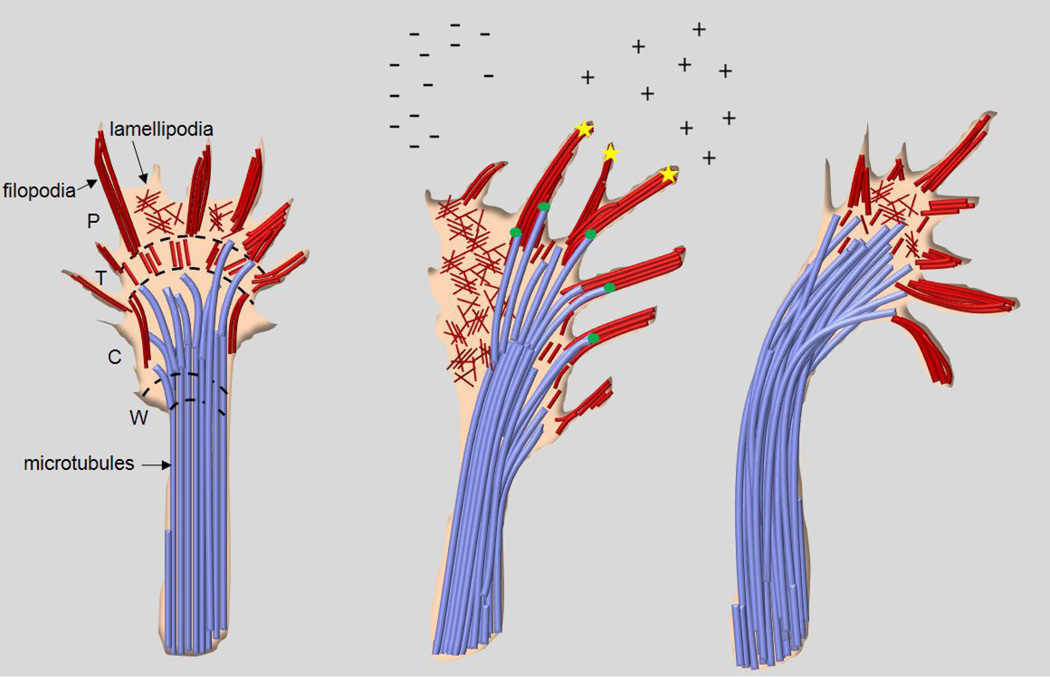
Tight regulation of the dynamic behavior and function of the microtubule cytoskeleton is essential for the development and survival of neurons. Microtubules are assembled from tubulin heterodimers, which contain different α- and β-tubulin isotypes each encoded by distinct genes [3]. Microtubules are polarized and, in neurons, their ‘minus-ends’ are usually oriented towards the centrosome in the cell body whereas their ‘plus-ends’ project towards the tips of axons [4]. Microtubule polarity serves important functions in both differentiating and adult neurons. First, the frequent transition between periods of growth and shortening at the dynamic ‘plus-ends’ permits differentiating neurons to extend or retract growing axons in response to guidance cues in order to maintain directional growth towards post-synaptic targets [5–7] (Figure 1). Second, dynein and kinesin motors transport protein vesicles and organelles towards the plus and minusends of microtubules, respectively, and are also necessary for the regulation of microtubule dynamics. Their activities are essential for cell migration, axon development, and guidance and are also required for the function and viability of adult neurons [8–11].
Figure 1. Dynamic microtubules regulate growth cone turning and axon guidance.
Illustration depicting an axon growth cone turning towards the direction of a positive guidance cue. Growth cones can be roughly divided into four zones: the wrist (W) which is found at the distal end of the axon shaft; the central domain (C); the transition zone (T); the peripheral domain consisting of F-actin rich lamellipodia and finger-like projections (filopodia) that are comprised of F-actin bundles. Microtubules are tightly bundled in the axon shaft and at the wrist (W) due to the stabilizing effects of MAPs, and their plus-ends are directed towards the Pdomain. In the growth cone C-domain, dynamic populations of growing and shortening microtubules splay apart from bundles in the wrist domain, and can sometimes interact with actin in the T- and P-domains. Their forward movement into the T- and P-domains is regulated by retrograde actin flow, and some evidence also suggests myosin-regulated contractions of actin arcs found at the edges of the C domain. Upon encountering a positive guidance cue, guidance receptors (yellow stars in middle panel) in the filopodia plasma membrane transduce signals to the cytoskeleton. This causes dynamic, growing microtubules to become stabilized alongside polymerizing F-actin in filopodia due to the actions of plus-end TIP binding proteins (green circles in middle panel). These proteins track along the plus-end tips of actin and microtubules coordinating their growth, permitting the growth cone to advance in the direction of the correct post-synaptic target. As growth cones turn towards a positive cue, microtubules rapidly advance into the former P-domain bringing with them essential organelles and protein vesicles. Rapidly invading microtubules are once again tightly bundled by MAPs forming a new wrist and Cdomain and consolidating the growth of the axon. Conversely, on the side of the growth cone facing the negative guidance cue, actin depolymerizes and retrograde flow prevents the advance of growing microtubules. This leads to growth cone retraction and causes an axon to steer away from inappropriate post-synaptic targets.
Recently, several congenital human neurological syndromes have been characterized that result from heterozygous missense mutations in genes encoding for α- and β-tubulin isotypes [12–15]. These syndromes emphasize the important functions of microtubules during nervous system development, as well as their role in the health and maintenance of neuronal circuits. Moreover, important phenotypic distinctions between each syndrome reveal that different α- and β-tubulin isotypes shape the development of the nervous system in distinct fashions, suggesting they are each necessary for specialized microtubule functions. Future studies that examine the role of each isotype in specific aspects of neuronal development will greatly impact our overall understanding of microtubule function and behavior, and may provide avenues for future therapeutic intervention.
Mutations in tubulin and associated proteins can cause cortical cell migration disorders
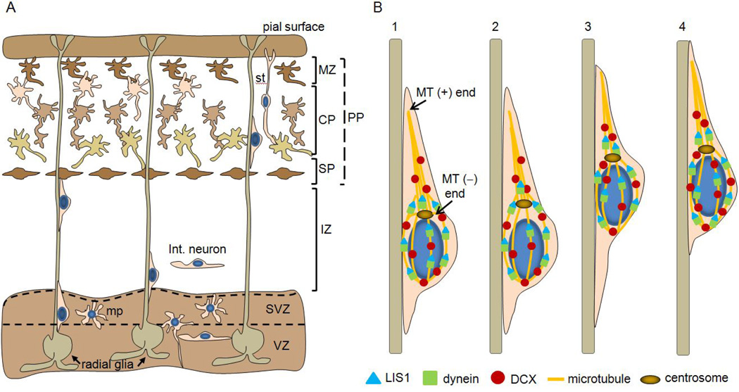
The cortex is a six layered structure comprised of neurons that originate in the proliferative zones of the dorsal telencephalon and the medial and lateral ganglionic eminences [16]. Cortical neurons travel considerable distances from their sites of origin in the ventricular zone to their final anatomical destinations, and can use distinct modes of migration that may depend on cell type and birth-date. Cortical layering proceeds in an inside-out fashion, such that early born neurons comprise deeper layers, whereas later born neurons migrate past these cells to form more superficial layers. Radial glial cells play an important role in cortical development because they divide in the ventricular zone to generate daughter neurons, and also because they extend long processes from the ventricular zone of the dorsal telencephalon to the pial surface (Figure 2a) [17–19]. Most neurons that will comprise cortical layers II-VI migrate along these processes in order to reach their final destinations using a mode of migration called locomotion (Figure 2b). In contrast, early born neurons that comprise the transient cortical preplate use soma translocation, whereby similar to radial glia, they extend a process from the ventricular zone to the pial surface. The soma migrates along the leading process which becomes progressively shorter while remaining anchored to the pial surface, effectively “pulling” the cell body to its final position as the trailing process retracts [20]. Inhibitory interneurons are born in the medial and lateral ganglionic eminences and use tangential migration to travel to the neocortex, hippocampus, and olfactory bulb [21, 22] (Figure 2a). Subpopulations of these tangentially migrating cells move dorsally towards the ventricles before co-migrating radially into the cortex with newborn projection neurons [23]. This may be a possible mechanism to organize excitatory projection neurons and inhibitory interneurons into defined cortical circuits.
Figure 2. Neuronal migration in the cerebral cortex is dependent upon microtubules and associated proteins.
A. Illustration depicting neuronal migration in the developing cerebral cortex. The immature cortex can be roughly divided into six zones (VZ, SVZ, IZ, SP, CP, MZ) from the bottom to top, respectively. The earliest migrating cells (beginning at ~e11 in mouse) form the transient preplate (PP), and the next wave of cell migration splits the preplate into the superficial marginal zone (MZ) which is future layer I, and the subplate (SP), thus forming a middle layer of cells called the cortical plate (CP). The cortical plate develops in an inside-out fashion as subsequent waves of migratory neurons pass deeper neurons to comprise layers II-VI. The subplate disappears postnatally, and is used mainly as an organizing zone for developing cortical axons. Newborn neurons are generated from radial glia which divide both asymmetrically and symmetrically to generate post-mitotic daughter neurons in the ventricular zone (VZ) and subventricular zone (SVZ). Post-mitotic neurons then commence migration towards the cortical plate through a complex series of movements and morphological changes. Projection neurons (excitatory) born in the dorsal VZ adopt a bipolar morphology and migrate radially along glia before pausing in the SVZ. There, these cells acquire a multi-polar (mp) morphology and frequently grow and retract processes. Next, most of these immature neurons in the SVZ grow a leading process that contacts the dorsal ventricle prior to moving towards the VZ, and this is also observed with neurons born in the SVZ. Finally, neurons migrate out of the VZ/SVZ by reversing polarity and extending a leading process towards the pial surface along radial glial fibers (locomotion, refer to B). The process that once contacted the ventricular surface becomes the retracting neurite during migration and is also the future axon. Migrating neurons are found in the space between the SVZ and subplate called the intermediate zone (IZ). As cells finish migrating along radial glia, they detach and extend a process towards the pial surface. The cell body then moves forward along the leading process in a mode of migration called soma translocation (st). Early born neurons that comprise the preplate are thought to use this mode of migration exclusively. Inhibitory interneurons are born in the ventral medial and lateral ganglionic eminences and migrate tangentially along the planes of the ventricles into the cortex. Some tangentially migrating neurons move dorsally towards the VZ/SVZ where they are observed to contact post-mitotic projection neurons before co-migrating radially towards the pial surface.
B. Illustration depicting cell locomotion along radial glia. The plus-ends of microtubules, which are nucleated at the centrosome, are found at the distal end of the leading process, and a cagelike network of microtubules also surrounds the nucleus in the soma. LIS1 and dynein are mainly found proximal to the centrosome (and at lower levels in the leading process) and along the cage structure that surrounds the nucleus, whereas DCX is also found along microtubules in the leading process. Migrating cells adopt a bipolar morphology with a leading process directed towards the pial surface, and a retracting edge that faces the ventricle. Migration is a step-wise event in which the centrosome is initially positioned in front of the nucleus (1) before moving forward into the leading process (2). The nucleus is then guided forward by the centrosome into the leading process (3), followed by retraction of the rear process (4). This cycle repeats as the leading process once again extends along the radial glial fibers. Studies suggest that the growing plus-ends of microtubules become stabilized in the leading process, exerting a pulling force on the centrosome. LIS1 is necessary for proper positioning of the centrosome within the leading neurite, and regulates dynein localization which in turn generates force on the microtubules through its minus-end directed motor activity, pulling the nucleus towards the centrosome. DCX may play a complementary role in this process by stabilizing the cage-like structure of microtubules surrounding the nucleus, allowing the nucleus to translocate cooperatively with the centrosome as dynein pulls on microtubules.
Neuronal migration along radial glial fibers is a step-wise process that relies on the coordinated activities of the microtubule cytoskeleton and its associated proteins [11, 24]. Post-mitotic neurons first acquire polarity and grow a leading neurite, a process that depends on spatial regulation of microtubule dynamics at the leading edge of the cell surface facing the pial basement membrane [1, 25, 26]. The centrosome, or microtubule organizing center, moves away from the soma into the leading neurite and directs nucleokinesis by “pulling” on a microtubule cage-like structure that surrounds the trailing nucleus. Microtubule associated proteins (MAPs) and dynein stabilize the cage structure and regulate the pulling force between the centrosome and nucleus [27–30]. Once the nucleus has moved into the leading neurite, the cell body can move forward as the trailing process retracts (Figure 2b). Similar types of migratory behavior have been observed in tangentially migrating interneurons, although the leading neurite undergoes frequent branching events while searching for possible guidance cues [31]. Other types of migratory behaviors have been observed in cerebellar granule neurons that suggest the nucleus and centrosome may move independently from one another in these cell types [32].
Lissencephaly, or “smooth brain”, encompasses a diffuse spectrum of congenital brain malformations that occur when post-mitotic neurons in the ventricular zones fail to migrate to their respective layers in the cortex [33–35]. As a result, the characteristic folds (gyri) and grooves (sulci) on the surface of the cerebral cortex do not develop properly. Lissencephaly can be broadly classified into two types according to the brain malformation. Type I or “classical” lissencephaly can range from complete absence of gyri (agyria) and sulci, giving the brain a smooth appearance, to a brain that has only simple, abnormally thick convolutions (pachygyria)[36]. In these patients, the normal six layer cortex has been condensed to four layers and the ventricles are enlarged. Subcortical bands of neurons that have prematurely arrested their migration are often present. Type II lissencephaly is characterized by the presence of numerous small gyri that are separated by shallow sulci (polymicrogyria), giving the brain a “cobblestone” like appearance [33]. Cortical lamination is severely disorganized and often absent, and can be accompanied by breaches in the pial basement membrane (brain surface) where neurons have ectopically settled in the overlying meningeal space [37]. Type II lissencephaly is less common than type I, and is thought to result from defects during the second wave of migration, thereby affecting cells that comprise the outer layers of the cortex. As discussed below, several gene mutations in tubulin and microtubule associated proteins are associated with these types of brain malformations.
MAPs, Tubulin, and Type I Lissencephaly
Heterozygous inactivating mutations in LIS1 or DCX, both encoding for MAPs, account for most cases of type I lissencephaly [38, 39]. LIS1 localizes to the perinuclear cage, centrosome, and plus-ends of microtubules in the leading neurite, and controls nucleokinesis during radial migration via the interaction with and regulation of dynein motor function and localization [27, 40, 41]. Inactivating or hypomorphic alleles of LIS1 hinder cell migration due to the uncoupling of the nucleus and centrosome [27], and the formation of the leading process is also affected [42]. Similarly, DCX also localizes to the perinuclear cage and the plus-ends of microtubules in the leading neurite, and is necessary for the coupling of the nucleus and centrosome during nucleokinesis [27, 43]. Some evidence suggests DCX can form a complex with LIS1 and dynein in vivo, and DCX overexpression restores nucleus-centrosome coupling defects in LIS1 deficient neurons [27, 44]. The mechanism for the latter observation is unclear, but DCX promotes the growth and bundling of microtubules, and this could stabilize the microtubules of the perinuclear cage that are coupled with the centrosome [30, 39, 44]. Thus, DCX may regulate nucleokinesis via the regulation of microtubule dynamics and normal LIS1/dynein localization and function. Some observations also suggest DCX might regulate neuronal polarity in the ventricular zones prior to migration [45]. Although LIS1 and DCX seem to have overlapping functions during neuronal migration, there are also likely some differences as LIS1 mutations usually result in more posterior gyral abnormalities, whereas DCX mutations predominately affect gyral patterning in the anterior cortex and are more commonly associated with cerebellar vermian hypoplasia [46].
Heterozygous missense mutations in TUBA1A, coding for an α-tubulin isotype that is highly expressed in post-mitotic differentiating neurons [47], cause a spectrum of cortical malformations that can resemble those resulting from LIS1 or DCX mutations [13, 48, 49] Affected individuals are microcephalic and have cortical malformations that range from agyria and posterior pachygyria in severe cases to perisylvian predominant pachygyria in the more common and less severe forms http://www.ncbi.nlm.nih.gov.ezp-prod1.hul.harvard.edu/pubmed?term=%22Morris-Rosendahl%20DJ%22%5BAuthor%5D&itool=EntrezSystem2.PEntrez.Pubmed.Pubmed_ResultsPanel.Pubmed_RVAbstract[48, 50]. Findings from brain autopsies of affected fetuses reveal abnormal cortical layering, hypoplastic and disorganized hippocampi, and clumps of poorly differentiated neurons interspersed with white matter [51]. The TUBA1A phenotype is somewhat distinct from LIS1 and DCX, however, because patients have additional brain malformations that are less commonly associated with LIS1 and DCX mutations, including cerebellar and brainstem hypoplasia, corpus callosum dysgenesis, and hypoplasia of the anterior limb of the internal capsule. This latter finding is one defining feature of TUBA1A mutations, and is associated with dysmorphic basal ganglia lacking clear separation between the caudate and putamen. Hypoplastic and disorganized white matter tracts suggest further errors in axon growth and guidance beyond cell migration. Overall, patients usually have severe neurological impairment, including mental retardation, spastic diplegia or tetraplegia, facial paralysis, and epilepsy.
Tubulin and Type II Lissencephaly
Heterozygous missense mutations in TUBB2B, a β-tubulin isotype also highly expressed in postmitotic differentiating neurons, cause a spectrum of cortical malformations that differ from TUBA1A and more closely resemble type II lissencephaly [15]. Affected individuals have microcephaly and bilateral, asymmetrical, and predominant anterior polymicrogyria. Neurohistopathological analysis of an affected fetal brain revealed disorganized cortical layering, heterotopic neurons in the white matter, and breaches in the pial basement membrane surface where ectopic clusters of neurons had migrated into the overlying leptomeningeal space. Radial glial cells were also disorganized and had failed to properly attach to the pial basement membrane. Similar to TUBA1A mutations, these malformations are often accompanied by brainstem and cerebellar hypoplasia, corpus callosum dysgenesis, and dysmorphic basal ganglia with hypoplasia of the anterior limb of the internal capsule. Neurological impairments are also similar to those of individuals harboring TUBA1A mutations.
Polymicrogyria can also result from homozygous splice-site mutations in the α-tubulin isotype TUBA8 [14]. Similar to TUBB2B, individuals harboring TUBA8 mutations also have agenesis or hypoplasia of the corpus callosum and dysgenic brainstems that lack a demarcated pontomedullary junction, and can have severe developmental delays and seizures. Unlike TUBB2B, however, these patients have diffuse bilateral polymicrogyria in both anterior and posterior regions of the brain and have hypoplastic optic nerves. Also, basal ganglia and internal capsule malformations, a hallmark of TUBB2B and TUBA1A mutations, have not been reported.
Cortical cell migration disorders can result from inactivating mutations in α and β-tubulin
Missense and splice-site mutations in TUBA1A, TUBB2B, and TUBA8 are predicted to diminish the formation of tubulin heterodimers and impair microtubule polymerization. In order to generate tubulin heterodimers that are capable of polymerizing, nascent α and β-tubulin peptides must first interact with a series of molecular chaperone proteins that are required for the folding and dimerization of α and β-tubulin monomers [52, 53]. Several mutations in both TUBA1A and TUBB2B resulting in lissencephaly can impair their interactions with chaperone proteins in vitro, resulting in the loss of TUBA1A and TUBB2B heterodimers [15, 54]. Moreover, reducing TUBB2B expression to ~60% of normal levels in the developing mouse brain by RNAi in-utero electroporation arrests the migration of neurons in a manner reminiscent of the human disorder [15]. Recessive splice-site mutations in TUBA8 were discovered to replace the full-length coding transcript with a shorter copy that lacked exon two [14]; removing the coding sequence for this exon would likely also alter TUBA8 folding and heterodimer formation.
Although missense mutations in TUBA1A and TUBB2B appear to primarily cause tubulin haploinsufficiency, other evidence suggests that some mutations might cause a dominantnegative effect on microtubule behavior. For example, despite the fact that TUBA1A mutations compromise folding and reduce overall protein levels, some mutant monomers still fold and form heterodimers that are capable of microtubule polymerization in mammalian cells [54]. This suggests that the resulting brain phenotypes may reflect the combined effects of tubulin loss and mutant heterodimer polymerization. Furthermore, some mutations in TUBB2B do not affect chaperone protein interactions and heterodimer formation, and instead, have been hypothesized to alter the function or behavior of microtubules by perturbing interactions with MAPs [15]. Thus, more studies are needed to determine if dominant effects of the mutations could influence the nature or variability of the brain malformations.
Mutations in TUBB3 can cause disorders of axon guidance and maintenance without affecting cortical cell migration
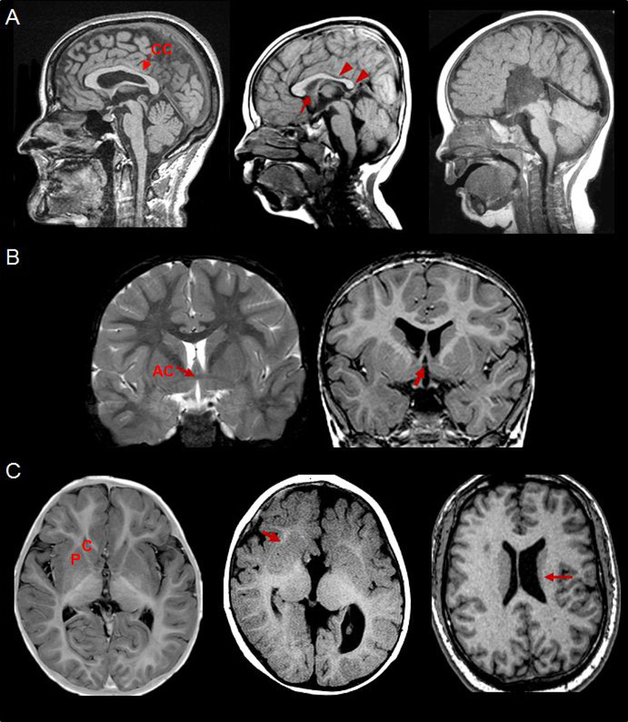
Although mutations in different α and β-tubulin isotypes can perturb the migration of cortical neurons, recent findings show this is not always the case. Remarkably, patients harboring heterozygous missense mutations in TUBB3, coding for the neuronal specific β-tubulin isotype III [55, 56], do not show radiological signs of cell migration defects. Instead, clinical and radiological findings point to a primary defect in the growth and/or guidance of axons in the brain and spinal cord [12]. The spectrum of nervous system malformations, called the TUBB3 syndromes, encompass eye movement restrictions, facial paralysis, spasticity, cognitive and behavioral impairments, and a later-onset progressive peripheral sensorimotor axonal polyneuropathy. Seizures are rarely reported, and overall neurological impairment is typically less severe than that associated with TUBA1A, TUBB2B, or TUBA8 mutations. Most patients have aberrant eye movements. In addition, several have synkinetic ptotic eyelid elevation and jaw movements (Marcus Gunn phenomenon), clinical manifestations of aberrant innervation of cranial musculature by the trigeminal nerve. Radiological findings reveal hypoplastic oculomotor nerves, dysmorphic basal ganglia with or without internal capsule hypoplasia, and agenesis or hypoplasia of the corpus callosum and anterior commissure (Figure 3). As discussed below, the extent of nervous system malformations and neurological impairments can depend on how each amino acid substitution alters the function of TUBB3.
Figure 3. Radiological findings in the TUBB3 syndromes.
A. Midline sagittal magnetic resonance images (MRI) of three patients harboring R262C (left panel), E410K (middle panel), and R380C (right panel) TUBB3 amino acid substitutions that demonstrate the spectrum of corpus callosum (CC) abnormalities. The corpus callosum is the largest band of commissural axons in the brain which cross the midline and project to homotypic neurons in the contralateral cortex. Those harboring R262C substitutions have a relatively normal corpus callosum with a trend towards mild thinning of the posterior body (red arrow). Individuals with E410K substitutions have more dysgenic corpus callosums, and the child shown here has agenesis of the rostrum (red arrow) and mild hypoplasia and the posterior body and splenium (arrow heads). The child on the right has a R380C amino acid substitution, and has corpus callosum agenesis.
B. Coronal images showing normal crossing of anterior commissural (AC) fibers above the third ventricle in a TUBB3+/+ individual (left). The child on the right harbors a D417N amino acid substitution and has anterior commissure hypoplasia. Agenesis or hypoplasia of the anterior commissure is one of the most consistent findings, along with corpus callosum dysgenesis, in the TUBB3 syndromes.
C. Axial images depicting various basal ganglia malformations in the TUBB3 syndromes. The bottom left image corresponds to the D417N patient in the middle panel, and demonstrates a well formed caudate head and putamen, with internal capsule fibers coursing between the two structures. The middle image is from the R380C patient in the top right panel, and demonstrates fusion of the caudate head and putamen (red arrow) resulting from hypoplasia of the internal capsule. The bottom right panel is from an individual harboring an R262C substitution. Almost all of these patients have hypoplasia of the left caudate body and tail (red arrow), a distinctive asymmetrical feature of R262C (and R262H) amino acid substitutions.
To further understand the nature of the nervous system malformations in humans, a mouse model harboring the most common amino acid substitution (R262C) was analyzed [12]. Heterozygous knock-in (KI) mice were viable and did not display external eye phenotypes, and brain development appeared normal with the exception of mild hypoplasia of the anterior commissure. Homozygous KI mice, however, died within hours of birth and displayed many phenotypes reminiscent of the human disease. The oculomotor and trigeminal nerves did not branch properly, and the oculomotor nerve often grew towards the wrong set of extraocular muscles. The anterior commissure was hypoplastic and often failed to cross the midline of the brain. Also, the corpus callosum was usually thin or absent, and when absent, bundles of callosal axons (Probst bundles) that had failed to cross the midline lined the lateral ventricles. Overall brain size was similar to wild-type and the architecture and layering in the cortex appeared normal. Thus, in contrast to TUBA1A, TUBB2B, or TUBA8 mutations, the underlying defects in the TUBB3 syndromes pertain to the growth, branching, and guidance of axons.
The TUBB3 syndromes result from dominant mutations that alter microtubule function and behavior
Similar to TUBA1A and TUBB2B mutations, missense mutations in TUBB3 also reduce the formation of mutant heterodimers in vitro. However, phenotype-genotype correlations present in the TUBB3 syndromes also suggest that mutations could alter microtubule function and behavior in a dominant fashion. For example, patients that harbor R62Q or R262C amino acid substitutions mainly have only isolated ocular motility restrictions. These mutations severely diminish heterodimer formation in vitro, and when expressed in mammalian cells, low levels of incorporation are observed throughout microtubules. In contrast, patients with R262H, E410K, or D417H substitutions can have additional neurological symptoms including facial paralysis and degeneration of peripheral motor and sensory axons. Interestingly, these mutations result in less severe reductions of heterodimer yield compared to R62Q or R262C substitutions, and mutant heterodimers cycle with native tubulin and incorporate into microtubules in mammalian cells at levels similar to wild-type. Moreover, R262C and R262H substitutions cause a mild and significantly more severe form of the TUBB3 syndromes, respectively. Thus, the segregation of more severe neurological impairments and/or brain malformations with specific amino acid substitutions may be due in part to higher incorporation levels of mutant heterodimers into microtubules [12].
Using budding yeast to model the dominant effects of TUBB3 mutations, it was discovered that all amino-acid substitutions stabilized microtubules by rendering them resistant to pharmacological induced depolymerization. Furthermore, the dynamic behavior and function of microtubules were altered in two distinct fashions. The first subset of mutations significantly attenuated the rate of microtubule growth and shortening, resulting in non-dynamic microtubules that were stuck in prolonged paused states, whereas a second subset reduced microtubule interactions with kinesin motor proteins. The second group of mutations (R262H, E410K, D417H/N) also result in facial paralysis and the progressive degeneration of peripheral motor and sensory axons. Thus, in addition to the overall levels of heterodimer incorporation, phenotypic variability in the TUBB3 syndromes can depend on how each amino acid substitution alters specific microtubule functions [12]. These findings might have important implications for understanding why variable phenotypic and functional correlations exist between some TUBB2B mutations.
Why do mutations in different tubulin isotypes cause distinct types of brain malformations?
Microtubules in mammalian cells are assembled from a heterogenous mixture of all available tubulin isotypes [3]. TUBA1A, TUBA8, TUBB2B, and TUBB3 all share similar expression patterns and are the major α and β-tubulin isotypes expressed in the developing brain and nervous system [12, 15, 47, 57]. Considering that microtubules in post-mitotic neurons should contain a mixture of these proteins, differences in the severity and types of brain malformations associated with each tubulin isotype are somewhat surprising. There are several possible explanations that may explain some of the noted phenotypic differences. First, simple loss-offunction versus altered function might explain why TUBA1A, TUBB2B, and TUBA8 mutations result in cortical cell migration defects, whereas TUBB3 mutations primarily affect axon growth and/or guidance. This explanation seems unlikely, however, since TUBB3 mutations that significantly compromise heterodimer formation do not cause lissencephaly or polymicrogyria, and some TUBB2B mutations do not affect the formation of heterodimers. Second, although the spatial and temporal expression patterns are similar for each isotype, there are some distinctions. For example, TUBB2B is expressed in radial glia and at high levels in cortical plate neurons during migration in the cortex, whereas TUBB3 expression is absent in radial glial cells and somewhat lower in the cortical plate versus other neurons [15, 58]. Third, the levels of microtubule incorporation could vary for each isotype during different modes of migration (i.e., locomotion versus soma translocation) or stages of development (i.e., polarization and migration versus axon guidance). This is particularly notable for TUBB3 because it is upregulated in microtubules as axons and dendrites continue to grow and mature [59, 60]. Microtubules in migrating neurons might contain lower amounts of TUBB3 versus other β-tubulin isotypes, explaining why it may be dispensable for cell positioning and cortical layering. Thus, as discussed below, each isotype may be required for specialized microtubule functions during different cellular events, which could explain fine phenotypic differences between each neurological syndrome.
The “multi-tubulin” hypothesis
It has been postulated that neurons might utilize different tubulin isotypes for distinct cellular functions in both the embryo and adult [61–63]. Prior to the identification and sequencing of tubulin genes, it was known that the structure of microtubules could vary during phases of cell division, in different intracellular compartments, and between cell types. This led to the “multitubulin” hypothesis which generally stated that different tubulin isotypes may be used to build specific microtubule structures necessary to support a diverse range of cellular functions [64]. Support for this hypothesis was bolstered upon the discovery that multiple genes encoded different α and β-tubulin isotypes in animals and other eukaryotes, and many of these displayed tissue-specific expression patterns [65–67]. Moreover, within humans, the protein sequences of different β-tubulins diverge mainly in two regions, the n-terminus and the extreme c-terminus tail, and these two regions are highly conserved within the same isotype found in other species [56] (Figures 4, 5). This conservation of isotype-specific sequences suggested that different tubulins may have retained important biochemical properties necessary to support the functions of different microtubule structures. However, other observations suggested that different tubulin isotypes could be functionally redundant; for example, it was found that microtubules in cultured mammalian cells were assembled from pools of all available isotypes, and that different α and β-tubulin isotypes in fungi were functionally interchangeable [3, 68].
Figure 4. Protein alignment of human β-tubulin isotypes.
Seven genes encode distinct β–tubulin isotypes in humans, and their protein sequences are remarkably conserved throughout evolution. The n- and c-terminal regions of sequence divergence are demarcated by blue and orange lines, respectively. Residues mutated in the TUBB2B and TUBB3 syndromes are denoted by green and red boxes, respectively. Note that all mutated residues are conserved among the seven different isotypes, and TUBB2B and TUBB3 share 90% protein sequence homology.
Figure 5. TUBB3 is highly conserved in vertebrates.
The amino acid sequence of TUBB3, including the n- and c-termini, is highly conserved among mammals and chicken, similar to other β-tubulin isotypes. In contrast to TUBB2B and TUBB4, both of which are also expressed in the mammalian brain, TUBB3 is not present in Xenopus; however, it has been reported in catfish, goosefish, the dogfish shark, and the Antarctic fish Chaenocephalus [61].
The first definitive evidence that tubulin isotypes could have divergent functions came from genetic studies in Drosophila [69]. Drosophila has three β-tubulin isotypes (β1, β2, β3), and only β2 is expressed in post-mitotic cells in the male germ line. β2 is required for the formation of meiotic spindles, cytoplasmic microtubules, and the axoneme of the sperm tail flagella. When β3 was ectopically expressed in male germ line cells over a β2 null background, assembly of the meiotic spindle and axoneme was deficient. Co-expression of β2 rescued the phenotype, but only when the levels of β3 were below a certain threshold. A follow-up study then showed that although the c-terminal tail of β2 was dispensable for microtubule assembly in the axoneme, it was specifically required for the organization of the 9+2 microtubule array that is characteristic of the axoneme [70]. Because the protein sequences of tubulins diverge mainly at the n-terminus and in the tail region found at the extreme c-terminus of the protein [56], these results demonstrated that the c-terminal tail could mediate isotype-specific functions, thus supporting the multi-tubulin hypothesis.
In neurons, the c-terminal tail of β-tubulin is required for numerous microtubule-MAP interactions, and these interactions regulate the dynamic behavior of microtubules and are necessary for neuron migration, differentiation, and axon guidance [71]. The c-terminal tail undergoes several types of post-translational modifications, some of which are unique to specific tubulin isotypes, and these modifications can also regulate specific MAP and motor protein interactions [72, 73]. Interestingly, in the presence of MAPs, microtubules assembled in vitro from purified pools of specific brain β-tubulins have different rates of assembly than when assembled from a heterogenous mixture of different brain β-tubulin isotypes [74]. Thus, upon binding MAPs, the divergent c-terminal tail of β-tubulin might confer unique structural or biochemical properties upon microtubules, thereby regulating their function or behavior in a spatio-temporal fashion during development. Post-translational modifications of α-tubulin that occur outside of the tail region, such as α-tubulin acetylation and tyrosination, can also regulate MAP and kinesin-microtubule interactions [75, 76]. Inactivating mutations in the enzymes that catalyze these modifications affect cortical cell migration and axon outgrowth [77, 78]. Interestingly, TUBA8 is an atypical α–tubulin because it is neither acetylated or tyrosinated [14], and the absence of these post-translational modifications could allow TUBA8 to regulate MAP interactions and microtubule dynamics in a manner distinct from other α-tubulin isotypes.
Tubulin isotypes themselves, in the absence of MAPs, also show intrinsic differences in the rates of microtubule assembly and the frequency of growth and shortening events, suggesting that cells can regulate microtubule dynamics by controlling the relative amounts of different tubulin isotypes [79]. In the absence of MAPs, isotypically homogenous microtubules polymerized in vitro from αβ3 (TUBB3) heterodimers were considerably more dynamic and spent less time in paused states than those composed of αβ2 (TUBB2), αβ4 (TUBB4), or a mixture of all three isotypes. Also, the mean growing and shortening rates of microtubules comprised solely from αβ3 heterodimers is nearly double that of microtubules polymerized from αβ2 or αβ4. By stark contrast, microtubules that contain equal amounts of αβ3 and αβ2 heterodimers are much less dynamic, and the frequency of growth and shortening events is similar to microtubules containing all three heterodimers. Interestingly, DCX has been shown to promote microtubule stability [39, 44], and similarly perhaps, TUBB2B may also be required to dampen microtubule dynamics and promote stability during neuronal polarization or migration. Conversely, axon guidance requires highly dynamic populations of microtubules in growth cones in order to mediate responses to extracellular guidance cues [80, 81]; drugs that dampen microtubule dynamics in cultured neurons perturb the directional growth and guidance of axons [6, 82–84]. Because TUBB3 is increasingly incorporated into the microtubule cytoskeleton as axons grow and elongate in culture [59], TUBB3 may endow microtubules with the dynamic properties needed for rapid responses to extracellular guidance cues. Mutations in TUBB3 that stabilize microtubules and reduce dynamic instability, either by reducing protein levels or altering its biochemical properties, thus might be expected to predominately affect axon growth or guidance rather than cell migration.
Concluding remarks
It has been nearly 35 years since the multi-tubulin hypothesis was first proposed by Fulton and Simpson and, now more than ever, the discoveries of the tubulin-related syndromes should allow further scientific breakthroughs on this subject matter. Because microtubules are absolutely essential for such a diverse range of cellular functions, it is imperative that scientists understand how different tubulin isotypes regulate their specialized functions for drug research and design. This is also relevant for cancer, because several types of malignant tumors are associated with the dysregulation of tubulin isotypes, especially TUBB3 [85]. Thus, by linking distinct cellular processes with the unique properties of different tubulin isotypes, we can begin to dissect the specialized functions of microtubules in both normal health and disease with the hopes of streamlining future therapeutic intervention strategies.
Acknowledgments
We would like to thank Judy Liu, Jay Tischfield and Victoria E. Abraira for helpful manuscript comments.
Work pertaining to the TUBB3 syndromes in this manuscript was supported by the National Institutes of Health [R01 EY012498], [R01 EY013583], [HD18655], [R01 GM061345] and the Howard Hughes Medical Institute.
References
- 1.LoTurco JJ, Bai J. The multipolar stage and disruptions in neuronal migration. Trends Neurosci. 2006;29:407–413. doi: 10.1016/j.tins.2006.05.006. [DOI] [PubMed] [Google Scholar]
- 2.Noctor SC, Martinez-Cerdeno V, Ivic L, Kriegstein AR. Cortical neurons arise in symmetric and asymmetric division zones and migrate through specific phases. Nat. Neurosci. 2004;7:136–144. doi: 10.1038/nn1172. [DOI] [PubMed] [Google Scholar]
- 3.Lopata MA, Cleveland DW. In vivo microtubules are copolymers of available beta-tubulin isotypes: localization of each of six vertebrate beta-tubulin isotypes using polyclonal antibodies elicited by synthetic peptide antigens. J. Cell Biol. 1987;105:1707–1720. doi: 10.1083/jcb.105.4.1707. [DOI] [PMC free article] [PubMed] [Google Scholar]
- 4.Gordon-Weeks PR. Microtubules and growth cone function. J. Neurobiol. 2004;58:70–83. doi: 10.1002/neu.10266. [DOI] [PubMed] [Google Scholar]
- 5.Dent EW, Kalil K. Axon branching requires interactions between dynamic microtubules and actin filaments. J. Neurosci. 2001;21:9757–9769. doi: 10.1523/JNEUROSCI.21-24-09757.2001. [DOI] [PMC free article] [PubMed] [Google Scholar]
- 6.Buck KB, Zheng JQ. Growth cone turning induced by direct local modification of microtubule dynamics. J. Neurosci. 2002;22:9358–9367. doi: 10.1523/JNEUROSCI.22-21-09358.2002. [DOI] [PMC free article] [PubMed] [Google Scholar]
- 7.Kalil K, Dent EW. Touch and go: guidance cues signal to the growth cone cytoskeleton. Curr. Opin. Neurobiol. 2005;15:521–526. doi: 10.1016/j.conb.2005.08.005. [DOI] [PubMed] [Google Scholar]
- 8.Nadar VC, Ketschek A, Myers KA, Gallo G, Baas PW. Kinesin-5 is essential for growth-cone turning. Curr. Biol. 2008;18:1972–1977. doi: 10.1016/j.cub.2008.11.021. [DOI] [PMC free article] [PubMed] [Google Scholar]
- 9.Hirokawa N, Takemura R. Molecular motors in neuronal development, intracellular transport and diseases. Curr. Opin. Neurobiol. 2004;14:564–573. doi: 10.1016/j.conb.2004.08.011. [DOI] [PubMed] [Google Scholar]
- 10.Chevalier-Larsen E, Holzbaur EL. Axonal transport and neurodegenerative disease. Biochim. Biophys. Acta. 2006;1762:1094–1108. doi: 10.1016/j.bbadis.2006.04.002. [DOI] [PubMed] [Google Scholar]
- 11.Ayala R, Shu T, Tsai LH. Trekking across the brain: the journey of neuronal migration. Cell. 2007;128:29–43. doi: 10.1016/j.cell.2006.12.021. [DOI] [PubMed] [Google Scholar]
- 12.Tischfield MA, Baris HN, Wu C, Rudolph G, Van Maldergem L, He W, Chan WM, Andrews C, Demer JL, Robertson RL, Mackey DA, Ruddle JB, Bird TD, Gottlob I, Pieh C, Traboulsi EI, Pomeroy SL, Hunter DG, Soul JS, Newlin A, Sabol LJ, Doherty EJ, de Uzcategui CE, de Uzcategui N, Collins ML, Sener EC, Wabbels B, Hellebrand H, Meitinger T, de Berardinis T, Magli A, Schiavi C, Pastore-Trossello M, Koc F, Wong AM, Levin AV, Geraghty MT, Descartes M, Flaherty M, Jamieson RV, Moller HU, Meuthen I, Callen DF, Kerwin J, Lindsay S, Meindl A, Gupta ML, Jr, Pellman D, Engle EC. Human TUBB3 mutations perturb microtubule dynamics, kinesin interactions, and axon guidance. Cell. 2010;140:74–87. doi: 10.1016/j.cell.2009.12.011. [DOI] [PMC free article] [PubMed] [Google Scholar]
- 13.Keays DA, Tian G, Poirier K, Huang GJ, Siebold C, Cleak J, Oliver PL, Fray M, Harvey RJ, Molnar Z, Pinon MC, Dear N, Valdar W, Brown SD, Davies KE, Rawlins JN, Cowan NJ, Nolan P, Chelly J, Flint J. Mutations in alpha-tubulin cause abnormal neuronal migration in mice and lissencephaly in humans. Cell. 2007;128:45–57. doi: 10.1016/j.cell.2006.12.017. [DOI] [PMC free article] [PubMed] [Google Scholar]
- 14.Abdollahi MR, Morrison E, Sirey T, Molnar Z, Hayward BE, Carr IM, Springell K, Woods CG, Ahmed M, Hattingh L, Corry P, Pilz DT, Stoodley N, Crow Y, Taylor GR, Bonthron DT, Sheridan E. Mutation of the variant alpha-tubulin TUBA8 results in polymicrogyria with optic nerve hypoplasia. Am. J. Hum. Genet. 2009;85:737–744. doi: 10.1016/j.ajhg.2009.10.007. [DOI] [PMC free article] [PubMed] [Google Scholar]
- 15.Jaglin XH, Poirier K, Saillour Y, Buhler E, Tian G, Bahi-Buisson N, Fallet-Bianco C, Phan-Dinh-Tuy F, Kong XP, Bomont P, Castelnau-Ptakhine L, Odent S, Loget P, Kossorotoff M, Snoeck I, Plessis G, Parent P, Beldjord C, Cardoso C, Represa A, Flint J, Keays DA, Cowan NJ, Chelly J. Mutations in the beta-tubulin gene TUBB2B result in asymmetrical polymicrogyria. Nat. Genet. 2009;41:746–752. doi: 10.1038/ng.380. [DOI] [PMC free article] [PubMed] [Google Scholar]
- 16.McConnell SK. Constructing the cerebral cortex: neurogenesis and fate determination. Neuron. 1995;15:761–768. doi: 10.1016/0896-6273(95)90168-x. [DOI] [PubMed] [Google Scholar]
- 17.Yokota Y, Kim WY, Chen Y, Wang X, Stanco A, Komuro Y, Snider W, Anton ES. The adenomatous polyposis coli protein is an essential regulator of radial glial polarity and construction of the cerebral cortex. Neuron. 2009;61:42–56. doi: 10.1016/j.neuron.2008.10.053. [DOI] [PMC free article] [PubMed] [Google Scholar]
- 18.Rakic P. Developmental and evolutionary adaptations of cortical radial glia. Cereb. Cortex. 2003;13:541–549. doi: 10.1093/cercor/13.6.541. [DOI] [PubMed] [Google Scholar]
- 19.Hatten ME. New directions in neuronal migration. Science. 2002;297:1660–1663. doi: 10.1126/science.1074572. [DOI] [PubMed] [Google Scholar]
- 20.Nadarajah B, Alifragis P, Wong RO, Parnavelas JG. Neuronal migration in the developing cerebral cortex: observations based on real-time imaging. Cereb. Cortex. 2003;13:607–611. doi: 10.1093/cercor/13.6.607. [DOI] [PubMed] [Google Scholar]
- 21.Austin CP, Cepko CL. Cellular migration patterns in the developing mouse cerebral cortex. Development. 1990;110:713–732. doi: 10.1242/dev.110.3.713. [DOI] [PubMed] [Google Scholar]
- 22.Metin C, Vallee RB, Rakic P, Bhide PG. Modes and mishaps of neuronal migration in the mammalian brain. J. Neurosci. 2008;28:11746–11752. doi: 10.1523/JNEUROSCI.3860-08.2008. [DOI] [PMC free article] [PubMed] [Google Scholar]
- 23.Nadarajah B, Alifragis P, Wong RO, Parnavelas JG. Ventricle-directed migration in the developing cerebral cortex. Nat. Neurosci. 2002;5:218–224. doi: 10.1038/nn813. [DOI] [PubMed] [Google Scholar]
- 24.Tsai LH, Gleeson JG. Nucleokinesis in neuronal migration. Neuron. 2005;46:383–388. doi: 10.1016/j.neuron.2005.04.013. [DOI] [PubMed] [Google Scholar]
- 25.Sapir T, Shmueli A, Levy T, Timm T, Elbaum M, Mandelkow EM, Reiner O. Antagonistic effects of doublecortin and MARK2/Par-1 in the developing cerebral cortex. J. Neurosci. 2008;28:13008–13013. doi: 10.1523/JNEUROSCI.2363-08.2008. [DOI] [PMC free article] [PubMed] [Google Scholar]
- 26.Reiner O, Sapir T. Polarity regulation in migrating neurons in the cortex. Mol. Neurobiol. 2009;40:1–14. doi: 10.1007/s12035-009-8065-0. [DOI] [PubMed] [Google Scholar]
- 27.Tanaka T, Serneo FF, Higgins C, Gambello MJ, Wynshaw-Boris A, Gleeson JG. Lis1 and doublecortin function with dynein to mediate coupling of the nucleus to the centrosome in neuronal migration. J. Cell Biol. 2004;165:709–721. doi: 10.1083/jcb.200309025. [DOI] [PMC free article] [PubMed] [Google Scholar]
- 28.Solecki DJ, Model L, Gaetz J, Kapoor TM, Hatten ME. Par6alpha signaling controls glial-guided neuronal migration. Nat Neurosci. 2004;7:1195–1203. doi: 10.1038/nn1332. [DOI] [PubMed] [Google Scholar]
- 29.Rivas RJ, Hatten ME. Motility and cytoskeletal organization of migrating cerebellar granule neurons. J. Neurosci. 1995;15:981–989. doi: 10.1523/JNEUROSCI.15-02-00981.1995. [DOI] [PMC free article] [PubMed] [Google Scholar]
- 30.Higginbotham HR, Gleeson JG. The centrosome in neuronal development. Trends Neurosci. 2007;30:276–283. doi: 10.1016/j.tins.2007.04.001. [DOI] [PubMed] [Google Scholar]
- 31.Metin C, Baudoin JP, Rakic S, Parnavelas JG. Cell and molecular mechanisms involved in the migration of cortical interneurons. Eur. J. Neurosci. 2006;23:894–900. doi: 10.1111/j.1460-9568.2006.04630.x. [DOI] [PubMed] [Google Scholar]
- 32.Umeshima H, Hirano T, Kengaku M. Microtubule-based nuclear movement occurs independently of centrosome positioning in migrating neurons. Proc. Natl. Acad. Sci. U S A. 2007;104:16182–16187. doi: 10.1073/pnas.0708047104. [DOI] [PMC free article] [PubMed] [Google Scholar]
- 33.Ross ME, Walsh CA. Human brain malformations and their lessons for neuronal migration. Annu. Rev. Neurosci. 2001;24:1041–1070. doi: 10.1146/annurev.neuro.24.1.1041. [DOI] [PubMed] [Google Scholar]
- 34.Barkovich AJ, Kuzniecky RI, Jackson GD, Guerrini R, Dobyns WB. A developmental and genetic classification for malformations of cortical development. Neurology. 2005;65:1873–1887. doi: 10.1212/01.wnl.0000183747.05269.2d. [DOI] [PubMed] [Google Scholar]
- 35.Kato M, Dobyns WB. Lissencephaly and the molecular basis of neuronal migration. Hum. Mol. Genet. 2003;12:R89–R96. doi: 10.1093/hmg/ddg086. Spec No 1. [DOI] [PubMed] [Google Scholar]
- 36.Gleeson JG. Classical lissencephaly and double cortex (subcortical band heterotopia): LIS1 and doublecortin. Curr. Opin. Neurol. 2000;13:121–125. doi: 10.1097/00019052-200004000-00002. [DOI] [PubMed] [Google Scholar]
- 37.Jaglin XH, Chelly J. Tubulin-related cortical dysgeneses: microtubule dysfunction underlying neuronal migration defects. Trends Genet. 2009;25:555–566. doi: 10.1016/j.tig.2009.10.003. [DOI] [PubMed] [Google Scholar]
- 38.Reiner O, Carrozzo R, Shen Y, Wehnert M, Faustinella F, Dobyns WB, Caskey CT, Ledbetter DH. Isolation of a Miller-Dieker lissencephaly gene containing G protein beta-subunit-like repeats. Nature. 1993;364:717–721. doi: 10.1038/364717a0. [DOI] [PubMed] [Google Scholar]
- 39.Gleeson JG, Lin PT, Flanagan LA, Walsh CA. Doublecortin is a microtubule-associated protein and is expressed widely by migrating neurons. Neuron. 1999;23:257–271. doi: 10.1016/s0896-6273(00)80778-3. [DOI] [PubMed] [Google Scholar]
- 40.Feng Y, Olson EC, Stukenberg PT, Flanagan LA, Kirschner MW, Walsh CA. LIS1 regulates CNS lamination by interacting with mNudE, a central component of the centrosome. Neuron. 2000;28:665–679. doi: 10.1016/s0896-6273(00)00145-8. [DOI] [PubMed] [Google Scholar]
- 41.Tsai JW, Bremner KH, Vallee RB. Dual subcellular roles for LIS1 and dynein in radial neuronal migration in live brain tissue. Nat. Neurosci. 2007;10:970–979. doi: 10.1038/nn1934. [DOI] [PubMed] [Google Scholar]
- 42.Youn YH, Pramparo T, Hirotsune S, Wynshaw-Boris A. Distinct dose-dependent cortical neuronal migration and neurite extension defects in Lis1 and Ndel1 mutant mice. J. Neurosci. 2009;29:15520–15530. doi: 10.1523/JNEUROSCI.4630-09.2009. [DOI] [PMC free article] [PubMed] [Google Scholar]
- 43.Schaar BT, Kinoshita K, McConnell SK. Doublecortin microtubule affinity is regulated by a balance of kinase and phosphatase activity at the leading edge of migrating neurons. Neuron. 2004;41:203–213. doi: 10.1016/s0896-6273(03)00843-2. [DOI] [PubMed] [Google Scholar]
- 44.Caspi M, Atlas R, Kantor A, Sapir T, Reiner O. Interaction between LIS1 and doublecortin, two lissencephaly gene products. Hum. Mol. Genet. 2000;9:2205–2213. doi: 10.1093/oxfordjournals.hmg.a018911. [DOI] [PubMed] [Google Scholar]
- 45.Bai J, Ramos RL, Ackman JB, Thomas AM, Lee RV, LoTurco JJ. RNAi reveals doublecortin is required for radial migration in rat neocortex. Nat. Neurosci. 2003;6:1277–1283. doi: 10.1038/nn1153. [DOI] [PubMed] [Google Scholar]
- 46.Dobyns WB, Truwit CL, Ross ME, Matsumoto N, Pilz DT, Ledbetter DH, Gleeson JG, Walsh CA, Barkovich AJ. Differences in the gyral pattern distinguish chromosome 17-linked and X-linked lissencephaly. Neurology. 1999;53:270–277. doi: 10.1212/wnl.53.2.270. [DOI] [PubMed] [Google Scholar]
- 47.Coksaygan T, Magnus T, Cai J, Mughal M, Lepore A, Xue H, Fischer I, Rao MS. Neurogenesis in Talpha-1 tubulin transgenic mice during development and after injury. Exp. Neurol. 2006;197:475–485. doi: 10.1016/j.expneurol.2005.10.030. [DOI] [PubMed] [Google Scholar]
- 48.Bahi-Buisson N, Poirier K, Boddaert N, Saillour Y, Castelnau L, Philip N, Buyse G, Villard L, Joriot S, Marret S, Bourgeois M, Van Esch H, Lagae L, Amiel J, Hertz-Pannier L, Roubertie A, Rivier F, Pinard JM, Beldjord C, Chelly J. Refinement of cortical dysgeneses spectrum associated with TUBA1A mutations. J. Med. Genet. 2008;45:647–653. doi: 10.1136/jmg.2008.058073. [DOI] [PubMed] [Google Scholar]
- 49.Poirier K, Keays DA, Francis F, Saillour Y, Bahi N, Manouvrier S, Fallet-Bianco C, Pasquier L, Toutain A, Tuy FP, Bienvenu T, Joriot S, Odent S, Ville D, Desguerre I, Goldenberg A, Moutard ML, Fryns JP, van Esch H, Harvey RJ, Siebold C, Flint J, Beldjord C, Chelly J. Large spectrum of lissencephaly and pachygyria phenotypes resulting from de novo missense mutations in tubulin alpha 1A (TUBA1A) Hum. Mutat. 2007;28:1055–1064. doi: 10.1002/humu.20572. [DOI] [PubMed] [Google Scholar]
- 50.Morris-Rosendahl DJ, Najm J, Lachmeijer AM, Sztriha L, Martins M, Kuechler A, Haug V, Zeschnigk C, Martin P, Santos M, Vasconcelos C, Omran H, Kraus U, Van der Knaap MS, Schuierer G, Kutsche K, Uyanik G. Refining the phenotype of alpha-1a Tubulin (TUBA1A) mutation in patients with classical lissencephaly. Clin. Genet. 2008;74:425–433. doi: 10.1111/j.1399-0004.2008.01093.x. [DOI] [PubMed] [Google Scholar]
- 51.Fallet-Bianco C, Loeuillet L, Poirier K, Loget P, Chapon F, Pasquier L, Saillour Y, Beldjord C, Chelly J, Francis F. Neuropathological phenotype of a distinct form of lissencephaly associated with mutations in TUBA1A. Brain. 2008;131:2304–2320. doi: 10.1093/brain/awn155. [DOI] [PubMed] [Google Scholar]
- 52.Lewis SA, Tian G, Vainberg IE, Cowan NJ. Chaperonin-mediated folding of actin and tubulin. J. Cell Biol. 1996;132:1–4. doi: 10.1083/jcb.132.1.1. [DOI] [PMC free article] [PubMed] [Google Scholar]
- 53.Tian G, Huang Y, Rommelaere H, Vandekerckhove J, Ampe C, Cowan NJ. Pathway leading to correctly folded beta-tubulin. Cell. 1996;86:287–296. doi: 10.1016/s0092-8674(00)80100-2. [DOI] [PubMed] [Google Scholar]
- 54.Tian G, Kong XP, Jaglin XH, Chelly J, Keays D, Cowan NJ. A pachygyria-causing alpha-tubulin mutation results in inefficient cycling with CCT and a deficient interaction with TBCB. Mol. Biol. Cell. 2008;19:1152–1161. doi: 10.1091/mbc.E07-09-0861. [DOI] [PMC free article] [PubMed] [Google Scholar]
- 55.Lee MK, Tuttle JB, Rebhun LI, Cleveland DW, Frankfurter A. The expression and posttranslational modification of a neuron-specific beta-tubulin isotype during chick embryogenesis. Cell Motil. Cytoskeleton. 1990;17:118–132. doi: 10.1002/cm.970170207. [DOI] [PubMed] [Google Scholar]
- 56.Sullivan KF, Cleveland DW. Identification of conserved isotype-defining variable region sequences for four vertebrate beta tubulin polypeptide classes. Proc. Natl. Acad. Sci. U S A. 1986;83:4327–4331. doi: 10.1073/pnas.83.12.4327. [DOI] [PMC free article] [PubMed] [Google Scholar]
- 57.Liu L, Geisert EE, Frankfurter A, Spano AJ, Jiang CX, Yue J, Dragatsis I, Goldowitz D. A transgenic mouse class-III beta tubulin reporter using yellow fluorescent protein. Genesis. 2007;45:560–569. doi: 10.1002/dvg.20325. [DOI] [PMC free article] [PubMed] [Google Scholar]
- 58.Menezes JR, Luskin MB. Expression of neuron-specific tubulin defines a novel population in the proliferative layers of the developing telencephalon. J. Neurosci. 1994;14:5399–5416. doi: 10.1523/JNEUROSCI.14-09-05399.1994. [DOI] [PMC free article] [PubMed] [Google Scholar]
- 59.Ferreira A, Caceres A. Expression of the class III beta-tubulin isotype in developing neurons in culture. J. Neurosci. Res. 1992;32:516–529. doi: 10.1002/jnr.490320407. [DOI] [PubMed] [Google Scholar]
- 60.Aletta JM. Phosphorylation of type III beta-tubulin PC12 cell neurites during NGF-induced process outgrowth. J. Neurobiol. 1996;31:461–475. doi: 10.1002/(SICI)1097-4695(199612)31:4<461::AID-NEU6>3.0.CO;2-7. [DOI] [PubMed] [Google Scholar]
- 61.Luduena RF. Are tubulin isotypes functionally significant. Mol. Biol. Cell. 1993;4:445–457. doi: 10.1091/mbc.4.5.445. [DOI] [PMC free article] [PubMed] [Google Scholar]
- 62.Cleveland DW. The multitubulin hypothesis revisited: what have we learned? J. Cell Biol. 1987;104:381–383. doi: 10.1083/jcb.104.3.381. [DOI] [PMC free article] [PubMed] [Google Scholar]
- 63.Wilson PG, Borisy GG. Evolution of the multi-tubulin hypothesis. Bioessays. 1997;19:451–454. doi: 10.1002/bies.950190603. [DOI] [PubMed] [Google Scholar]
- 64.Fulton C, Simpson PA. The multi-tubulin hypothesis. Cold Spring Harbor: Cold Spring Harbor Press; 1976. Selective synthesis and utilization of flagellar tubulin. [Google Scholar]
- 65.Lopata MA, Havercroft JC, Chow LT, Cleveland DW. Four unique genes required for beta tubulin expression in vertebrates. Cell. 1983;32:713–724. doi: 10.1016/0092-8674(83)90057-0. [DOI] [PubMed] [Google Scholar]
- 66.Cleveland DW, Hughes SH, Stubblefield E, Kirschner MW, Varmus HE. Multiple alpha and beta tubulin genes represent unlinked and dispersed gene families. J. Biol. Chem. 1981;256:3130–3134. [PubMed] [Google Scholar]
- 67.Sullivan KF. Structure and utilization of tubulin isotypes. Annu. Rev. Cell Biol. 1988;4:687–716. doi: 10.1146/annurev.cb.04.110188.003351. [DOI] [PubMed] [Google Scholar]
- 68.Schatz PJ, Solomon F, Botstein D. Genetically essential and nonessential alpha-tubulin genes specify functionally interchangeable proteins. Mol. Cell Biol. 1986;6:3722–3733. doi: 10.1128/mcb.6.11.3722. [DOI] [PMC free article] [PubMed] [Google Scholar]
- 69.Hoyle HD, Raff EC. Two Drosophila beta tubulin isoforms are not functionally equivalent. J. Cell Biol. 1990;111:1009–1026. doi: 10.1083/jcb.111.3.1009. [DOI] [PMC free article] [PubMed] [Google Scholar]
- 70.Fackenthal JD, Turner FR, Raff EC. Tissue-specific microtubule functions in Drosophila spermatogenesis require the beta 2-tubulin isotype-specific carboxy terminus. Dev. Biol. 1993;158:213–227. doi: 10.1006/dbio.1993.1180. [DOI] [PubMed] [Google Scholar]
- 71.Wade RH. On and around microtubules: an overview. Mol. Biotechnol. 2009;43:177–191. doi: 10.1007/s12033-009-9193-5. [DOI] [PubMed] [Google Scholar]
- 72.Hammond JW, Cai D, Verhey KJ. Tubulin modifications and their cellular functions. Curr. Opin. Cell Biol. 2008;20:71–76. doi: 10.1016/j.ceb.2007.11.010. [DOI] [PMC free article] [PubMed] [Google Scholar]
- 73.Ikegami K, Heier RL, Taruishi M, Takagi H, Mukai M, Shimma S, Taira S, Hatanaka K, Morone N, Yao I, Campbell PK, Yuasa S, Janke C, Macgregor GR, Setou M. Loss of alpha-tubulin polyglutamylation in ROSA22 mice is associated with abnormal targeting of KIF1A and modulated synaptic function. Proc. Natl. Acad. Sci. U S A. 2007;104:3213–3218. doi: 10.1073/pnas.0611547104. [DOI] [PMC free article] [PubMed] [Google Scholar]
- 74.Banerjee A, Roach MC, Trcka P, Luduena RF. Preparation of a monoclonal antibody specific for the class IV isotype of beta-tubulin. Purification and assembly of alpha beta II, alpha beta III. alpha beta IV tubulin dimers from bovine brain. J. Biol. Chem. 1992;267:5625–5630. [PubMed] [Google Scholar]
- 75.Reed NA, Cai D, Blasius TL, Jih GT, Meyhofer E, Gaertig J, Verhey KJ. Microtubule acetylation promotes kinesin-1 binding and transport. Curr. Biol. 2006;16:2166–2172. doi: 10.1016/j.cub.2006.09.014. [DOI] [PubMed] [Google Scholar]
- 76.Konishi Y, Setou M. Tubulin tyrosination navigates the kinesin-1 motor domain to axons. Nat. Neurosci. 2009;12:559–567. doi: 10.1038/nn.2314. [DOI] [PubMed] [Google Scholar]
- 77.Erck C, Peris L, Andrieux A, Meissirel C, Gruber AD, Vernet M, Schweitzer A, Saoudi Y, Pointu H, Bosc C, Salin PA, Job D, Wehland J. A vital role of tubulin-tyrosine-ligase for neuronal organization. Proc. Natl. Acad. Sci. U S A. 2005;102:7853–7858. doi: 10.1073/pnas.0409626102. [DOI] [PMC free article] [PubMed] [Google Scholar]
- 78.Creppe C, Malinouskaya L, Volvert ML, Gillard M, Close P, Malaise O, Laguesse S, Cornez I, Rahmouni S, Ormenese S, Belachew S, Malgrange B, Chapelle JP, Siebenlist U, Moonen G, Chariot A, Nguyen L. Elongator controls the migration and differentiation of cortical neurons through acetylation of alpha-tubulin. Cell. 2009;136:551–564. doi: 10.1016/j.cell.2008.11.043. [DOI] [PubMed] [Google Scholar]
- 79.Panda D, Miller HP, Banerjee A, Luduena RF, Wilson L. Microtubule dynamics in vitro are regulated by the tubulin isotype composition. Proc Natl Acad Sci U S A. 1994;91:11358–11362. doi: 10.1073/pnas.91.24.11358. [DOI] [PMC free article] [PubMed] [Google Scholar]
- 80.Geraldo S, Gordon-Weeks PR. Cytoskeletal dynamics in growth-cone steering. J Cell Sci. 2009;122:3595–3604. doi: 10.1242/jcs.042309. [DOI] [PMC free article] [PubMed] [Google Scholar]
- 81.Dent EW, Gertler FB. Cytoskeletal dynamics and transport in growth cone motility and axon guidance. Neuron. 2003;40:209–227. doi: 10.1016/s0896-6273(03)00633-0. [DOI] [PubMed] [Google Scholar]
- 82.Tanaka E, Ho T, Kirschner MW. The role of microtubule dynamics in growth cone motility and axonal growth. J Cell Biol. 1995;128:139–155. doi: 10.1083/jcb.128.1.139. [DOI] [PMC free article] [PubMed] [Google Scholar]
- 83.Williamson T, Gordon-Weeks PR, Schachner M, Taylor J. Microtubule reorganization is obligatory for growth cone turning. Proc Natl Acad Sci U S A. 1996;93:15221–15226. doi: 10.1073/pnas.93.26.15221. [DOI] [PMC free article] [PubMed] [Google Scholar]
- 84.Challacombe JF, Snow DM, Letourneau PC. Dynamic microtubule ends are required for growth cone turning to avoid an inhibitory guidance cue. J Neurosci. 1997;17:3085–3095. doi: 10.1523/JNEUROSCI.17-09-03085.1997. [DOI] [PMC free article] [PubMed] [Google Scholar]
- 85.Ferlini C, Raspaglio G, Cicchillitti L, Mozzetti S, Prislei S, Bartollino S, Scambia G. Looking at drug resistance mechanisms for microtubule interacting drugs: does TUBB3 work? Curr Cancer Drug Targets. 2007;7:704–712. doi: 10.2174/156800907783220453. [DOI] [PubMed] [Google Scholar]


